
Nimbadon lavarackorum Hand et al., 1993
|
publication ID |
https://doi.org/ 10.1206/666.1 |
|
persistent identifier |
https://treatment.plazi.org/id/03EA87CF-AC1A-C43E-FD49-FF2E1072FCE2 |
|
treatment provided by |
Carolina |
|
scientific name |
Nimbadon lavarackorum Hand et al., 1993 |
| status |
|
Nimbadon lavarackorum Hand et al., 1993
HOLOTYPE: QM F23141, a left maxillary fragment containing M2–3, and buccal half of P3.
TYPE LOCALITY AND AGE: Henk’s Hollow Locality, Riversleigh World Heritage Area, Lawn Hill National Park, northwestern Queensland, Australia. Henk’s Hollow is stratigraphically near the top of the Faunal Zone C sequence and is interpreted to be middle Miocene in age ( Travouillon et al., 2006).
REFERRED SPECIMENS AND LOCALITIES: From the AL90 local fauna: QM F30552, mandible with complete left and right tooth rows; QM F30736, RM 3; QM F30751, left partial dentary with m1 (broken), m2–4 and associated M3; QM F30775, juvenile skull with left and right I1–3, P3, M1–3, M4 encrypted; QM F30781, LP 3; QM F30784, juvenile left dentary with i1, dp3, m1–3; QM F30793, right partial dentary with p3, m1–2; QM F30794, left partial maxilla with M1–2; QM F30795, right partial maxilla with P3–M2 (lacking buccal margins); QM F30834, Lm1; QM F30835, Rm4; QM F30856, right dentary fragment with p3–m4; QM F30903, LM2, LM3, RM 3, Lm2, Lm3 + molar fragments; QM F30904, periotic; QM F30905, associated RM 3–4, LM2, LM4, Rm1; QM F31498, dentary fragment with m2–4; QM F40337 View Materials , mandible with L and R p3–m4; QM F40338 View Materials , right dentary with m1–4; QM F40340 View Materials , RM 1, RM 2, RM 3; QM F40341 View Materials , palate with L and R P3–M4; QM F40344 View Materials , RI1, P3, M1, M2 fragment, M4 fragment; QM F40345 View Materials , skull with LI1, P3–M4, RP3–M3; QM F41101 View Materials , left partial maxilla with M1 + isolated dP3 + P3 fragment +?M3; QM F41115 View Materials , juvenile skull with left and right I1, LP 3–M2,M4, RP3–M4, right periotic and basioccipital detached; QM F41122 View Materials a–e, associated Li1, Ri1, Lm1, Rm1, Lm3; QM F41124 View Materials , right dentary with i1, p3– m3, partial m4; QM F41190 View Materials , RI1; QM F41191 View Materials , juvenile RI1; QM F41192 View Materials , juvenile right dentary with i1, m1–2; QM F41205 View Materials , juvenile skull with LP 3, M1–4, RM 2–4; QM F41233 View Materials , R i1; QM F41263 View Materials , left juvenile partial maxilla with?M4; QM F41280 View Materials , right partial dentary with m2–4; QM F41282 View Materials , right partial dentary with p3, m1, partial m2–3; QM F41293 View Materials , juvenile right partial dentary with m2–3 encrypted; QM F42677 View Materials , subadult skull with left and right P3–M4; QM F42678 View Materials , mandible with broken left and right i1, p3– m3, partial left and right m4; QM F42679 View Materials , juvenile cranial fragments with RM 1 and RM 3 in maxilla and left basicranium; QM F50508 View Materials , Lp3; QM F50509 View Materials , Li1; QM F50548 View Materials , left partial dentary with p3–m4; QM F50684 View Materials a, right dentary with i1, p3, m1–4; QM F50684 View Materials b, right premaxilla fragment with I1; QM F50694 View Materials , RP3; QM F50695 View Materials , RP3; QM F50696 View Materials , RP3; QM F50697 View Materials , LP 3; QM F50698 View Materials , RI1; QM F50699 View Materials , RM 1; QM F50700 View Materials , RM 2; QM F50701 View Materials , RM 4; QM F50702 View Materials , LM2; QM F50703 View Materials , Ri1; QM F50704 View Materials , Lp3; QM F50705 View Materials , Rp3; QM F50706 View Materials , Lm1; QM F50707 View Materials , Rm1; QM F50708 View Materials , Lm2; QM F50709 View Materials , Lm3; QM F50710 View Materials , Rm2; QM F50745 View Materials , LP 3; QM F50746 View Materials , RP3; QM F50747 View Materials , Lm1; QM F53642 View Materials , skull with LP 3, M1–4, RP3; QM F53647 View Materials , mandible with Lp3, m1–4, partial Rp3–m1, m2–4; QM F53648 View Materials a, right juvenile premaxilla/maxilla with I1, dP3 (isolated), P3, M 1–3; QM F53648 View Materials b, right dentary with i1, m1–3; QM F53648 View Materials c, left squamosal; QM F53648 View Materials d, right squamosal; QM F53648 View Materials e, basisphenoid; QM F53649 View Materials , mandible with Li1,p3, m1–3; Ri1, p3, m1–2; QM F53650 View Materials , LP 3; QM F53651 View Materials , LP 3; QM F53652 View Materials , LP 3; QM F53653 View Materials , LP 3; QM F53654 View Materials , LP 3; QM F53655 View Materials , LP 3–M1; QM F53656 View Materials , RP3; QM F53657 View Materials , LP 3; QM F53658 View Materials , LP 3; QM F53659 View Materials , RP3; QM F53660 View Materials , LP 3; QM F53784 View Materials , RP3; QM F53785 View Materials , RP3 missing lingual margin; QM F53786 View Materials , right maxilla with P3, M1; QM F53787 View Materials , RP3; QM F53788 View Materials , left basicranium; QM F53789 View Materials , juvenile right premaxilla with I1; QM F53791 View Materials , juvenile left premaxilla with I1; QM F53792 View Materials , juvenile partial skull with left and right dentaries, left and right I1–2, P3, M1–4, i1, p3, m1–4; QM F53793 View Materials , articulated skull and mandible with left and right I1–3, P3, M1–4 and left and right i1, p3, m1–4; QM F53794 View Materials , partial left dentary with p3,m1–4; QM F53795 View Materials , partial right maxilla with M1–2; QM F53796 View Materials , left maxilla with P3, M1–3; QM F53797 View Materials , RM 1; QM F53798 View Materials , LM1; QM F53799 View Materials , partial left dentary retaining ascending ramus and condyle; QM F53800 View Materials , partial left maxilla with M1–2; QM F53801 View Materials , partial left maxilla with P3, M1; QM F53802 View Materials , Lp3; QM F53807 View Materials , associated p3, m1, m2, m3; QM F53808 View Materials , RM 1; QM F53809 View Materials , Lm1; QM F53810 View Materials , Lm1; QM F53811 View Materials , Lm1; QM F53812 View Materials , Lp3; QM F53813 View Materials , Lp3; QM F53814 View Materials , Rm1; QM F53815 View Materials , Rm1; QM F53816 View Materials , Rp3; QM F53817 View Materials , Rp3; QM F53818 View Materials , Rp3; QM F53819 View Materials , Rp3; QM F53820 View Materials , Rp3; QM F53821 View Materials , Rp3; QM F53822 View Materials , partial left dentary with m1–2, m3 (isolated); QM F52823 View Materials , Rp3, m1–2, m3 (isolated); QM F53824 View Materials , mandible with left and right p3, m1–4; QM F53841 View Materials , dp3; QM F53842 View Materials , left periotic; QM F53844 View Materials , right periotic; QM F53845 View Materials , left periotic; QM F53846 View Materials , left periotic; QM F53847 View Materials , left periotic; QM F53848 View Materials , left periotic; QM F53850 View Materials , juvenile Ri1, dp3; QM F53851 View Materials , left periotic; QM F53852 View Materials , juvenile left periotic; QM F53866 View Materials , right periotic; QM F53867 View Materials , RM 3. From Dome Site local fauna: QM F23308, left partial maxilla with P3, M1–2; QM F24049, right partial maxilla with P3, M1; QM F53843 View Materials , mandible with Li1, Ri1, Lp3, Rp3, Rm1–4. From Gag Site Dwornamor local fauna: QM F23160, RM 3–4; QM F53862 View Materials , RM 1; QM F53863 View Materials , Rm4. From Henk’s Hollow local fauna: QM F53855 View Materials , LM4; QM F53856 View Materials , LM2; QM F53857 View Materials , Lm4; QM F53858 View Materials , LM2; QM F53859 View Materials , RM 1. From Jim’s Carousel local fauna: QM F16918, LM3; QM F20595, Rm1; QM F22777, LP 3; QM F23423, fused mandibular symphyseal region with Li1, Ri1; QM F23895 View Materials , LM4; QM F23896 View Materials , LM4; QM F53860 View Materials , right partial dentary with m3–4; QM F53861 View Materials , partial right maxilla with M2–4. From Last Minute Site local fauna: QM F53854 View Materials , Rm2. From Cleft of Ages local fauna: QM F30561, LM3; QM F53849 View Materials , LM3; QM F53853 View Materials , Rm2.
REFERRED LOCALITIES AND AGES: Gag Site and Last Minute Site occur on the northern section of the Gag Plateau and are stratigraphically near the base (188m) of the Faunal Zone C sequence ( Creaser, 1997) and are interpreted to be middle Miocene in age. The AL90, Dome Site, Cleft, of Ages and Jim’s Carousel local faunas are interpreted to be Faunal Zone C deposits (or middle Miocene) based on biocorrelation of their fossil faunas (Black, 1997).
REVISED SPECIES DIAGNOSIS: The diagnosis for the species is that for the genus until other taxa are known.
SUPPLEMENT TO THE ORIGINAL DESCRIPTION
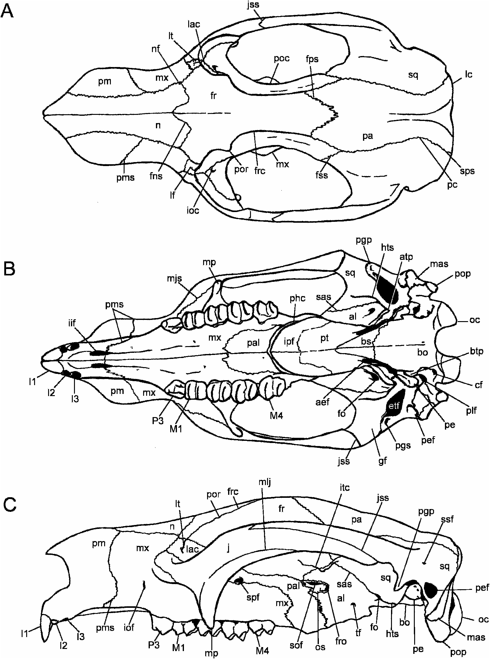
The following description of the adult cranium of Nimbadon lavarackorum is based on QM F31541 (fig. 2), unless otherwise stated. Previously unknown elements of the dentition are also described. Figure 1 View Fig is a stylized drawing of a N. lavarackorum cranium (based on QM F31541, QM F31377 and QM F53642 View Materials ) highlighting the major features of the skull.
GENERAL: QM F31541 (fig. 2) is a partially preserved cranium missing the right zygomatic arch, the central portion of the left zygomatic arch, the left pterygoid, the anterior of the parietals and the middorsal section of the frontals. Both mastoid-paroccipital processes are broken and the right petrosal is only partially preserved. Although not as complete as some other Nimbadon lavarackorum skulls available for description, QM F31541 is used as a basis for cranial description due to the excellent state of preservation of its features and the clearly discernable sutural relationships of the skull. The lack of preservation of the middorsal roof of the cranium allows investigation of the braincase and sinus development. In addition, closer analysis of the side wall of the braincase and orbital region is made possible by the absence of the right zygomatic arch.
QM F31541 is a relatively short broad skull, its maximum width occurring across the lateral glenoid eminences of the jugal. The highest point of the cranium is indeterminate because most of the dorsal surfaces of the frontals are not preserved; however, in QM F50470 View Materials (fig. 3) and QM F53642 View Materials the highest point of the skull occurs just anterior to the frontoparietal suture. In lateral profile, the skull is domelike and convex at this point. The basicranium of QM F31541 is elevated relative to the cheek tooth row but this feature was found to vary between skulls.
ROSTRUM: The rostrum is deep, broad, and short, comprising 33% of total skull length. It is constricted above the infraorbital foramen but flares laterally at the premaxilla resulting in an anteriorly bulbous snout. The nasals are inflated dorsal to the anterior orbital margin resulting in a convex rostrum in lateral profile, although the degree of convexity varies between skulls. The highest point of the rostrum is level with interproximal P3–M1.
The nasals are elongate and narrow, flaring both anteriorly and posteriorly resulting in an hourglass shape. They are bounded laterally by the premaxilla and maxilla and posteriorly by the frontals, and are generally confined to the dorsal surface of the rostrum with a slight ventral extension posterolaterally along the nasomaxillary suture. The nasals terminate posteriorly in line with the anterior margin of the orbit. Anteriorly, they terminate in a sharp point above I1. The frontonasal suture is Wshaped (when viewed posterodorsally) with the frontals forming a wedge between the nasals. In QM F50470 View Materials , however, the frontonasal suture is semicircular and broadly concave. The ventral surfaces of the nasals are visible within the narial aperture and are dominated by a ridge of bone that runs parallel along either side of the nasal septum. On their dorsal surface the nasals are perforated by two small (, 1 mm in diameter) foramina. The first lies 5 mm anterior to the frontonasal suture and 2 mm medial to the nasomaxillary suture. This foramen is confined wholly within the nasals in QM F31541, but its position varies in other skulls. The second foramen lies 7.7 mm anterior to the first, on or just medial to the nasomaxillary suture. The narial aperture is much wider (30.7 mm) than it is high (23.3 mm) due to the flaring premaxillary walls. The anterior palatal fenestrae (interincisive foramina) open into the floor of the narial aperture from the premaxillary palate. The floor of the narial aperture is deep and troughlike and is bordered laterally by thick premaxillary walls.
On the lateral face of the rostrum, the premaxilla-maxilla suture is nearly vertical except for a concave expansion posteriorly toward the infraorbital foramen. On the dorsal surface of the rostrum, the suture extends posteriorly to meet the nasals at a point in line with the infraorbital foramen. Ventrally, the premaxilla-maxilla suture extends onto the occlusal surface of the palate 25.6 mm anterior to the root of P3. It then extends 9.6 mm posteriorly, following the lateral border of the palate to a small nutrient foramen. In QM F50470 View Materials a small canine was present at this point (but was broken during preparation). The premaxilla-maxilla suture then extends 16.8 mm anteromedially to a point approximately midway along the length of the lateral margin of the interincisive foramen.
The maxillae comprise just over 50% of the rostrum in lateral view. A narrow infraorbital foramen (5.9 mm high; 2.7 mm wide) lies 15.6 mm posterior to the premaxilla-maxilla suture, vertically in line with the anterior border of P3. The maxilla is best described in terms of its sutural relationships with adjacent bones. The maxillofrontal suture is short (right 5.6 mm; left 7.2 mm) and extends posteroventrally from the junction of the nasal, frontal, and maxilla. Contact between the maxilla and frontal is limited in all referred skulls and is at its maximum length in QM F53642 View Materials at 9.9 mm. The maxillolacrimal suture originates at the point of most lateral extension of the maxillofrontal suture and extends anteroventrally for 14.0 mm, terminating at the junction of the maxilla, lacrimal, and jugal bones at the anterior margin of the orbit. Within the orbit, the maxillolacrimal suture extends 17 mm posteriorly from the junction of the maxilla, lacrimal, and jugal bones.
The maxillopalatine suture originates from the midline of the palate at a point opposite the posterior root of M2. It curves posterolaterally, then posteriorly, running parallel with the tooth row, into the posterior palatine foramen. From here it ascends sharply for approximately 22 mm then continues anteriorly along the suborbital shelf for approximately 30 mm, curving dorsally once more and continues (for 8.5 mm) to meet the ventral border of the lacrimal. Within the orbit the maxillojugal suture curves laterally, following the curve of the anterior root of the zygomatic process then descends along the masseteric process to within 6 mm of its tip. It then curves around the lateral face of the masseteric process and continues anterodorsally, following the line of the maxillolabial fossa, eventually terminating at the anteromedial orbital margin at the maxilla-jugal-lacrimal suture.
PALATE: The maxillary palate is relatively shallow and open in QM F31541 but varies in depth among the referred skulls. It is perforated by numerous foramina, the largest of which lie 5.5 mm and 7.9 mm medial to the left and right M1, respectively, and 4.6 mm medial and 6.2 mm anteromedial to the left and right P3. The frequency and position of the many small foramina are highly variable between the referred skulls. There is also significant variation within the left and right sides of an individual with some foramina present on one side and not the other, or asymmetrically distributed. The foramina most consistently present are those medial to the M1 and anteromedial to P3. The tooth rows are relatively linear in QM F31541 running parallel to one another.
The medial palatal sulcus originates opposite the anterior root of M2 and becomes wider and deeper as it grades into the interincisival fossa anteriorly. The premaxillary palate is relatively narrow and 45.5 mm in length. The interincisival fossa is bordered anteriorly by the narrow U-shaped incisor arcade (19.6 mm long, 20.8 mm wide). The interincisive foramina are long and narrow (left 18.3 X 2.2 mm; right 19.7 X 2.3 mm) but their dimensions vary considerably within the skulls available for study. The diastema is 62.0 mm long and 37.6 mm wide across the anterior bases of the premolars and 25.6 mm wide across the most posterior extension of the premaxilla-maxilla suture. In lateral profile, the diastema is slightly arched.
The linear midline palatine suture is 20 mm long and dominated by a moderately developed ridge. This ridge is variably expressed in the referred skulls. The posterior border of the palate is defined by a moderately developed, arcuate, pharyngeal crest that forms the anterior border of the interpterygoid fossa. In QM F31541, the left palatine is perforated by three small foramina, and the right by a single foramen.
The posterior palatine foramen (or posterolateral palatine foramen sensu Archer, 1976) is found at the posterior end of a deep canal formed between the pharyngeal crest (medially) and the most posterior extension of the maxilla on the palate (laterally). It is much larger, deeper, and more trenchant in QM F50470 View Materials . It opens on the lateral surface of the interpterygoid fossa on the maxillopalatine suture.
The large interpterygoid fossa (35 mm wide, 25 mm deep) is bordered anteriorly and anterolaterally by the palatine and posterolaterally by the pterygoid and alisphenoid. The relationship of the pterygoid is difficult to discern in all the adult crania. In the subadult skull QM F42677 View Materials , however, the pterygoids are thin bony laminae that overlie the presphenoid and basisphenoid (on the roof of the interpterygoid fossa) and the palatine and alisphenoid (on the medial walls of the interpterygoid fossa). Elongate, tapering, winglike projections of the pterygoids extend posteriorly toward the tympanic processes of the basioccipital where they terminate. In more complete skulls (e.g., QM F53642 View Materials ) the pterygoids form a bony floor to the entocarotid canal.
ORBIT AND ZYGOMATIC ARCH: The lacrimal is roughly triangular in shape and forms a wedge between the frontals and maxillae posteriorly. The lacrimal is generally confined within the orbit, extending only 3 mm onto the rostrum. A moderately developed lacrimal tuberosity is situated at the anterodorsal border of the lacrimal and is perforated by small foramina on the left side of the skull. A very small (, 1 mm diameter) lacrimal foramen is situated at the anteroventral base of the tuberosity. Within the orbit there is a broad (8 mm) connection between the lacrimal and palatine bones due to the deep extent of the lacrimal into the orbit.
Only a narrow wedge of the orbitosphenoid is visible in lateral profile. The fronto-orbitosphenoid suture extends 13 mm anteroventrally to meet the palatine at a point 10 mm directly anterior to the sphenorbital fissure. The orbitosphenoid-palatine suture extends posteriorly from this point to the sphenorbital fissure. A break in the bones surrounding the sphenorbital fissure reveals the lateral aspect of the presphenoid, which appears to form a bridge across the sphenorbital fissure.
The anteriorly oriented orbit has a semiovate opening in anterior view. The presence of a distinct postorbital process of the frontal cannot be determined for QM F31541 and is a variable feature in the referred skulls. It is absent in QM F53645 View Materials and QM F30833, but it is well developed in QM F50470 View Materials (fig. 3), QM F53642 View Materials , QM F40345 View Materials and QM F53643 View Materials a. The postorbital process is most prominent and winglike in QM F50470 View Materials , lying at the posterior end of a 12.5 mm crest at the anterodorsal border of the orbit. The postorbital process of the jugal is a weak, roughened area for muscle attachment on the dorsal surface of the jugal at a point in line with the masseteric process. In QM F50470 View Materials , the distance between the postorbital processes of the jugal and frontal is 27.8 mm.
The suborbital shelf is narrow and slopes steeply ventrally. A large (4.5 mm diameter), round infraorbital canal sits 4 mm ventral to the maxillolacrimal suture, 12 mm posterior to the anterior orbital margin. A smaller anteroposteriorly ovate sphenopalatine foramen lies approximately 16 mm posterior to the infraorbital canal and extends ventrally into the palatine. A number of smaller foramina perforate the suborbital shelf on either side of the skull, supplying vessels to the alveolar region.
Description of the zygomatic arch is based on QM F50470 View Materials (fig. 3) because this region is not preserved in QM F31541. The zygomatic arch is composed almost equally of jugal and squamosal. It is deepest through the masseteric processes. The elongate zygomatic process of the jugal tapers posteroventrally and terminates at the anterolateral border of the glenoid fossa. In ventral view, the jugal is ridgelike along its lateral border. The squamosal process of the zygomatic arch tapers anterodorsally and terminates 45 mm posteri- or to the anterior orbital margin. The lateral jugosquamosal suture extends anteroventrally for 10 mm, then curves in the opposite direction extending posteroventrally for 73 mm. Ventrally, the jugosquamosal suture is short (15 mm) and runs diagonally (posterolaterally) across the posterior base of the zygomatic arch. The masseteric line of the jugal is a deep facet for the attachment of the masseter lateralis profundus muscle. It originates on the masseteric process and curves posterodorsally then posteroventrally, following the curve of the dorsal border of the zygomatic arch before attenuating along the inferior margin of the jugal. The masseteric process is well developed, elongate, slender, and rounded at its tip. In lateral view, the tip of the process curves posteriorly and terminates approximately 5 mm ventral to the occlusal surface of the molar row.
The dorsal surface of the frontals and the frontoparietal suture are not preserved in QM F31541 revealing the internal structure of the frontals (fig. 2). The frontals are dominated internally by two bilaterally symmetrical sinuses separated by a thin, transverse septum. They are confluent posteriorly with a network of sinuses within the alisphenoid, squamosal, and parietal bones. These sinuses separate the inner table of the braincase from the outer table of the skull both dorsally and laterally. The inner table of the braincase is a thinwalled bony capsule scarred by numerous nutrient channels and measures 50 mm (mediolaterally) at its widest point (fig. 2). Many of the septa separating the endocranial sinuses are missing from QM F31541, but analysis of partial crania (e.g., QM F53788 View Materials ) indicate the following sinuses are present: separate anteri- or and posterior parietal sinuses; an anterolateral parietal sinus that is confluent with a lateral squamosal/alisphenoid sinus; and lateral squamosal sinus that is confluent with a series of epitympanic sinuses surrounding the middle ear and invading the squamosal root of the zygomatic arch.
Description of the external relationships of the frontals is based on QM F53642 View Materials . Dorsally, the frontals are broadly triangular and bound- ed by the nasals anteriorly and the parietals posteriorly. A shallow frontal depression occupies the anterior two-thirds of the frontals and is emarginated laterally by moderately developed frontal crests. They are 39 mm apart at their point of origin at the postorbital processes of the frontal, converging to within 17 mm of each other at a point in line with the anterior root of M4. From here they diverge, fading toward the highest point on the cranium at the anteriormost point of the frontoparietal suture. In QM F50470 View Materials (fig. 3), the frontal crests extend beyond the frontparietal suture, converging 35 mm behind this point, at the dorsal midline of the cranium where they become continuous with a welldeveloped sagittal crest.
The frontals measure 83 mm at their midline and are partially fused with only 34 mm of the midline suture evident anteriorly. The convoluted frontoparietal suture, however, remains highly visible. The frontals form a wedge between the parietals dorsally and the parietal and squamosal laterally. In QM F50470 View Materials , however, the dorsal frontoparietal suture is relatively linear mediolaterally (such that the frontals do not form a wedge between the parietals), although a lateral frontal wedge is still evident.
In QM F31541, from the lateral junction of the frontal, parietal, and squamosal bones, the frontosquamosal suture descends anterolaterally along the wall of the orbit to its juncture with the alisphenoid. A rugose tuberosity, the infratemporal crest, lies at this point. The fronto-alisphenoid suture continues ventrally and slightly posteriorly from this point toward the sphenorbital fissure. Its junction with the orbitosphenoid occurs, 5 mm dorsal to the sphenorbital fissure.
The squamoso-alisphenoid suture extends posteroventrally from its contact with the frontal across the rugose infratemporal crests to the hypotympanic swelling, which is situat- ed medial to the glenoid fossa. The squamosoalisphenoid suture extends posteriorly across the lateral third of the bulla to its posterior margin then curves dorsomedially around the alisphenoid tympanic process, disappearing from view at the anterolateral tip (superior periotic process) of the petrosal. A full description of the basicranium is given below.
The squamosal comprises approximately two-thirds of the side wall of the braincase, which descends relatively steeply from the midline parietal suture. In QM F53642 View Materials , the parietals are constricted to a minimum width of 12 mm, at a point 16 mm anterior to the lambdoid crest, expanding to their maximum width (52 mm) at the lambdoid crest. A subsquamosal foramen is consistently present at the posterior base of the squamosal root of the zygomatic arch; however, in QM F31541 there is a secondary foramen 2 mm posteromedial to the primary subsquamosal foramen (but only on the right side of the skull).
CRANIAL BASE: The cranial base is elevated with respect to the alveolar row. Its maximum width (and indeed the maximum width of the skull) occurs through the glenoid process of the jugal at the posterior root of the zygomatic arch. The minimum width of the cranial base occurs across the postzygomatic constriction of the squamosal. The cranial base of QM F53642 View Materials is flat along the plane of the basioccipital and basisphenoid except for the shallow sulci lateral to the median crest of the basioccipital. In QM F31541, the basioccipital is inflated anteriorly where it contacts the basisphenoid (33 mm anterior to the foramen magnum). In QM F53642 View Materials , a distinct median basioccipital crest extends anteriorly from the foramen magnum for 13 mm before fading out. The median basioccipital crest is unclear in QM F31541 and the basioccipital sulci are much shallower.
A short basioccipital tympanic process extends 3 mm ventral to the periotic. The maximum width of the basioccipital (37 mm) occurs at this point. Two condylar foramina occur posterior to the basioccipital tympanic process. The small (1 mm) anterior and larger (2.5 mm) posterior condylar foramina are confluent with the foramen magnum internally. The basisphenoid is 30 mm long and narrows anteriorly to 8 mm wide at its junction with the presphenoid. In more complete specimens, the presphenoid-basisphenoid suture is covered by the bony lamina of the pterygoid.
The pterygoids are broken posteriorly in QM F31541, thus exposing the groove for the entocarotid artery. This narrow (4 mm wide, 17 mm long) groove lies lateral to the basioccipital and basisphenoid bones and medial to the pterygoid crests. At its anterior margin lies the anterior entocarotid foramen, which enters the basisphenoid 8 mm anterior to the basisphenoid-basioccipital suture in QM F31541 (11 mm anterior in QM F53642 View Materials ). In QM F53642 View Materials , the pterygoids are partially preserved and overlay the groove for the entocarotid artery lateral to the basioccipital. The groove remains open ventrally along its basisphenoid portion. In QM F42677 View Materials (in which the pterygoids are complete) the entocarotid groove and the anterior entocarotid foramen are not visible and only a small triangular wedge of the basisphenoid is exposed ventrally.
The glenoid fossa is broad and flat and composed entirely of squamosal. It is open laterally and terminates medially at the base of the hypotympanic swelling. The postglenoid sulcus (5 mandibular fossa sensu Aplin, 1987) is an elongate, straight, mediolateral groove that runs the length of the glenoid fossa. It is bordered anteriorly by a relatively flat articular eminence. The postglenoid process is a slightly inflated, roughened nodule that lacks a postglenoid foramen; however, this feature varied markedly between skulls (see below).
AUDITORY REGION: The middle ear is open ventrally. The ectotympanic was unfused and is not preserved in any of the skulls under study. The hypotympanic swelling (‘‘bulla’’) is comprised mostly of the alisphenoid (fig. 4) with the squamosal contribution varying from 25%–33% in referred skulls. The degree of inflation of the hypotympanic swelling also varies from relatively flat (e.g., QM F40345 View Materials ) to more sinus inflated and ‘‘bulla-like’’ (e.g., QM F31541 and QM F50470 View Materials ). A rugose posterior extension or ‘‘wing’’ of the alisphenoid, the alisphenoid tympanic process, projects 5 mm from the floor of the hypotympanic swelling, in the direction of the paroccipital process. In QM F53642 View Materials (fig. 4), the alisphenoid tympanic process measures 5.2 mm at its maximum width and is composed of a central groove bordered by thin irregular ridges. The squamoso-alisphenoid suture follows the lateral border of the alisphenoid tympanic process, then curves dorsomedially around the posterior base of the hypotympanic swelling to meet the basisphenoid at the opening of the anterior entocarotid foramen. In QM F53788 View Materials , howev- er, the squamoso-alisphenoid suture extends more sharply posteromedially across the hypotympanic swelling resulting in a squamosal tympanic process (rather than an alisphenoid tympanic process). In QM F31541, the floor of the hypotympanic swelling is broken revealing the bilaminate internal structure wherein the alisphenoid overlays the squamosal medioventrally. The roof of the hypotympanic sinus is produced solely by the squamosal.
The size of the epitympanic fenestra varies between skulls. It is generally a moderate to large obliquely oriented fenestra. In QM F53642 View Materials (fig. 4), it measures 12 mm anteroposteriorly, 15 mm posteromedially and 12 mm mediolaterally. QM F31541 exhibits a proportionately larger, squarer epitympanic fenestra measuring 16 mm mediolaterally and anteroposteriorly. In all skulls, the epitympanic fenestra opens into a large epitympanic sinus, which is divided into smaller sinuses by a series of bony struts. A large posterior epitympanic fossa is positioned posterolaterally to the epitympanic fenestra and recessed within the squamosal in the roof of the paroccipital/mastoid process. It is clearly seen in lateral view as an oblique ovoid fossa behind the zygomatic arch. In QM F53642 View Materials it is 11 mm high and 5 mm wide. It is much smaller in QM F31541 (8 mm X 3 mm). It is not confluent with the large network of epitympanic sinuses.
The foramen ovale is located fully within the alisphenoid, but its structure is subject to considerable variation (fig. 17). In QM F53642 View Materials (fig. 4), the foramen ovale is 3 mm in diameter and opens into two shallow anteromedially and anterolaterally directed channels. In QM F30833, these grooves are covered by a thin lamina of the alisphenoid resulting in two secondary foramina, anteromedial and anterolateral, to the primary foramen ovale.
The median lacerate foramen is most evident in QM F42677 View Materials and is positioned at the anteromedial tip of the petrosal, dorsal to the posterior opening of the entocarotid foramen. It is a short (3 mm), slitlike opening directed anterodorsally into the braincase. A larger, rounded posterior lacerate foramen lies posterior to the petrosal at the anteromedial base of the paroccipital/mastoid process.
The deep stylomastoid groove (for facial nerve VII) originates at the posterolateral corner of the petrosal, just lateral to the fenestra cochlearis, attenuating as it descends the anterior surface of the mastoid process. It is almost canallike in QM F53642 View Materials (fig. 4) wherein an anterior mastoid tympanic process overhangs the groove anteriorly. The lateral border of the stylomastoid groove is defined by the crista tympanica, which is a thin bony crest originating from the squamosal at the posteromedial corner of the epitympanic fenestra.
The tympanic recess that holds the periotic is roughly triangular in outline. It is bounded anteriorly by the hypotympanic swelling, posteriorly by the mastoid/paroccipital process and exoccipital, laterally by the epitympanic fenestra and medially by the basioccipital. The long axis of the petrosal is aligned anteromedially in ventral view and is partially floored by the basioccipital tympanic process. In QM F53642 View Materials the pars petrosus measures 13 mm long by 9 mm wide.
PETROSAL: Description of the petrosal (fig. 5) is facilitated by isolated specimens QM F53844 View Materials –QM F53848 View Materials . The pars cochlearis is comprised of a smooth and flattened promontorium on its anterolateral face, and a roughened anteromedial flange. The promontorium can be either broad and rounded (e.g., QM F53642 View Materials ) or more flattened and pyramidial (e.g., QM F53844 View Materials ). In QM F53846 View Materials the promontorium is rugose rather than smooth.
The promontorium tapers anteromedially into an irregular, blunt anteromedial flange (or superior periotic process sensu Stirton, 1967). The anteromedial flange terminates anteriorly at the median lacerate foramen. The rostral tympanic process of the petrosal is a weak, blunt bulge at the posteroventral border of the promontorium that abuts the basioccipital tympanic process in some specimens (e.g., QM F53788 View Materials ).
A small 2 mm X 1.5 mm fenestra cochlearis (fenestra rotundum sensu Stirton, 1967) opens onto the posterolateral face of the petrosal. Anterodorsal to it lies the smaller (1.3 mm diameter) fenestra vestibularis (fenestra ovalis sensu Stirton, 1967). The fenestra cochlearis and fenestra vestibularis are separated by a 1.8 mm rounded crest (cristainterfenestralis). Dorsolateral to the fenestra vestibularis is a short (5.1 mm long), anteroposteriorly orient- ed groove that houses a slitlike secondary facial foramen that is confluent with the internal auditory meatus on the cerebellar face of the periotic. The secondary facial foramen carried the chorda tympani branch of the facial nerve. In QM F53844 View Materials it lies 2.9 mm anterior to the fenestra vestibularis. In QM F53845 View Materials the groove is covered by a thin bony lamina resulting in the development of a narrow 4.7 mm long canal.
Posterolateral and slightly dorsal to the fenestra cochlearis lies a deep pocket that is the stapedial fossa. In correct anatomical position, the facial nerve would travel posteriorly, lateral to the fenestra vestibularis, ventral to the stapedial fossa and onto the deep perpendicular stylomastoid groove in the anterior face of the mastoid.
The cerebellar (internal) side of the periotic is dominated by two large fossae: a dorsolateral subarcuate fossa and a deeper, ventromedial internal auditory meatus. The depth of the subarcuate fossa varies considerably between specimens and in some specimens (e.g., QM F53845 View Materials , QM F53848 View Materials ) it is perforated by a foramen (fig. 21). The oval-shaped internal auditory meatus of QM F53844 View Materials has a maximum diameter of 3.9 mm. Two channels are evident within the internal auditory meatus: the more anterodorsal (foramen acousticum superius) carries the facial nerve and is confluent with the facial foramen on the tympanic face of the periotic, and the posteroventral channel (foramen acousticum inferious) that carries the acoustic (vestibulocochlear) nerve into the substance of the periotic.
Posteroventral to the subarcuate fossa in the rugose area of the sigmoid sinus lies the small, slitlike aqueductus vestibulae. A similarly small and slitlike aqueductus cochleae lies on the rugose basioccipital articular surface (posteromedial face) of the periotic, 6 mm dorsal to the rostral tympanic process.
OCCIPITAL REGION: The shape of the occiput in posterior view is highly variable in Nimbadon skulls (figs. 6, 19). In QM F31377, the occipital region is semicircular and relatively flat dorsoventrally except for the deep fossae on either side of the midline of the supraoccipital (for the attachment of the rectus capitus major muscle). The median crest of the supraoccipital is relatively indistinct, running dorsoventrally from the lambdoid to the foramen magnum. The distinct, semicircular lambdoid crest overhangs the occipital region, and is more pronounced on the median dorsal surface of the skull. The occipital region is inflated along the squamosal-mastoid suture, which, although irregular, generally parallels the lambdoid crest ventrally. The mastoid is a thin (4 mm) wedge of bone between the squamosal and the exoccipital. It extends 24 mm dorsally to a point in line with the dorsal margin of the occipital condyle. Lateral to the fossa for the rectus capitus major and dorsal to the occipital condyles lie the smaller, shallower, subtriangular fossae for the rectus capitus minor muscle. In QM F31377 (fig. 6) the foramen magnum has a distinct supraoccipital notch on its dorsal border, but in QM F53642 View Materials it is a broad (24 mm wide X 20 mm high; compared with 21 mm X 16 mm in QM F31541, which is a larger skull overall), laterally ovate structure bordered by large, subovate occipital condyles. The shape and dimensions of the foramen magnum and occipital condyles vary considerably between referred skulls. The intercondylar notch is broadly U-shaped.
The mastoid-paroccipital processes are large, rugose, sinus-inflated structures. The mastoid contribution generally terminates in line with the ventralmost point of the occipital condyles. The paroccipital process continues for approximately 10 mm ventral to this point and its structure varies between narrow, mediolaterally compressed processes (e.g., QM F53642 View Materials ) to blunt, rounded processes (e.g., QM F31377). Anteriorly, there is a rugose mastoid tympanic process that extends to within 1 mm of the periotic at its dorsal extreme. Laterally, this process underlies the stylomastoid groove. In some more complete specimens, the mastoid tympanic process actually contacts the periotic (e.g., QM F53642 View Materials ).
In QM F31541 the supraoccipital (or possibly the interparietal) extends 8 mm onto the dorsal surface of the skull preventing contact between the parietals and the lambdoid crest. In dorsal view, the lambdoid crest has a distinct inverted U-shaped notch (9 mm deep) at its midline (fig. 19B) where it meets the sagittal crest. In most skulls, however, the lambdoid crest is generally arcuate.
MANDIBLE: Description of the mandible is based on QM F40337 View Materials (figs. 7–8), a mostly complete mandible with left and right i1, p3, m1–4. It is missing the coronoid processes, the right articular condyle, the floor of the right pterygoid fossa, and the posterior tip of the left angular process. Description is based on the left dentary. The dentaries of QM F40337 View Materials are fused at the mandibular symphysis limiting photographic access to their medial side. Instead, the unfused dentary QM F53792 View Materials is figured in medial view (fig. 9). The horizontal ramus is generally flat, bulging laterally below m2. The alveolar border of the dentary is straight, whereas the inferior border is convex. The greatest depth of the dentary occurs below the level of the protolophid of m3 and the horizontal ramus tapers anteriorly from this point. The diastemal crests are distinct and curve slightly laterally along their dorsal margin. The mandibular symphysis is well fused on its dorsal margin, the suture line only evident posterior to the posterior root of p3. However, the suture is clearly evident on the ventral surface, becoming wider (i.e., more open) posteriorly along the transverse torus. Two distinct genial pits are found on the posteroventral border of the symphysis. The bone is rugose at this point. The sublingual fossa is relatively shallow (although much deeper in QM F53824 View Materials ) and slopes posteroventrally. A small mental foramen (2.5 mm diameter) lies 4.0 mm ventral to the occlusal surface of the diastema and 7.5 mm anterior to the anterior root of p3. A smaller, secondary mental foramen (, 1.0 mm diameter) lies 4.0 mm posterior to the first. An additional posterior mental foramen lies 19.0 mm ventral to the interloph valley of m1, but it is not present on the right dentary. The anterior border of the ascending ramus rises at an angle of 75 ° to the horizontal plane and is compressed mediolaterally such that it is very crestlike. It arises opposite the protolophid of m4 where it flares laterally at this point. The masseteric fossa is relatively shallow and fades out onto the horizontal ramus anteriorly. There is no masseteric foramen. The masseteric eminence (submasseteric crest) flares laterally almost to the lateral width of the articular condyle. The condyle is situated 37.0 mm above the level of the alveolar border of the cheek tooth row. The articular surface of the condyle is deepest (anteroposteriorly) laterally, tapering medially. It is flattened and anterolaterally inclined laterally, becoming convex medially, and tipping ventrally at its most medial point. The mediolateral width of the condyle is 27.8 mm. The mandibular notch is broad (21.1 mm) with relatively linear ventral and anterior borders. A deep and rugose pterygoid fossa is housed within a well-developed angular process of the dentary. A moderate (4.1 mm diameter) mandibular foramen opens posteriorly in line with the postalveolar shelf. A thin, shallow digastric fossa runs anteriorly from below the level of the postalveolar process, fading out below m3. A digastric process is not evident. The postalveolar shelf is short (10.2 mm) and terminates in a small but distinct postalveolar process. The cheektooth rows are relatively linear with a slight concavity opposite the protolophid of m2. The occlusal surfaces of m3–4 on each tooth row tilt lingually toward each other.
DENTITION: Description of the P3, M1–4 of Nimbadon lavarackorum can be found in Hand et al. (1993). What follows is a description of previously unknown teeth dP3, I1–3, i1, and dp3 (fig. 10).
I1: Large, curved, open-rooted tooth flattened mesially and concave laterally (fig. 10D–F). Enamel is restricted to the anterolateral surface. Tooth wear initially occurs only on the ventral tips, yet older individuals exhibit wear along the entire exposed posterior surface. In situ, the left and right incisors converge at their tips. In QM F31541 there is a 2.3 mm gap separating the incisors where they emerge from the premaxilla. The length of I1 from the alveolus to its tip is 15.5 mm. Of this, 10.3 mm is covered in enamel. An isolated I1 showing a similar degree of wear and enamel coverage to that in QM F31541 is 47.7 mm long (measured in a straight line from the distal tip to the proximal end of the root). Because the incisor is open rooted and continues to grow throughout the animals life, incisor length varies greatly between specimens.
I2: Small, subovate crowned tooth (4.9 mm long, 3.5 mm wide) that abuts I1 anteromedially. All surfaces are enamel covered, with tooth wear resulting in only an enamel outline around the occlusal surface. A shallow pocket is formed by wear in the dentine of the occlusal surface. I2 and I3 are rarely preserved in situ in both adult and juvenile skulls.
I3: Small, subtriangular tooth in occlusal outline. The apex of the triangle occurs at its anteromesial tip. Only one skull, QM F30833, preserves I3. The worn occlusal surface of I3 has an enamel perimeter and lies along the same plane as I2, which is 15.1 mm above the ventral tip of I1.
DECIDUOUS P3: Description is based on QM F53648 View Materials a (fig. 10A–C). The dP3 is a small, three-cusped (paracone, metacone, protocone), triangular tooth. It is aligned in an anterolingual direction with respect to M1. A weak longitudinal crest runs posterobuccally from a subcentral paracone (tallest cusp), through the metacone to the posterobuccal cingulum. A small posterolingually positioned protocone is the smallest cusp. There is a weak swelling in the anterior parastylar region of the tooth. A weak posterior cingulum runs lingually from its junction with the main longitudinal crest, to the posterior base of the protocone.
I1: Slender, elongate, lanceolate tooth flattened mesially and concave laterally (fig. 10G–I). Enamel is restricted to the ventral and lateral surfaces. In occlusal view, i1 is generally straight along its length with a medial curvature at its tip. In lateral view, the lower incisor projects at a slight angle to the generally horizontal plane of the dorsal diastemal surface as it follows the ventral curvature of the horizontal ramus anteriorly. In unworn specimens, the lateral occlusal enamel surface is ridgelike. Medial to the enamel ridge is a shallow sulcus in the dentine that runs the length of i1.
DECIDUOUS P3: Description is based on QM F30784, a juvenile left dentary with i1, dp3, m1–3 (m3 encrypted). The dp3 (fig. 10J– L) is a small (4.8 mm long X 3.5 mm wide), two-rooted tooth, similar in most respects to p3 but on a smaller scale. The tooth is wider posteriorly and tapers anteriorly. In position in the dentary, dp3 is directed anteromedially, whereas m1 is positioned anterolaterally. The occlusal surface of dp3 is dominated by a central protoconid that shows a large degree of wear relative to its size. A small cuspate swelling lies at the posterior border of the tooth. A short crest originates from the protoconid apex and fades down its steep anterolingual face. The posterolateral corner of dp3 abuts the anterior terminus of the paracristid of m1.
| QM |
Queensland Museum |
| RM |
McGill University, Redpath Museum |
| LP |
Laboratory of Palaeontology |
| R |
Departamento de Geologia, Universidad de Chile |
No known copyright restrictions apply. See Agosti, D., Egloff, W., 2009. Taxonomic information exchange and copyright: the Plazi approach. BMC Research Notes 2009, 2:53 for further explanation.
|
Kingdom |
|
|
Phylum |
|
|
Class |
|
|
Order |
|
|
Family |
|
|
Genus |