
Hermanellopsis arsia Savage & Peters, 1983
|
publication ID |
https://doi.org/ 10.11646/zootaxa.4768.4.10 |
|
DOI |
https://doi.org/10.5281/zenodo.3794565 |
|
persistent identifier |
https://treatment.plazi.org/id/03EA87DD-FF82-7516-2EF4-FA3ECBE4F9E6 |
|
treatment provided by |
Plazi |
|
scientific name |
Hermanellopsis arsia Savage & Peters, 1983 |
| status |
|
Hermanellopsis arsia Savage & Peters, 1983 View in CoL
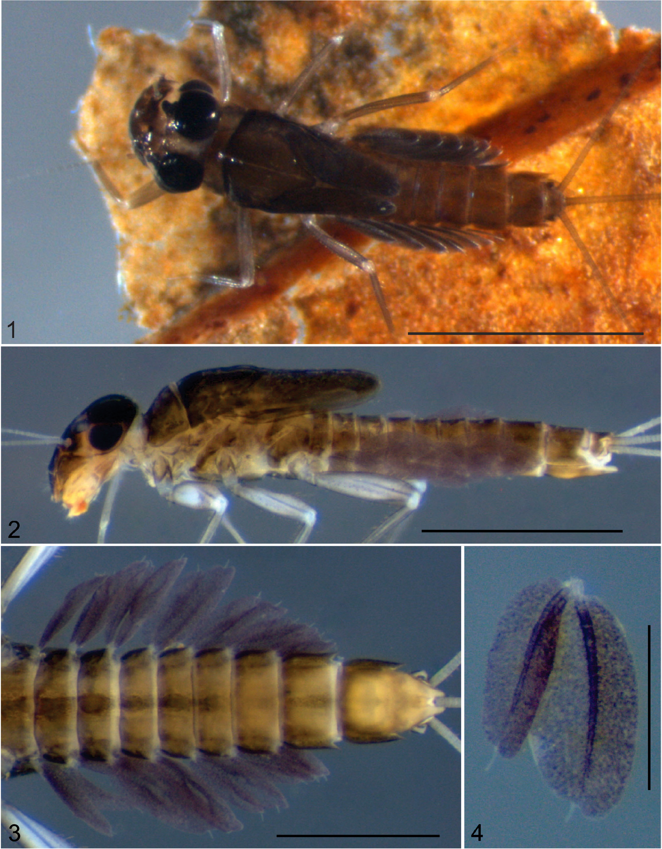
( Figs 1–14 View FIGURES 1–4 View FIGURES 5–15 )
Diagnosis. Nymph ( Figs 1–2 View FIGURES 1–4 ): 1) width of labrum equal to width of clypeus, lateral margins rounded ( Fig. 5 View FIGURES 5–15 ); 2) outer margin of mandibles smoothly curved with few setae medially ( Figs 6–7 View FIGURES 5–15 ); 3) glossae of labium ventrally curved; 4) gills on abdominal segments I–VII oval, each with very short median apical projection and two shorter lateral lobes ( Figs 3– 4 View FIGURES 1–4 ); dorsal portion of gills smaller than ventral portion; 5) posterolateral spines present on abdominal segments V–IX, more developed on VII–IX ( Fig. 3 View FIGURES 1–4 ); 6) denticles on tarsal claws progressively larger apically, apical denticle largest ( Fig. 11 View FIGURES 5–15 ).
Description. Mature nymph.
Lengths. Body: 4.54–4.82 mm. Foreleg: 2.30–2.32 mm; middle leg: 2.37–2.40 mm; hind leg: 2.75–2.76 mm (coxa and trochanter not included). Medial caudal filament: 7.10–7.49 mm. Cerci: 6.21–6.85 mm. Ratios. Mouthparts: mandibles with width 0.6 x length; labial palp with width of segment I 0.8 x its length; segment III length 0.4 x segment II length; segment III length 0.6 x segment I length; segment II length 1.5 x segment I length; maxillary palp with segment III length 0.4 x segment II length; segment II length 0.9 x segment I length. Abdomen: dorsal gill IV with length of medial filament 0.1 x length of body of gill.
Coloration and Morphology. Body. brown washed with orange ( Figs 1–2 View FIGURES 1–4 ). Head: brownish; pale between upper and lower portion of compound eye, around lateral ocelli and anterior to medial ocellus; antenna translucent yellow; compound eye reddish brown; ocelli blackish. Labrum yellowish with apical and subapical rows of bristles present, medially excavated and five small denticles ventrally; lateral margins rounded; ventral surface with two median rows of robust, median bristles and dorsal surface with several, long bristles scattered ( Fig. 5 View FIGURES 5–15 ). Mandibles light brown, with row of setae on outer margin extending to middle region; setae long on distal half and short on proximal half; molar area with serrated ridges; prosthecae well-developed; left mandible with three denticles on inner and outer incisors ( Fig. 6 View FIGURES 5–15 ); right mandible with two apical denticles on inner incisor and three on outer incisor ( Fig. 7 View FIGURES 5–15 ). Maxilla pale brown, palpi with few and scattered bristles on segment I, and segments II–III covered with bristles scattered on inner margin and outer margin ( Fig. 8 View FIGURES 5–15 ). Hypopharynx pale brown, with superlinguae bearing long setae on apical margin; lingua with medial deep cleft and anterolateral apex pointed, ventral portion covered by short setae ( Fig. 9 View FIGURES 5–15 ). Labium pale brown; glossae curved ventrally, densely covered by bristles; paraglossae densely covered by bristles on apical third; submentum light brown ( Fig. 10 View FIGURES 5–15 ).
Legs: greyish. Coxae and trochanters washed with brown. Femora uniformly washed with brown (except fore femur with medial area unpigmented) and with blackish line on anterior surface; inner margin with short bristles, outer margin with long bristles, varying in size, concentrate on apical half ( Figs 12–14 View FIGURES 5–15 ). Tibiae uniformly washed with brown with long and simple bristles on outer margins and short bristles on inner margins (hind tibiae with short spine-like setae on inner margin), with concentration of long spines at apex ( Figs 12–14 View FIGURES 5–15 ). Tarsal claws with 13 denticles progressively larger apically with apical denticle largest ( Fig. 11 View FIGURES 5–15 ).
Abdomen: light brown. Terga I–X with dark brown marks on lateral and medial regions ( Fig. 1 View FIGURES 1–4 ). Posterolateral spines present on abdominal segments V–IX, more developed on VII–IX ( Fig. 3 View FIGURES 1–4 ). Gills on abdominal segments I–VII, oval, with very short median projection on apex and two shorter lateral lobes; dorsal portion smaller than ventral one and main trachea unbranched; membrane purplish and trachea darker ( Fig. 4 View FIGURES 1–4 ).
Biology. The nymphs were collected inhabiting leaves in a stream 3 m wide, with moderate current, and under a closed canopy.
Life cycle association. Male imago reared from nymph.
Distribution. Brazil: states of Amazonas ( Savage & Peters 1983) and Roraima ( Raimundi et al. 2017). New record from Maranhão State: Caxias municipality.
Material examined. Three mature male nymphs and one male imago (reared, exuviae mounted on slides), Brazil, Maranhão State, Caxias municipality, Inhamum Municipal Environmental Protection Area (APA Inhamum), Riacho Areia Branca , 4°53’34.9”S / 43°26’11.1”W, 22.viii–04.x.2019, Nascimento, S. R.S. col. (Two nymphs in LEAq; one nymph and male imago reared in UFVB—EP00090 and EP00091) GoogleMaps .
Discussion. With the description of the nymph of Hermanellopsis arsia , it will be possible to improve understanding characteristics of the genus and its relationship with the four genera of the Miroculis- lineage proposed by Savage & Peters (1983): Miroculis Edmunds, 1963 ; Microphlebia Savage & Peters, 1983 ; Hermanellopsis Demoulin, 1955 ; and Miroculitus Savage & Peters, 1983 . The nymph described here has similarities with Microphlebia , such as a labrum with rounded lateral margins, glossae of the labium ventrally curved, and gills on abdominal segments I–VII oval, with dorsal portions smaller than ventral portions. However, the most important characteristic that distinguishes the nymph of H. arsia is the presence of gills each with a very short median apical projection and two shorter posterolateral projections on segments I–VII (absent in Microphlebia ). Besides this, the lingua of the hypopharynx has a very deep medial cleft, the cleft being more shallow in Microphlebia .

| R |
Departamento de Geologia, Universidad de Chile |
No known copyright restrictions apply. See Agosti, D., Egloff, W., 2009. Taxonomic information exchange and copyright: the Plazi approach. BMC Research Notes 2009, 2:53 for further explanation.
|
Kingdom |
|
|
Phylum |
|
|
Class |
|
|
Order |
|
|
Family |
|
|
Genus |