
Sphaerobelum spinatum, Wesener, 2019
|
publication ID |
https://doi.org/10.11646/zootaxa.4563.2.1 |
|
publication LSID |
lsid:zoobank.org:pub:CF79B01B-8B5F-4B3A-B642-2CADE4B339AF |
|
DOI |
https://doi.org/10.5281/zenodo.5934528 |
|
persistent identifier |
https://treatment.plazi.org/id/03EB5506-E31E-8C23-A2ED-FC43FAE2FA61 |
|
treatment provided by |
Plazi |
|
scientific name |
Sphaerobelum spinatum |
| status |
sp. nov. |
Sphaerobelum spinatum new species
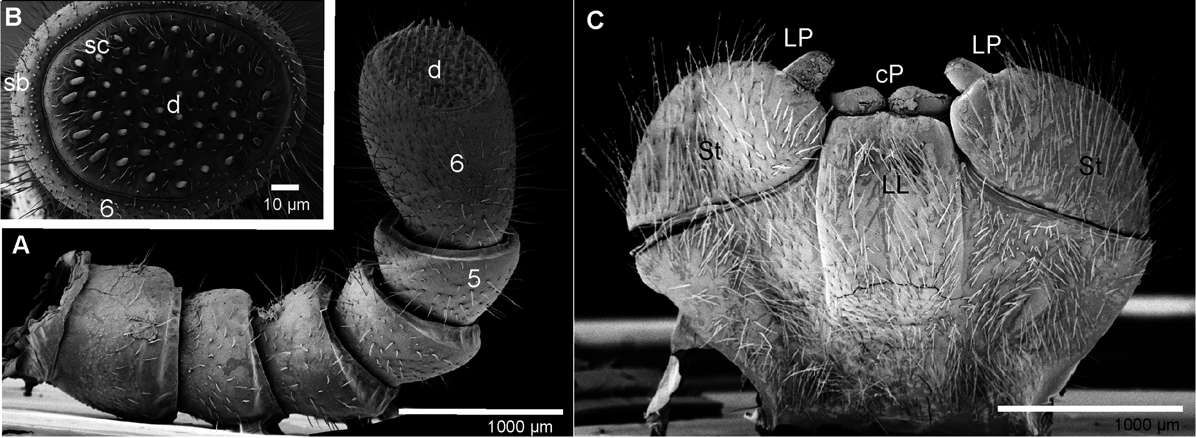
Figures 6D View FIGURE 6 , 9C View FIGURE 9 , 14 View FIGURE 14 , 15 View FIGURE 15 , 16 View FIGURE 16
Material examined: Type specimens. 1 M holotype (ZMUC00040258) from Laos, Vientiane Province, Phou Khao Khouay ( N18°20.369' N, 102°48.523'E), 700–800 m, strongly disturbed primary evergreen forest, leg. 26- 31.V.2008, A. Solodovnikov & J. Pedersen; 1 F paratype; ZMUC00040259; same data as paratype; 1 M, 1 F paratype ( ZFMK MYR8101 About ZFMK & MYR8102), same data as holotype View Materials .
Diagnosis: S. spinatum n. sp. belongs to a group of Sphaerobelum species in which the mesal margin of the femur is extended with several teeth ( Fig. 15B View FIGURE 15 ). S. spinatum n. sp. shares only with S. cattiense and S. konkakinhense a straight telopoditomere 4 ( Figs 16E, F View FIGURE 16 ). S. spinatum n. sp. differs in several characters from both other species: mesal margin of prefemur with indentations like the femur ( Fig. 15B View FIGURE 15 ), male antenna with <60 apical cones (> 70 in S. denticulatum n. sp.), tergites shiny, but setose (tergites covered with setose pits in the other species), first laterotergite strongly projecting, extended into a sharp tip (first laterotergite only weakly projecting in the other two species).
Description. Based on holotype and both paratypes.
Measurements: Body length: holotype male: length ca 26.9 mm. Width, of thoracic shield = 13.6 mm (= broadest). Height, of thoracic shield = 7.5 mm (= highest). Female: length ca 32.5 mm. Width, of thoracic shield = 14.5 mm, of tergite 7 = 15.7 mm (=broadest). Height, of thoracic shield = 7.7 mm, of tergite 9 = 9.1mm (= highest).
Coloration: in preserved specimens black, paratergite depressions and groove of thoracic shield dark green ( Fig. 6D View FIGURE 6 ). Antennae brown-orange, legs light brown, but prefemur and femur green.
Head: Eyes with>60 ocelli. Aberrant ocellus located inside antennal groove. Antennae short, with rounded joints, extending posteriorly to leg-pair 5. Lengths of antennomeres: 1>2=3=4=5<<6 ( Fig. 14A View FIGURE 14 ). All antennomeres densely pubescent, sensilla basiconica surrounding apical disc. Shape of antennae sexually dimorphic, cylindrical in female, thickened, apically widened and slightly flattened in male ( Fig. 14B View FIGURE 14 ). Apical disc with ca 52/52 apical cones (male) ( Fig. 14b View FIGURE 14 ), or 30–36 (female). Organ of Tömösváry located inside antennal groove. Gnathochilarium: Damaged. Structure typical of the order ( Fig. 14C View FIGURE 14 ). Palpi with sensory cones arranged in single field. Rudimentary lateral palps not visible.
Mandibles: not dissected.
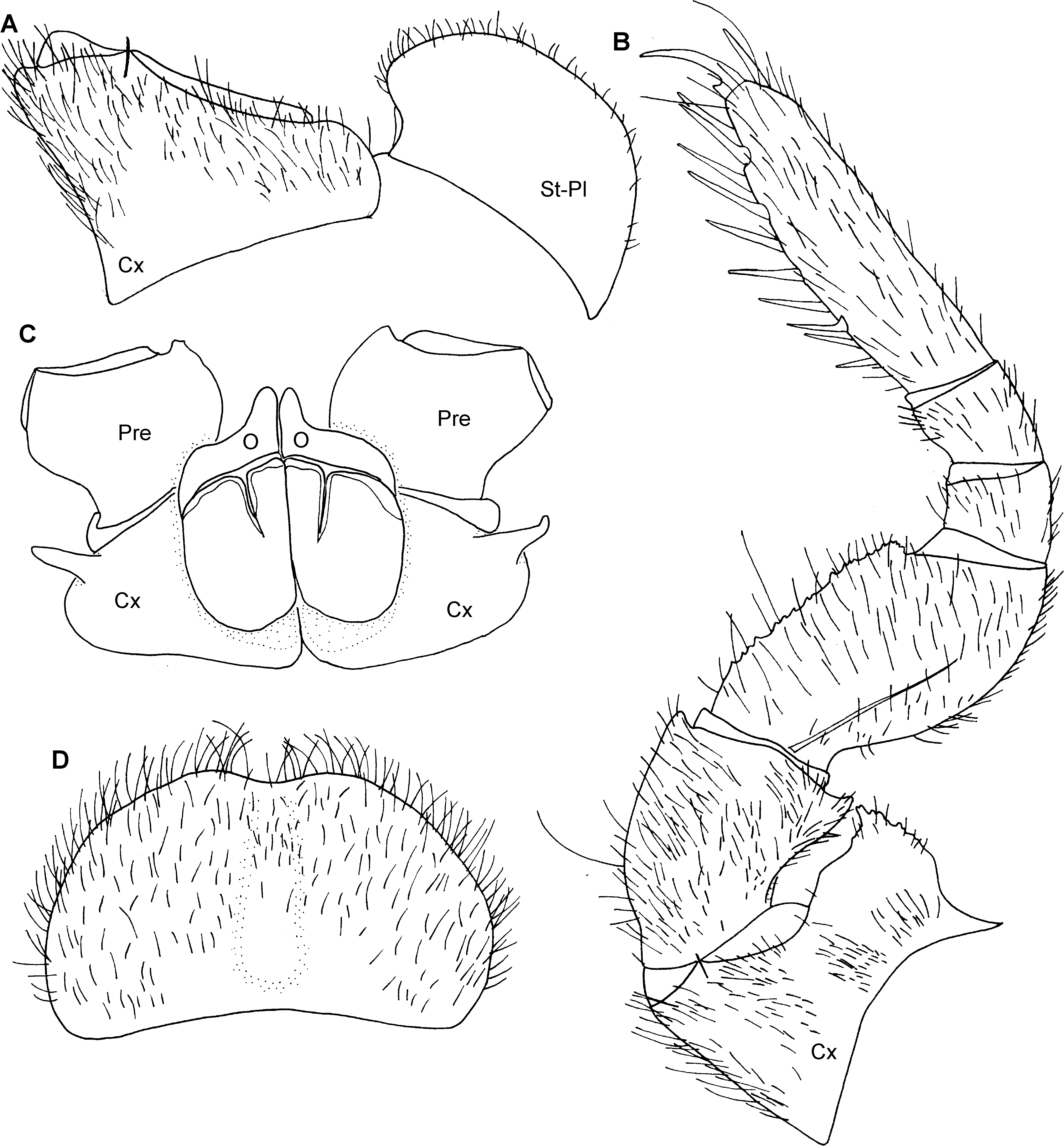
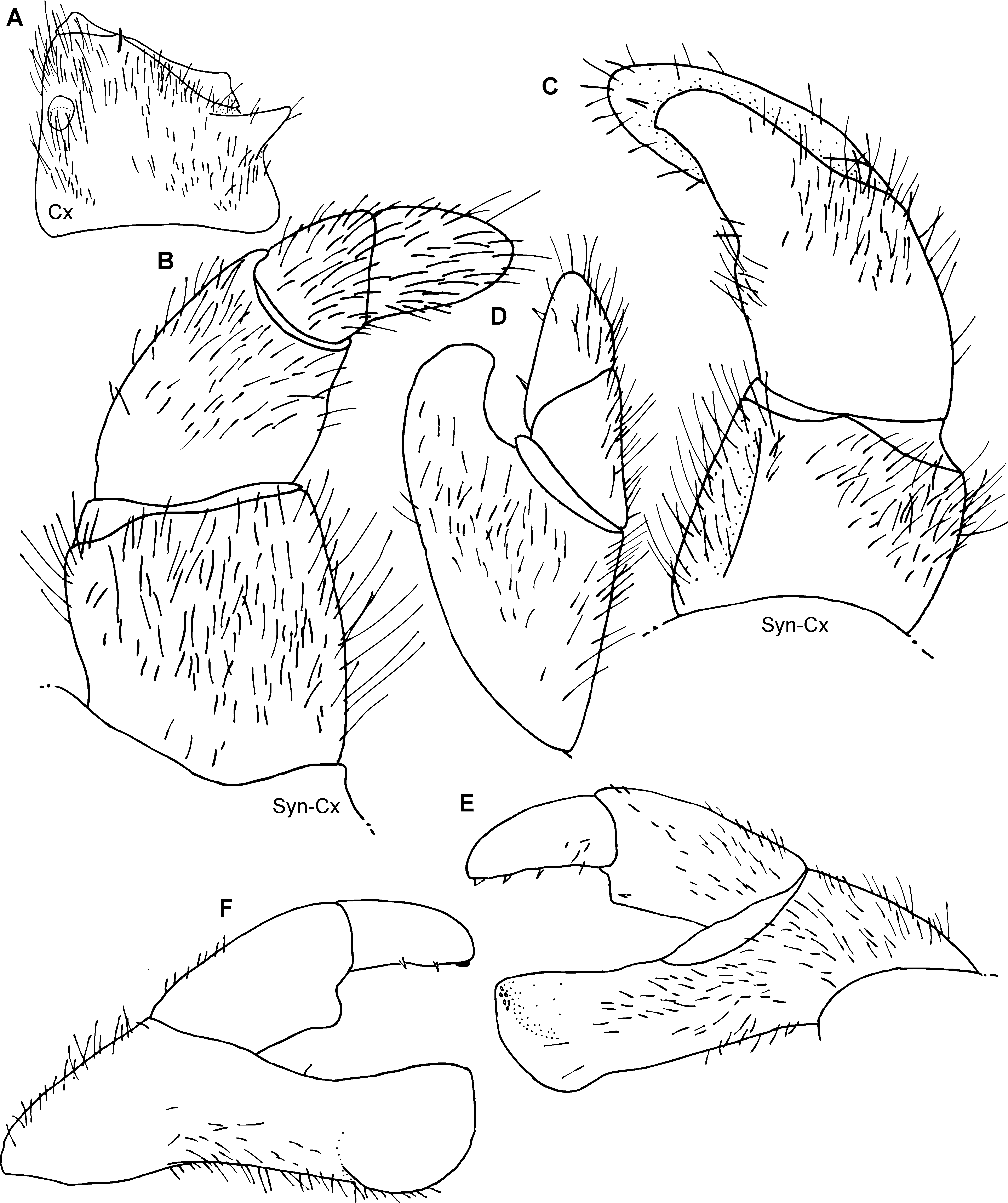
Stigmatic plates: first stigmatic plate rounded, apex well-rounded, straight towards coxa 1 ( Fig. 15A View FIGURE 15 ). Laterotergites: laterotergite 1 strongly projecting into a sharp tip. Laterotergite 2 with a broad, stout, much shorter projection. Collum: with glabrous surface, margins with few isolated setae. Thoracic shield: surface glabrous but covered with tiny pits, setae only in grooves. Slope towards groove without anterior keels but with 4 or 5 posterior keels. Tergites: surface anterior half densely setose with short setae, posterior half glabrous, shiny. Tips of paratergites of midbody tergites projecting posteriorly ( Fig. 6D View FIGURE 6 ). Endotergum: inner section lacking any spines or setae. Middle area with a single row of large, sparse, elliptical, cuticular impressions. Distance between impressions twice as wide as their diameter. Apically, 2-3 dense rows of long marginal bristles, the tips of the longest setae slightly protruding beyond tergal margin ( Fig. 9C View FIGURE 9 ). Bristles not smooth, but with numerous small spinicles. Anal shield: large, in both sexes well-rounded. Surface in both sexes completely covered by tiny setae located in small pits. Underside with a single, short, black, locking carina, located close to last laterotergite. Legs: leg-pair 1 with 2 or 3 ventral spines, leg-pair 2 with 6, leg-pair 3 with 7 or 8. First two leg-pairs without an apical spine. Leg-pairs 4–21 with 10 ventral spines and one dorso-apical spine. In leg 9, femur 1.6 times, tarsus 3.7 times longer than wide ( Fig. 15B View FIGURE 15 ). All podomeres densely setose. Coxa with a large and marginally toothed process. Coxa process absent at first leg ( Fig. 15A View FIGURE 15 ) and sharply projecting at second ( Figs 15C View FIGURE 15 , 16A View FIGURE 16 ). Prefemur at apical margin with a projection laterally and mesally. Lateral projection triangular and sharply edged, juxtaposed to coxal process. Femur extended mesally into a dentate margin featuring 12–14 teeth.
Female sexual characters: female gravid. Vulva large, covering 2/3 of coxa, extending mesally to anterior third of prefemur ( Fig. 15C View FIGURE 15 ). Operculum rounded, very slightly invaginated medially, mesal margin strongly projecting into a well-rounded lobe twice as high as remaining operculum. Subanal plate: large and wide, divided by a suture into two halves. Densely setose ( Fig. 15D View FIGURE 15 ).
Male sexual characters: gonopore covered with a single, undivided, circular, sclerotized plate ( Fig. 16A View FIGURE 16 ). Anterior telopods ( Figs 16 View FIGURE 16 B–D): consisting of 4 telopoditomeres above syncoxite. Telopoditomere 1 rectangular, as long as wide. Telopoditomere 2 large, as long as telopoditomere 1. Process of telopoditomere 2 located posteriorly, not visible in anterior view. Process of telopoditomere 2 wide, well-rounded, projecting to half of telopoditomere 3. Telopoditomere 3 small, slightly shorter than telopoditomere 4. telopoditomere 4 cylindrical, well-rounded, posterior side with two small spines. All telopoditomeres covered with long setae. Posterior telopods ( Figs 16E, F View FIGURE 16 ): telopoditomere 1 short, half as long as wide. Immovable finger (process of telopoditomere 2) as long as movable finger, consisting of telopoditomeres 3 and 4. Immovable finger with a characteristic, distally swollen apex, well rounded apically, apex only slightly wider than base. Telopoditomere 3 rectangular, well rounded, apically with a well-rounded extension carrying a small spine. Telopoditomere 4 only slightly shorter and slightly more slender than telopoditomere 3, 2.1 times longer than wide, apically weakly tapering, straight, not curved, with 3 small spines at margin towards immovable finger. In anterior view telopoditomere 1–3 covered by setae, in posterior view telopoditomeres 2–4 mostly glabrous except for a few setae at the margins.
Derivatio nominis: spinatum, noun in apposition, after the sharp process of the first laterotergite.


| ZFMK |
Zoologisches Forschungsmuseum Alexander Koenig |
No known copyright restrictions apply. See Agosti, D., Egloff, W., 2009. Taxonomic information exchange and copyright: the Plazi approach. BMC Research Notes 2009, 2:53 for further explanation.
|
Kingdom |
|
|
Phylum |
|
|
Class |
|
|
Order |
|
|
Family |
|
|
Genus |