
Homeryon armarium Galil, 2000
|
publication ID |
https://doi.org/ 10.11646/zootaxa.3764.3.3 |
|
publication LSID |
lsid:zoobank.org:pub:F1B0E174-89C5-4A9E-B7DA-C5E27AF624D3 |
|
DOI |
https://doi.org/10.5281/zenodo.6132357 |
|
persistent identifier |
https://treatment.plazi.org/id/03EB595D-FF8F-756C-FF66-F9AA4E84FBB2 |
|
treatment provided by |
Plazi |
|
scientific name |
Homeryon armarium Galil, 2000 |
| status |
|
Homeryon armarium Galil, 2000 View in CoL
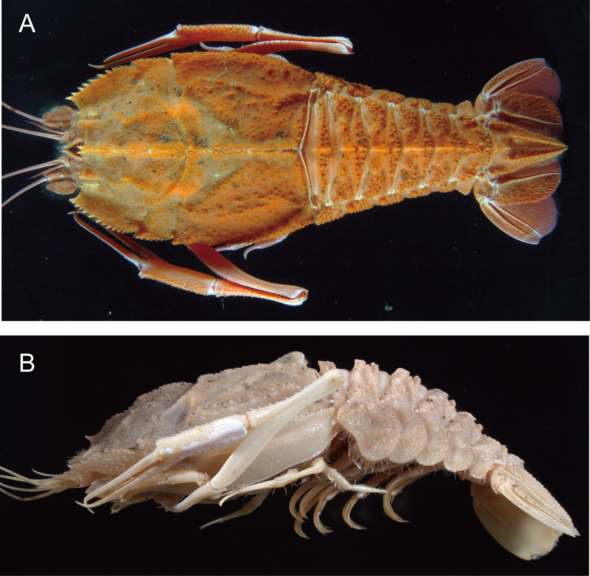
( Figs. 6–11 View FIGURE 6 View FIGURE 7 View FIGURE 8 View FIGURE 9 View FIGURE 10 View FIGURE 11 , 30 View FIGURE 30 A)
Polycheles View in CoL sp.— Baba et al. 1986: 157, 283, pl. 108.
Homeryon armarium Galil, 2000: 297 View in CoL , fig. 5 [type locality: Kyushu-Palau Ridge, 700 m].— Ahyong 2009: 382, 387, 388, fig. 2D, 3D.— Chan 2010: 162 (list).
Material examined. RV “Natsushima”, NT10-13 cruise, ROV “Hyper-Dolphin”, dive #1165, Northeast Nikko Seamount, 23°06.608’N, 142°21.831’E, 604 m, 31 July 2010, 1 ovigerous female (cl 65.0 mm), JAMSTEC 0 81538.
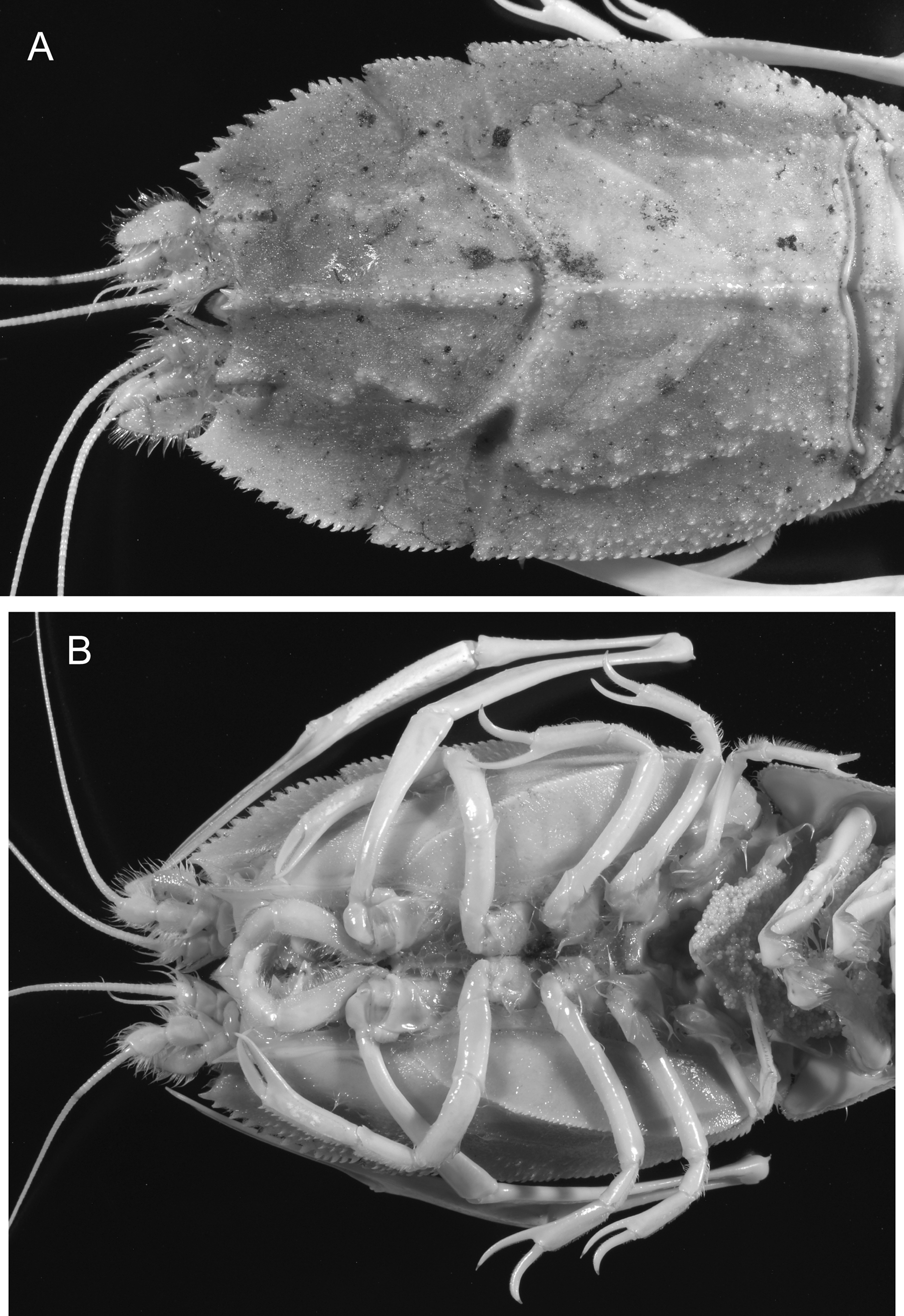
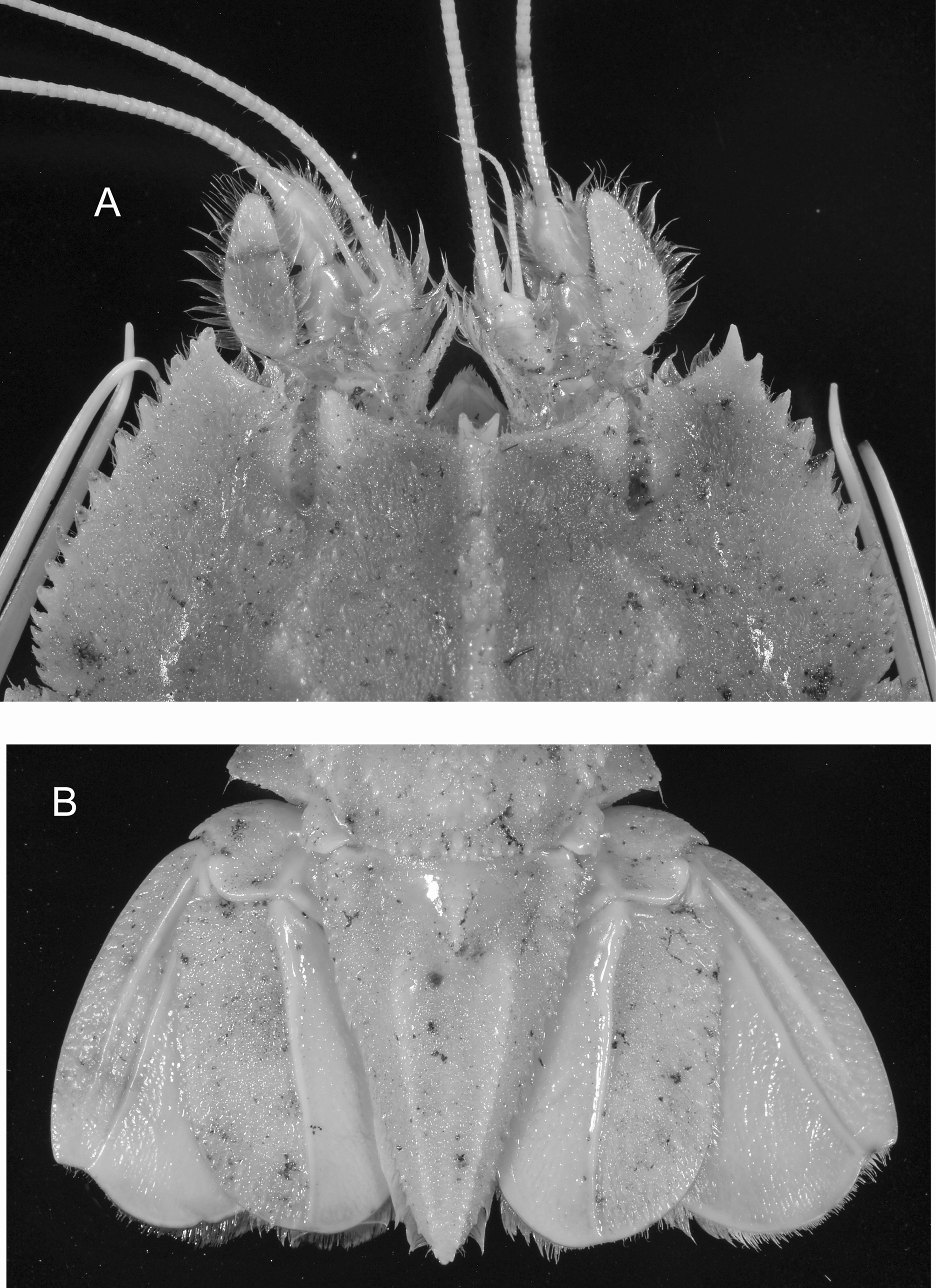
Description of female specimen from Nikko Seamount. Carapace ( Figs. 6 View FIGURE 6 A, B, 7A, B) subovate with lateral margins gently convex; dorsal surface covered with short, dense pubescence (granules or spinules naked); gastroorbital region sloping anteriorly, with sinuous, blunt postorbital carina and scattered antrorse spinules often obscured by pubescence; frontal margin generally concave, with 2 small rostral spines and few minute spinules near internal angle of dorsal orbital sinus. Pterygostomial angle produced into prominent acute tooth, supported by sharp submarginal carina extending posteriorly. Dorsal orbital sinus narrow, slit-like ( Fig. 9 View FIGURE 9 A); outer margin with row of 6 or 7 small spines including one at external angle; ventral side of orbit deeply depressed, bordered with sharp keel ( Fig. 10 View FIGURE 10 A). Lateral margins of carapace with 12–15 spines arranged in a single distinct row on anterior part anterior to cervical incision, middle part between cervical and postcervical incisions with several spinules arranged in irregular double row, posterior part posterior to postcervical incision with numerous spinules arranged in irregular double row. Cervical and postcervical incisions with spinulose margins. Cervical and postcervical carinae lined with row of spinules. Median postrostral carina blunt but prominent, ornamented by antrorse spinules arranged in irregular longitudinal rows, divided in three parts. Median postcervical carina also blunt but prominent, ornamented by spinules or granules arranged in 2 irregular longitudinal rows, divided into some parts by slight constrictions. Branchial carina low, not sharply delimited, converging posteriorly, bearing antrorse spinules arranged in irregular longitudinal rows. Branchial groove shallow, carina defining this groove in line with row of spinules. Posteromarginal carina sharply delimited. Lateral surface of carapace with scattered numerous spinules dorsally and with trace of ridge on midline, becoming distinct and spinulose in posterior 0.3; ventrolateral carina sharply delimited ( Fig. 7 View FIGURE 7 B), with minutely serrated margin.
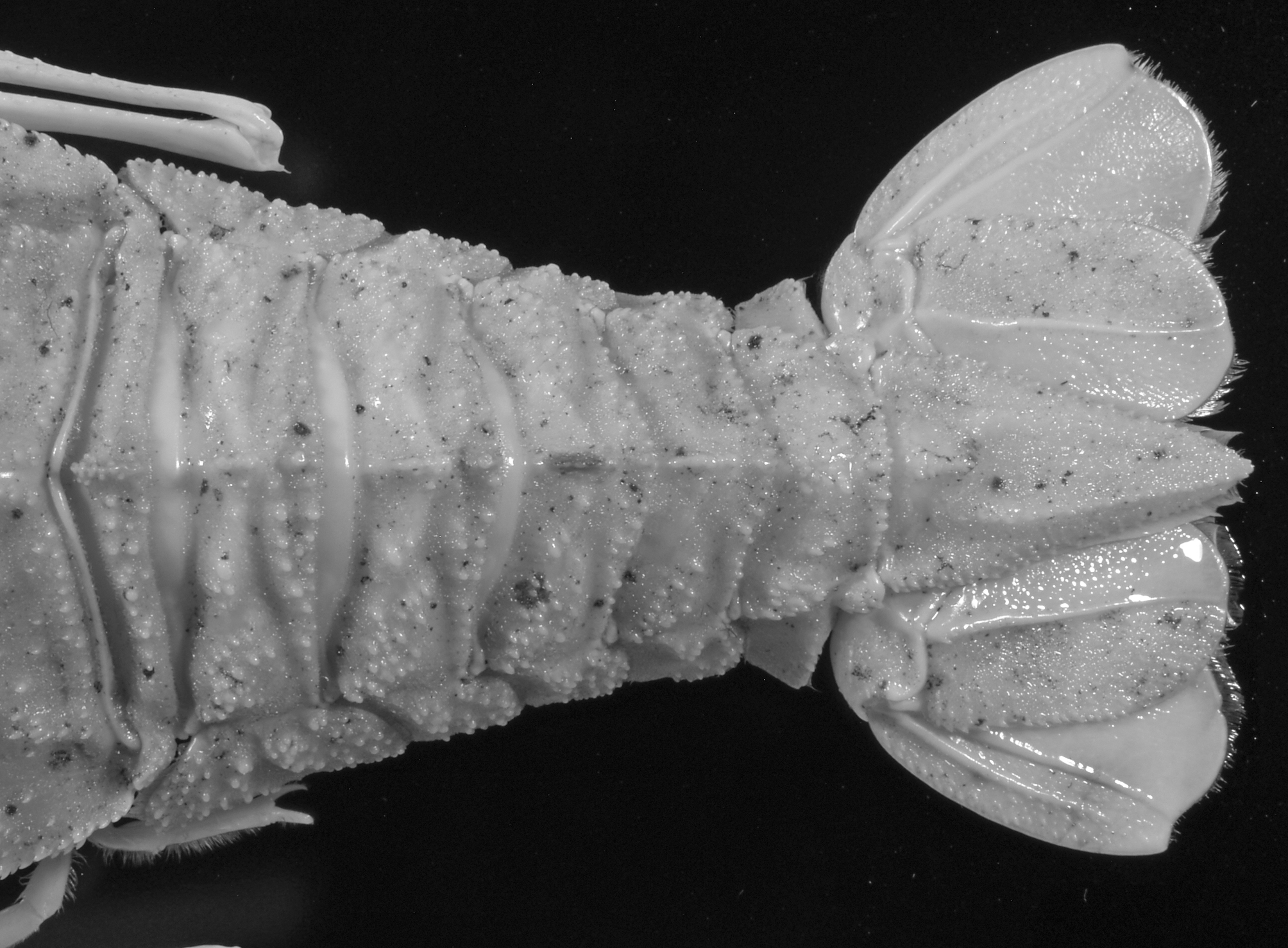
Pleon ( Figs. 6 View FIGURE 6 B, 8) well sculptured, tuberculate, medially carinate in anterior 5 pleomeres; shallow transverse groove present on second to fifth tergites. Middorsal carina of first pleomere entire, with anterior tip slightly produced; those on second to fifth pleomeres divided in 2 parts by transverse groove, not spiniform, each anterior part larger than posterior part, granulose. Sixth tergite with clusters of rounded granules medially and laterally. First pleuron reduced, hidden by second pleuron; second to fifth pleura with sparse granules on surfaces, posterior margins bordered by granules; second pleuron reniform with sinuous anteroventral margin, with short, vertical groove medially; third to fifth pleura each with low, granulate protuberance mediodorsally. Telson ( Fig. 9 View FIGURE 9 B) lanceolate, terminating in acute tip; basal median tubercle low, granulate; submedian carinae and lateral margins lined with small, posteriorly-directed spiniform tubercles.
Eyestalk ( Fig. 10 View FIGURE 10 A) unarmed, tapering to bluntly pointed apex.
Antennular peduncle ( Fig. 9 View FIGURE 9 A) nearly reaching midlength of antennal peduncle. Basal segment with distolateral angle terminating in tiny process; distomesial process prominent, overreaching distal margin of second segment, vertically compressed, terminating in sharp spine, armed with few spinules on dorsal margin. Second segment widened distally, distomesial angle with distinct spine. Distal segment with small distomesial spine and blunt protuberance-like process on distal margin medially. Outer flagellum short, only slightly overreaching antennal scaphocerite; inner flagellum subequal in length to carapace.
Antennal peduncle ( Figs. 9 View FIGURE 9 A, 10B) flattened dorsoventrally. First segment with prominent, tubular renal process fitting into hollow on ventral surface of basal segment of antennular peduncle. Second segment unarmed. Third segment with prominent distomesial spine not reaching midlength of fourth segment. Fourth segment suboval in shape, with small distomesial spine. Fifth segment with slightly produced distomesial margin, but spineless. Scaphocerite lanceolate, setose, reaching distal margin of fifth segment; margins unarmed, lateral margin strongly convex proximally, mesial margin gently convex.
Third maxilliped ( Fig. 10 View FIGURE 10 C) pediform, broad, flattened dorsoventrally. Ischium distinctly narrowing proximally, bearing small distomesial spine and short row of minute tubercles on distal 0.2. Merus slightly longer than carpus-propodus combined, unarmed. Dactylus slightly longer than propodus, strongly incurved, terminating in sharp corneous spine.
First pereopod ( Fig. 11 View FIGURE 11 A) subequal in length to body, fairly compressed; when folded, merus not reaching posteriorly to midpoint of second abdominal pleuron. Coxa with small distolateral tooth directed laterally. Basis and ischium fused though suture still discernible; ischium broadened distally. Merus about 1.6 times longer than ischium; regularly convex flexor margin with row of spinules in distal 0.6, sinuous extensor margin with some minute denticles. Carpus about 0.6 times as long as merus, widened distally, upper (extensor) margin with 2 rows of spinules increasing in size distally, lower (flexor) margin unarmed except for a few spinules on distal margin. Chela about 1.9 times longer than carpus; palm with numerous scattered spinules on upper (dorsal) half; lower (ventral) margin carinate, with row of minute spinules; fingers hooked distally; cutting edges of both fingers each bordered by row of overlapping flexible platelets, that of fixed finger with shallow concavity proximally; dactylus about 1.2 times longer than palm, with short row of some minute spinules proximally upper (dorsal) surface.
Second pereopod ( Fig. 11 View FIGURE 11 B) with basis, ischium and merus clearly articulated, not fused; merus with few spines on outer distal margin and spinules arranged in 2 irregular rows on inner margin. Carpus with extensor facet clearly defined by sharply carinate dorsal margin and ventral margin with irregular double row of spinules; ventral surface with median row of setae. Palm slightly widened distally, with extensor facet defined by dorsal carina and ventral row of spinules; ventral surface with 2 lines of short setae (one submedian and one adjacent to flexor margin extending onto fixed finger); flexor surface rounded, not carinate. Fingers hooked distally; cutting edges each with closely set, flexible platelets; fixed finger slightly deflexed; dactylus about 0.7 times as long as palm.
Third and fourth pereopods ( Fig. 11 View FIGURE 11 C, D) similar in structure, though latter slightly shorter than former. Ischium and merus fused, bearing scattered minute spinules or granules on outer surface. Carpus with spinule (third) or tubercle (fourth) on extensor distal margin. Palm with carinate extensor margin. Fingers strongly recurved; cutting edges pectinate with row of minute corneous spinules.
Fifth pereopod chelate ( Fig. 11 View FIGURE 11 E). Basis and ischium clearly articulated, but ischium and merus fused, demarcation between ischium and merus not discernible. Fingers subequal in length; fixed finger slightly recurved, with row of closely set, minute corneous spinules on cutting edge; dactylus slightly incurved, with narrowly spaced corneous spinules on cutting edge.
Second to fifth pleopods each with long appendix interna.
Uropod ( Fig. 8 View FIGURE 8 B) with protopod flattened, with small posterolateral spine. Endopod with sharp median carina on dorsal surface, surface lateral to median carina with scattered small spines and setae, surface mesial to median carina smooth, glabrous; nearly straight lateral margin bordered by row of small spines; posterior margin rounded. Exopod with 2 longitudinal carinae laterally on dorsal surface, surface lateral to carinae with scattered spinules, remainder glabrous; slightly convex lateral margin unarmed; obtuse posterolateral tooth present.
Eggs small, about 0.6 × 0.6 mm.
Coloration. Body and appendages generally orange-brown; boundary between abdominal tergites and pleura whitish; cheliped orange in general, whitish on dorsal side of each segment; longitudinal carinae on uropods white. See Fig. 6 View FIGURE 6 A.
Distribution. Previously known only from Kyushu-Palau Ridge, 520–700 m ( Galil 2000). Now from the Northeast Nikko Seamount, 604 m.
Ecology. During the dive, we encountered three individuals of this species, two of which were successfully collected with slurp gun. All of the three individuals were found to rest on the bedrock ( Fig. 29 View FIGURE 29 A). One individual avoided to being sucked by slurp gun by using tail flap.
Remarks. As noted above, during the dive, two specimens of Homeryon armarium were collected. One of the two specimens was kept alive in aquarium, and transferred to the Tokyo Sea Life Park for further rearing. Then only a single specimen was available for this study.
The present specimen agrees well with the descriptions of Homeryon armarium by Baba et al. (1986; as Polycheles sp.) and Galil (2000). The original description by Galil (2000) is sufficient for species recognition, but omitted many morphological details of possible diagnostic significance. Therefore, we provide a detailed description accompanied by illustrations herewith for better understanding of the morphology of this rarely collected species.
The genus Homeryon is represented only by two species, H. armarium (type species) and H. asper (Rathbun, 1906) . Differences between these two species are discussed by Galil (2000).

No known copyright restrictions apply. See Agosti, D., Egloff, W., 2009. Taxonomic information exchange and copyright: the Plazi approach. BMC Research Notes 2009, 2:53 for further explanation.
|
Kingdom |
|
|
Phylum |
|
|
Class |
|
|
Order |
|
|
Family |
|
|
Genus |
Homeryon armarium Galil, 2000
| Komai, Tomoyuki & Tsuchida, Shinji 2014 |
Homeryon armarium
| Chan 2010: 162 |
| Ahyong 2009: 382 |
| Galil 2000: 297 |
Polycheles
| Baba 1986: 157 |