
Phrikoceros mopsus ( Marcus 1952 ) Quiroga, Bolaños & Litvaitis 2004
|
publication ID |
https://doi.org/ 10.5281/zenodo.191231 |
|
DOI |
https://doi.org/10.5281/zenodo.6224076 |
|
persistent identifier |
https://treatment.plazi.org/id/03EB7608-D665-FFE8-33ED-9684FBD3A11F |
|
treatment provided by |
Plazi |
|
scientific name |
Phrikoceros mopsus ( Marcus 1952 ) Quiroga, Bolaños & Litvaitis 2004 |
| status |
|
Species: Phrikoceros mopsus ( Marcus 1952) Quiroga, Bolaños & Litvaitis 2004 View in CoL
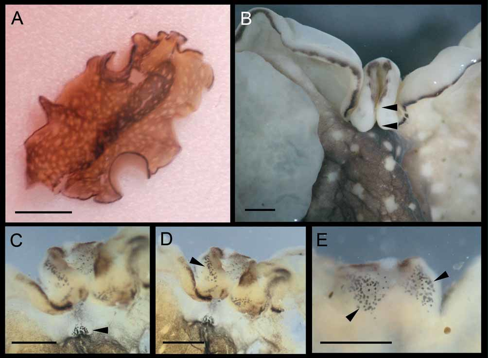
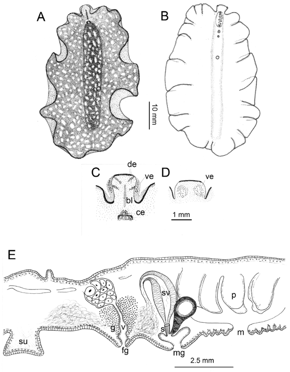
( Figs. 1–2 View FIGURE 1 View FIGURE 2 )
Synonyms: Pseudoceros mopsus Marcus 1952
Material examined and localities. One mature specimen preserved in 70% ethanol (50 mm x 20 mm) MPL 5955. Collected 26 November 2003 at Puerto Pirámides (42º 34 32.70 S, 64º 17 15.82 W), Península Valdés, Chubut, Argentina. Two mature specimens as serial sagittal sections MPL 5956. Collected 1st March 2005 at the same locality.
One mature specimen as whole mount (25 mm x 12 mm) MPL 5957. Collected in March 2005 at Puerto Madryn (42º 46 36.04 S, 65º 0 2 12.89 W), Chubut, Argentina.
Distribution. The species is widely distributed throughout the Caribbean (SQ, personal observation) and has been described from Brazil ( Marcus 1952) and Colombia ( Quiroga et al. 2004).
Description. External features
Color: Dorsally, live specimens are brown with white spots over the entire surface. Along the mid-dorsal region the brown color is darker forming a longitudinal band. The margin is bordered by a thin black line which is interrupted along the apical region of the pseudotentacles. Anterior to the cerebral eye cluster there is a short longitudinal black line which disappears before the anterior end of the body. The ventral surface is beige ( Fig. 1 View FIGURE 1 B).
Form: Live specimens are oval with undulated body edges. The sucker is large (1 mm diameter) located in the anterior half of the body, approximately 4.3 mm posterior to the female gonopore ( Fig. 2 View FIGURE 2 B, E).
Tentacles: The pseudotentacles are square-shaped, formed by the folds of the anterior margin ( Figs. 1 View FIGURE 1 , 2 View FIGURE 2 A–D).
Eyes: The cerebral eyes in live specimens are arranged in a triangular-shaped cluster of about 40–50 eyes ( Figs. 1 View FIGURE 1 A, C, D, 2A, C). The dorsal pseudotentacular eyes are arranged in four longitudinal clusters of about 50 eyes each ( Figs. 1 View FIGURE 1 C, D, 2C). The ventral pseudotentacular eyes occur in four clusters of about 80–100 eyes each ( Figs. 1 View FIGURE 1 C–E, 2C, D).
Pharynx: The pharynx is ruffled with six simple folds ( Fig. 2 View FIGURE 2 E), located in the anterior part of the body. The mouth is placed at the center of the pharynx about 5 mm from the anterior end of the body.
Gonopores: There is only one male gonopore immediately posterior to the pharynx. The female gonopore is separated by 2.3 mm from the male pore.
Internal features. Body wall: The epidermis is well developed on both surfaces of the body even though the ventral epithelium is taller (20–25 µm). In the dorsal epidermis there are conspicuous grains of black pigment. The body musculature is organized in three fine layers; the external one is formed by longitudinal fibers followed by circular muscles and the innermost is made up of diagonal fibers. The height of the ventral and dorsal musculature is 40–60 µm and 25 µm respectively.
Reproductive anatomy: Male reproductive system – single male system with an oblong, large and highly muscular seminal vesicle (610 µm long, 360 µm wide). The free prostatic vesicle is rounded and located anterior to the seminal vesicle (260 µm in diameter). The penis papilla bears a short, sclerotised stylet (120– 150 µm long). The prostatic and ejaculatory ducts join at the penis papilla ( Fig. 2 View FIGURE 2 E). Female reproductive system – The short vagina is ciliated and surrounded by numerous cement glands. The cement pouch is a shallow depression of the vagina next to the female gonopore ( Fig. 2 View FIGURE 2 E). In mature specimens, usually the oviducts are expanded with a great number of eggs inside.
Taxonomic remarks. Phrikoceros was described by Newman and Cannon (1996) who distinguish the genus from the rest of pseudocerotids mainly by the possession of both a simple ruffle pharynx as in Pseudobiceros Faubel 1984 , and a single male reproductive system as in Pseudoceros Lang 1884 . Among other characteristics they mentioned the presence of a horseshoe-shaped cerebral eye cluster which differs from the one in the species described herein. However, this characteristic can be variable among pseudocerotids. Phrikoceros mopsus had been described based on material from Isla de São Sebastião in Brazil, it was recorded from the Caribbean cost of Colombia by Quiroga et al. (2004). These authors also recognized that the species belonged to the genus Phrikoceros and formed a new combination.
The difference in the dorsal pattern of coloration (dotted with white spots, and net-like shape) between the Argentine flatworms and the others is due to the nutritional status of the animals (M. Litvaitis, personal communication). The reproductive differences, such as a considerably bigger and more muscular seminal vesicle and a longer stylet, are due to the larger size of the Argentine specimens.
| MPL |
Musee de Port Louis |
No known copyright restrictions apply. See Agosti, D., Egloff, W., 2009. Taxonomic information exchange and copyright: the Plazi approach. BMC Research Notes 2009, 2:53 for further explanation.
|
Kingdom |
|
|
Phylum |
|
|
Class |
|
|
Order |
|
|
Family |
|
|
Genus |