
Prionomeion gaullei ( Konow, 1906 )
|
publication ID |
https://doi.org/ 10.11646/zootaxa.4127.3.7 |
|
publication LSID |
lsid:zoobank.org:pub:997EEE83-1C91-49BA-84C3-C6483AF3FF57 |
|
DOI |
https://doi.org/10.5281/zenodo.5317088 |
|
persistent identifier |
https://treatment.plazi.org/id/03EB7C14-960F-0543-18DE-8D983174ECD5 |
|
treatment provided by |
Plazi |
|
scientific name |
Prionomeion gaullei ( Konow, 1906 ) |
| status |
|
Prionomeion gaullei ( Konow, 1906)
Figs 1A–D View FIGURES 1 A – H , 2A–F View FIGURES 2 A – I , 3A–D View FIGURES 3 A – H , 4A–E View FIGURES 4 A – J , 5A–B View FIGURES 5 A – F , 6A–B, E–F View FIGURES 6 A – I. A – D , 7A, C–D View FIGURES 7 A – F. A , 8A–C View FIGURES 8 A – H
Lophyrus Gaullei [sic] Konow, 1906: 123.
Lophyrus gaullei: Benson, 1939: 341 ; Quinlan, 1974: 223; Oehlke & Wudowenz, 1984: 384; Abe & Smith, 1991: 69; Liston, 2015.
Diprion gaullei: Forsius, 1930: 7 .
Prionomeion gaullei: Gussakovskij, 1947: 146 , 223; Smith, 1975: 410; Chevin, 1987: 17 –19; Taeger et al., 2010: 210; Liston, 2015.
Redescription. Female (holotype). Length 6.0 mm. Black, without metallic reflections ( Figs 1A–B View FIGURES 1 A – H ). Labrum black brown. Mandible apically red brown widely. Palps dark brown. Legs brown to dark brown on fore tibia and tarsus; hind first tarsomere basally brown narrowly. Wings clear hyaline; veins brown to black; vein C distinctly paler than vein R; stigma dark brown. Abdomen orange on second to sixth segments and anterior and lateral parts of seventh tergum.
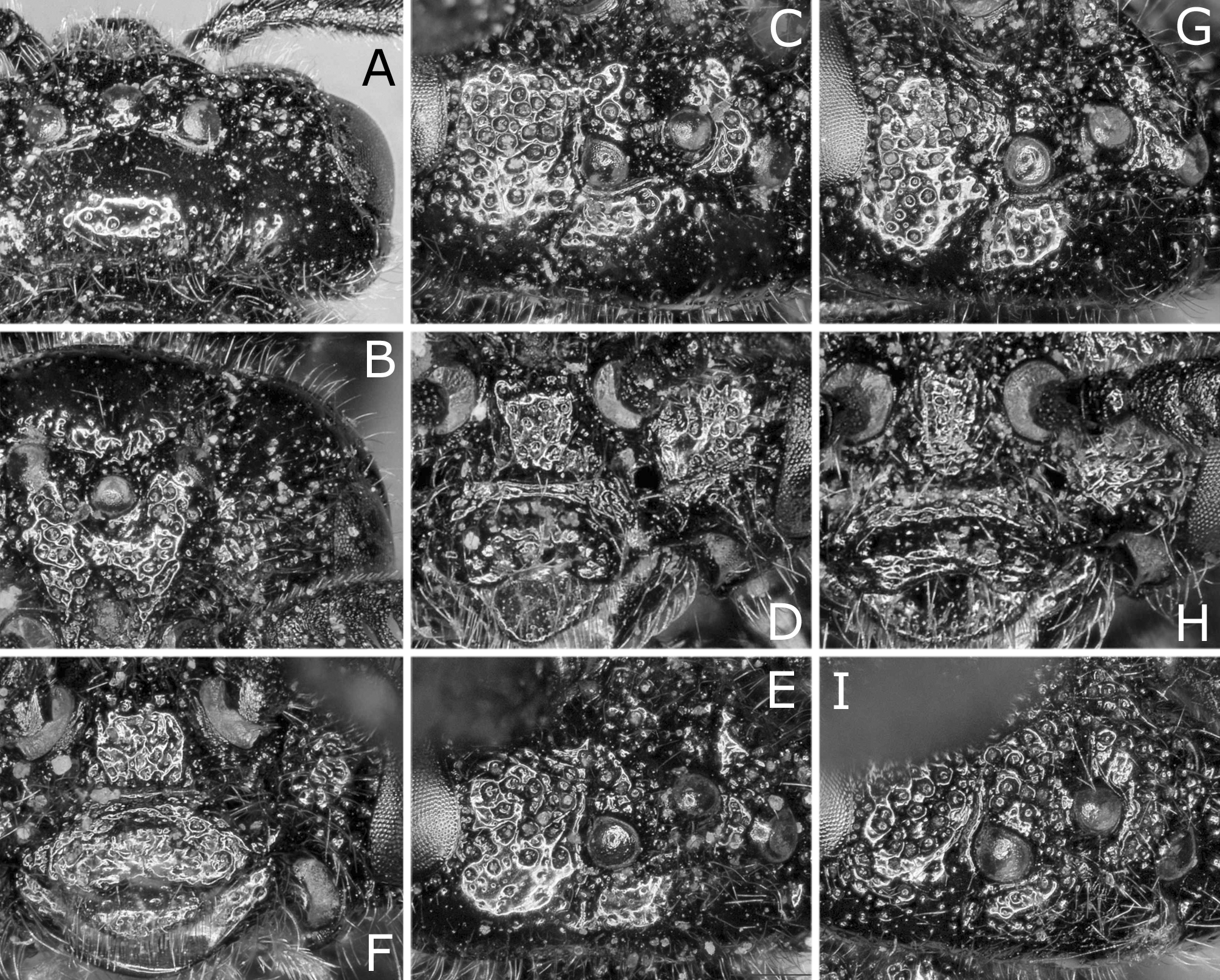
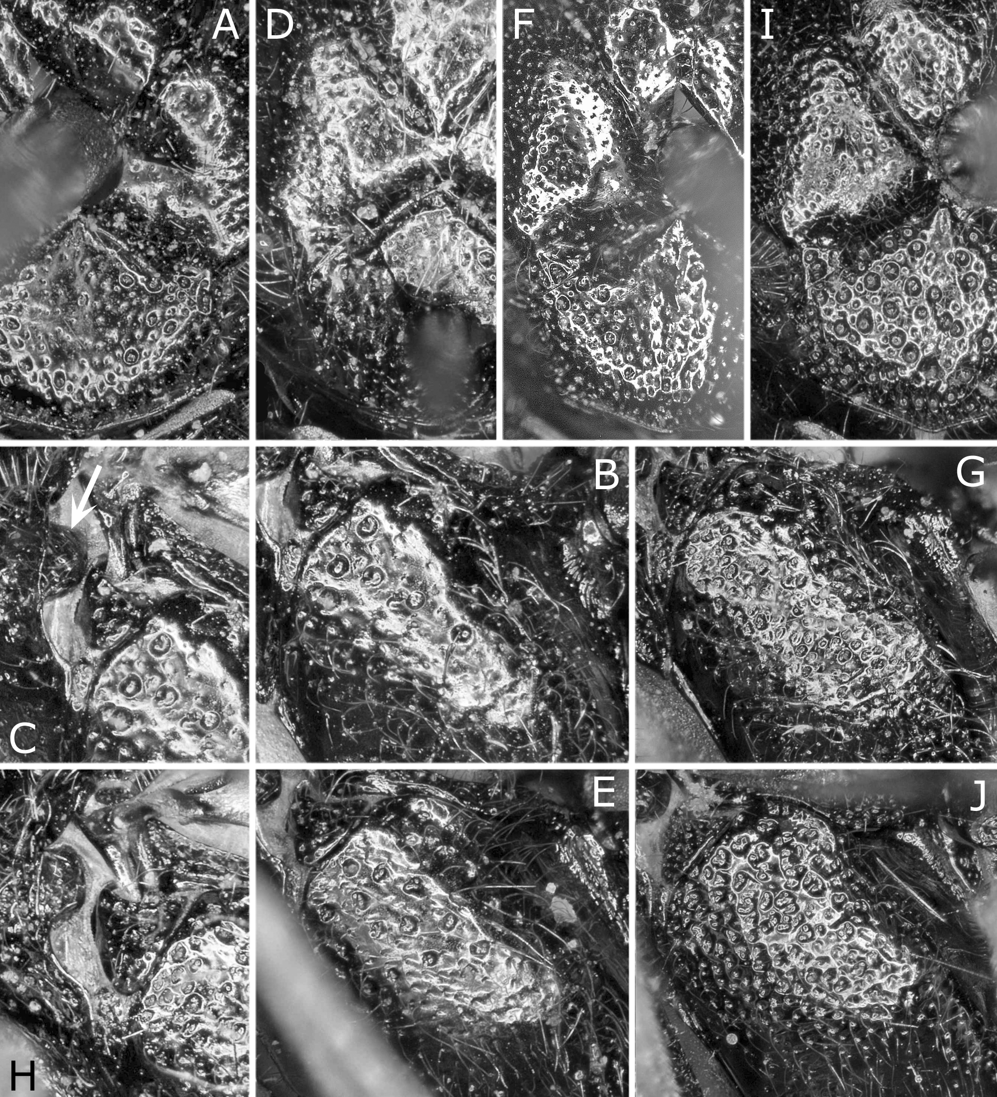
Head and thorax with punctures predominantly small and separated, and interspaces smooth and shiny ( Figs 2A–D View FIGURES 2 A – I , 4A–C View FIGURES 4 A – J , 5A View FIGURES 5 A – F ); punctures predominantly large and contiguous on anterior part of head; labrum sparsely and finely punctured ( Fig. 2D View FIGURES 2 A – I ); posterior part of pronotum, postspiracular sclerite, and lateral and apical parts of mesoscutellum with sparse large distinct punctures; dorsal part of mesepisternum shallowly and indistinctly punctured, and with sparse large distinct punctures ( Fig. 4B View FIGURES 4 A – J ); anepimeron densely punctured; metascutellum rough.
Distance between eyes 1.8 × eye height. Postocellar area moderately convex, width 1.6 × length ( Figs 2A–C View FIGURES 2 A – I ). Ratio of distances between eye and lateral ocellus, between lateral ocelli and between lateral ocellus and posterior margin of head 1.0:1.0:1.1; ratio of distances between eye and lateral ocellus and between lateral ocellus and posterior margin of head 0.9:1.0. Distance between torulus and eye 1.1 × distance between toruli. Malar space 0.8 × width of median ocellus. Antenna ( Figs 3A–D View FIGURES 3 A – H ) with 20 antennomeres; first flagellomere in lateral view with length along dorsal margin 1.0 × apical breadth except for ramus, and ramus length 1.1 × its flagellomere length; middle flagellomeres each with slender ramus 1.7 × as long as its associated flagellomere and with distinct inner projection. Maxillary palp short; apical palpomere about as long as median ocellus width.
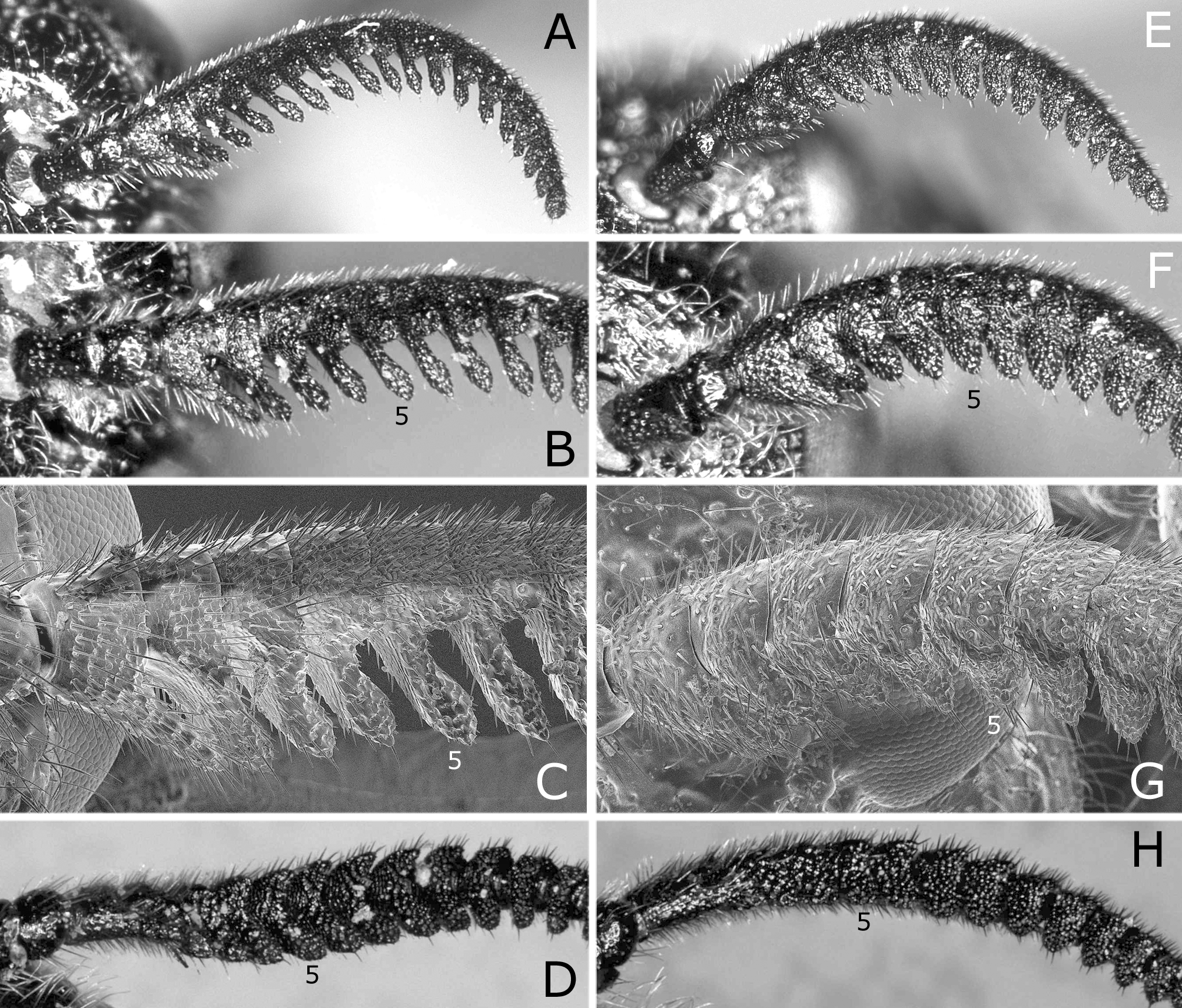
Prepectus distinct, length (= short axis) about 1/3 length of postspiracular sclerite ( Figs 4B–C View FIGURES 4 A – J ). Pronotum with posterolateral lobe separated from its main part by sharp ridge throughout ( Fig. 4C View FIGURES 4 A – J ). Anterior edge of mesoscutellum angled about 100° ( Fig. 4A View FIGURES 4 A – J ). Hind leg ( Fig. 6A View FIGURES 6 A – I. A – D ) with posterior tibial spur 0.9 × as long as first tarsomere exclusive of pulvillar pad, 1.2 × breadth of tibia in lateral view; first tarsomere length 1.4 × tibia breadth in lateral view; second and third tarsomeres combined 1.3 × as long as first. In hind wing, section of vein 1A between crossveins a and cu-a 1.5 × width of cell 1A.
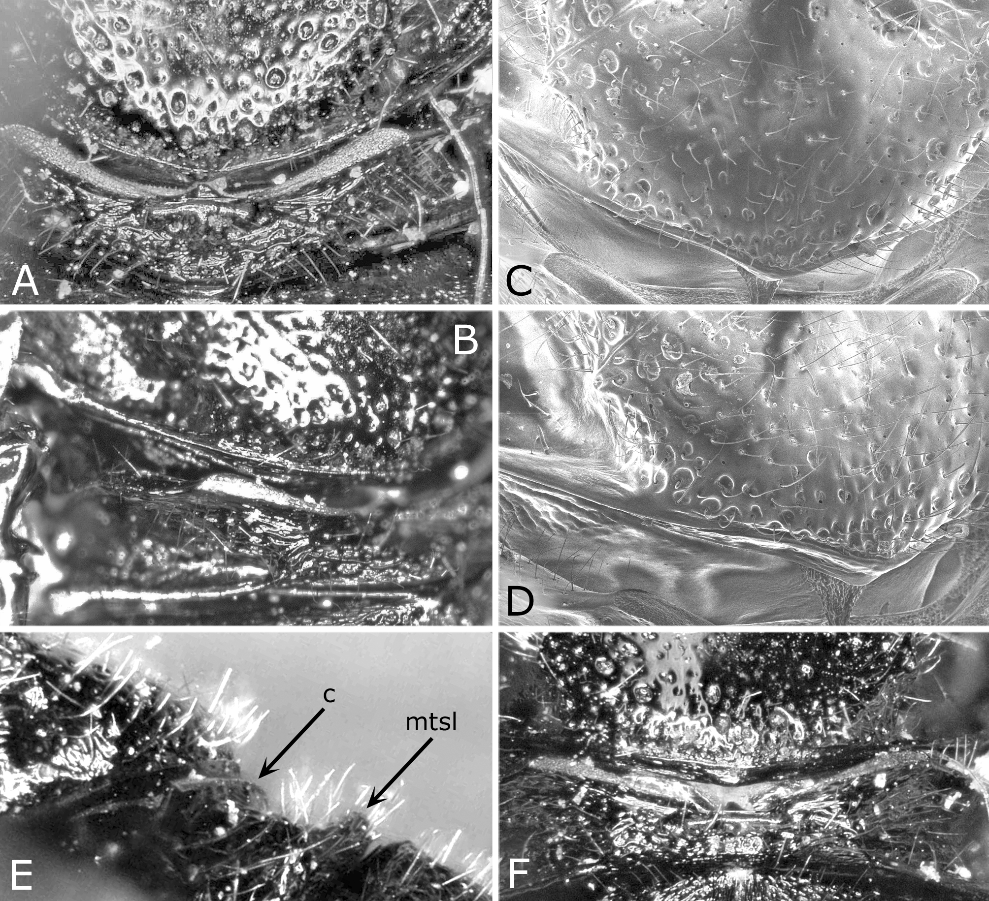
Ovipositor sheath ( Figs 7C–D View FIGURES 7 A – F. A ) in dorsal view about 3.0 × as wide as cercus, in posterior view with lateral margin gently roundly convex. Lance ( Figs 8A–B View FIGURES 8 A – H ) with short posterior projection of processus articularis and eight annular sutures. Lancet ( Fig. 8C View FIGURES 8 A – H ) with 11 annuli, with length from apex to ventral end of first row of spines 2.9 × maximum width; sclerotized area present basal to first annulus; first row of spines slightly arched basally and its ventral end widely separated from ventral margin of lancet; second and third rows of spines each arched; serrula of second annulus distinctly convex on ventral margin; serrulae of third to fifth annuli each angularly convex on ventral margin; middle serrulae simple; apical two serrulae each angulating on ventral margin.
Male. Length 6.0 mm ( Figs 1C–D View FIGURES 1 A – H ). As in female, but differing as follows except for usual sexual differences.
Palps yellow, basally darkened. Legs yellow on wide apex of fore femur, narrow apices of middle and hind femora, tibiae, tibial spurs, tarsi and claws. Abdomen entirely black.
Distance between eyes 1.7 × eye height. Postocellar area width 2.0 × length. Ratio of distances between eye and lateral ocellus, between lateral ocelli and between lateral ocellus and posterior margin of head 1.0:1.0:0.8; ratio of distances between eye and lateral ocellus and between lateral ocellus and posterior margin of head 1.3:1.0 ( Fig. 2E View FIGURES 2 A – I ). Distance between torulus and eye 0.9 × distance between toruli ( Fig. 2F View FIGURES 2 A – I ). Antenna with 22 antennomeres. Anterior edge of mesoscutellum angled about 90° ( Fig. 4D View FIGURES 4 A – J ). Hind leg ( Fig. 6B View FIGURES 6 A – I. A – D ) with posterior tibial spur 0.8 × as long as first tarsomere, 1.4 × breadth of tibia in lateral view; first tarsomere length 1.8 × tibia breadth in lateral view; second and third tarsomeres combined 1.1 × as long as first. In hind wing, section of vein 1A between crossveins a and cu-a 1.1 × width of cell 1A. Subgenital plate in ventral view with apical margin rounded ( Fig. 1C View FIGURES 1 A – H ).
Genitalia not examined (missing in the male examined; penis valve in lateral view was figured by Chevin 1987 in fig. 9).
Material examined. Holotype ( Figs 1A–B View FIGURES 1 A – H ): ♀, labelled “ Lophyrus Gaullei Konow. Algeria.” “ Algerie Djidjelli” “Coll. Konow” “Holo = Typus ” “ Holotypus ” and “GBIF-GISHym 4667”, deposited in the Senckenberg Deutsches Entomologisches Institut, Müncheberg (see also Liston 2015).
Konow (1906) did not designate the holotype and did not state how many specimens he had. However, there is no indication suggesting the presence of more than one type specimen in his description. The above female was considered the holotype of Lophyrus gaullei Konow, 1906 by Oehlke & Wudowenz (1984) and Chevin (1987).
Other material: 1♂, Mts. Djurdjura, Tikjda, 1475m, 19. V. 1976, J. M. Maldes, CBGP _ INRA _HYM_000020_0101, deposited in the UMR Centre de Biologie pour Gestion des Populations, Montferrier-sur-Lez (cited by Chevin 1987).
Distribution. Algeria.
Remarks. This species is distinguished from P. maghrebense sp. nov. mainly by the characters given in the key. For more differences, see under the remarks of the latter species.
Fabre & Chevin (1991) gave the host plant record and the description of the larva for P. gaullei . However, I consider that the reared material of Fabre & Chevin (1991) (one male) is not P. gaullei but P. maghrebense , although the difference between the two is small in the male.


| INRA |
Institut National de la Recherche Agronomique |
No known copyright restrictions apply. See Agosti, D., Egloff, W., 2009. Taxonomic information exchange and copyright: the Plazi approach. BMC Research Notes 2009, 2:53 for further explanation.
|
Kingdom |
|
|
Phylum |
|
|
Class |
|
|
Order |
|
|
Family |
|
|
Genus |
Prionomeion gaullei ( Konow, 1906 )
| Hara, Hideho 2016 |
Prionomeion gaullei:
| Taeger 2010: 210 |
| Chevin 1987: 17 |
| Smith 1975: 410 |
| Gussakovskij 1947: 146 |
Lophyrus gaullei:
| Abe 1991: 69 |
| Oehlke 1984: 384 |
| Quinlan 1974: 223 |
| Benson 1939: 341 |
Diprion gaullei:
| Forsius 1930: 7 |
Lophyrus
| Konow 1906: 123 |