
Pseudoanthidium kunesense, Niu & Luo & Griswold & Zhu, 2021
|
publication ID |
https://doi.org/10.11646/zootaxa.4996.1.5 |
|
publication LSID |
lsid:zoobank.org:pub:843F2FBA-4A9B-4D31-B6F0-FFDEFD310370 |
|
persistent identifier |
https://treatment.plazi.org/id/03EB8799-FFED-FF6A-D6EF-2349FA6DF88F |
|
treatment provided by |
Plazi (2021-07-05 08:16:17, last updated 2023-11-03 05:17:32) |
|
scientific name |
Pseudoanthidium kunesense |
| status |
|
Key to Chinese species of Pseudoanthidium View in CoL
(The females of P. tenellum ( Mocsáry, 1880) , P. ludingense ( Wu, 1993) , as well as the males of P. circinatum (Wu, 2004) , P. xinjiangense (Wu, 2004) , P. kunesense , sp. nov. and P. chenggongense , sp. nov. are unknown and not covered by the key)
1 Flagellum 10-segmented ( Figs 4b View FIGURE 4 , 7b View FIGURE 7 , 8b View FIGURE 8 , 10b View FIGURE 10 , 11b View FIGURE 11 ), female................................................... 2
- Flagellum 11-segmented ( Figs 3b View FIGURE 3 , 5b View FIGURE 5 , 9a View FIGURE 9 ), male............................................................ 7
2 Paraocular area maculated up to top of eye ( Figs 4b View FIGURE 4 , 6b View FIGURE 6 , 8b View FIGURE 8 , 10b View FIGURE 10 )............................................... 3
- Paraocular area maculated only up to the level of antennal socket ( Figs 2b View FIGURE 2 , 7b View FIGURE 7 , 11b View FIGURE 11 )................................ 5
3 Clypeus yellow, with two black longitudinal belt-shaped markings ( Figs 8b View FIGURE 8 , 10b View FIGURE 10 ).................................. 4
- Clypeus entirely yellow, without black marking ( Figs 4b View FIGURE 4 , 6b View FIGURE 6 )....................... P. (P.) orientale ( Bingham, 1897) View in CoL
4 Genal area yellow marking not separated from that on vertex ( Fig. 8c View FIGURE 8 ); mandible with six teeth ( Fig. 8d View FIGURE 8 )....................................................................................... P. (P.) yanruae Niu & Zhu View in CoL , sp. nov.
- Genal area yellow marking separated from that on vertex ( Fig. 10c View FIGURE 10 ); mandible with five teeth ( Fig. 10d View FIGURE 10 )...................................................................................... P. (P.) kunesense Niu & Zhu View in CoL , sp. nov.
5 Frons with an oval yellow spot ( Fig. 11b View FIGURE 11 )................................ P. (P.) chenggongense Niu & Zhu View in CoL , sp. nov.
- Frons black, without yellow spot ( Figs. 2b View FIGURE 2 , 7b View FIGURE 7 )............................................................. 6
6 Clypeus yellow except central and apical parts black ( Fig. 7b View FIGURE 7 )........................ P. (P.) xinjiangense (Wu, 2004) View in CoL
- Clypeus yellow, with a round-shaped black marking at center ( Fig. 2b View FIGURE 2 )................... P. (P.) circinatum (Wu, 2004) View in CoL
7 Mandible with 3 teeth ( Fig. 9b View FIGURE 9 ).......................................................................... 8
- Mandible with 4 teeth ( Fig. 3f View FIGURE 3 )................................................... P. (P.) ludingense ( Wu, 1993) View in CoL
8 Paraocular area maculated up to top of eye ( Fig. 9a View FIGURE 9 )......................................................... 9
- Paraocular area maculated only up to the level of antennal socket ( Fig. 1b View FIGURE 1 )............. P. (P.) tenellum ( Mocsáry, 1880) View in CoL
9 Supraclypeal area yellow; shape of S8 as heart of playing cards..................... P. (P.) orientale ( Bingham, 1897) View in CoL
- Supraclypeal area only with a little yellow marking ( Fig. 9a View FIGURE 9 ); S8 sub-trapezoid-shaped ( Fig. 9g View FIGURE 9 ).............................................................................................. P. (P.) yanruae Niu & Zhu View in CoL , sp. nov.
Bingham, C. T. (1897) The Fauna of British India Including Ceylon and Burma, Hymenoptera. Vol. I. Wasps and Bees. Taylor and Francis, London, xxix + 577 pp., 4 pls.
Mocsary, A. (1880 - 1881 1879 ) Data nova ad Faunam Hymenopterologicam Hungariae meridionalis comitatu Temesiensis. Termeszettudomanyi Kozlony, 16, 1 - 70.
Wu, Y. R. (1993 1992 ) Hymenoptera: Apoidea (I). In: Chen, S. X. (Ed.), Insects of the Hengduan Mountains Region. Vol. II. Science Press, Beijing, pp. 1378 - 1421.

FIGURE 1. Pseudoanthidium (Pseudoanthidium) tenellum (Mocsáry, 1880), male. a: Body in lateral view; b: Head in frontal view; c: Head in lateral view; d: Mesosoma in dorsal view; e: Metasoma in dorsal view; f: Genitalia in dorsal view; g: S7 in ventral view; h: S8 in ventral view. Scale bar: a, b, c, d, e, 1 mm; f, g, h, 0.5 mm.

FIGURE 2. Pseudoanthidium (Pseudoanthidium) circinatum (Wu, 2004), female, holotype. a: Body in lateral view; b: Head in frontal view; c: Head in lateral view; d: Mesosoma in dorsal view; e: Metasoma in dorsal view; f: Propodeum in dorsolateral view, showing the propodeal spiracle; g: Mandible in lateral view; h: Claw in frontal view. Scale bar: a, b, c, d, e, f, 1 mm; g, h, 0.5 mm.

FIGURE 3. Pseudoanthidium (Pseudoanthidium) ludingense (Wu, 1993), male, holotype. a: Body in lateral view; b: Head in frontal view; c: Head in lateral view; d: Mesosoma in dorsal view; e: Metasoma in dorsal view; f: Mandible in lateral view. Scale bar: a, b, c, d, e, 1 mm; f, 0.5 mm.

FIGURE 4. Pseudoanthidium (Pseudoanthidium) orientale (Bingham, 1897), female, holotype of Anthidium kryzhanovskii Wu, 1962. a: Body in lateral view; b: Head in frontal view; c: Mandible in lateral view; d: Mesosoma in dorsal view; e: Metasoma in dorsal view; f: Propodeum in dorsolateral view, showing the propodeal spiracle. Scale bar: a, b, d, e, 1 mm; c, f, 0.5 mm.

FIGURE 5. Pseudoanthidium (Pseudoanthidium) orientale (Bingham, 1897), male. a: Body in lateral view; b: Head in frontal view; c: Mandible in lateral view; d: T7 in dorsal view; e: Mesosoma in ventral view, showing S3; f: Mesosoma in ventral view, showing S5 with black combs on the apicolateral projections; g: Genitalia in dorsal view; h: Genitalia in lateral view. Scale bar: a, b, e, f, 1 mm; c, d, g, h, 0.5 mm.
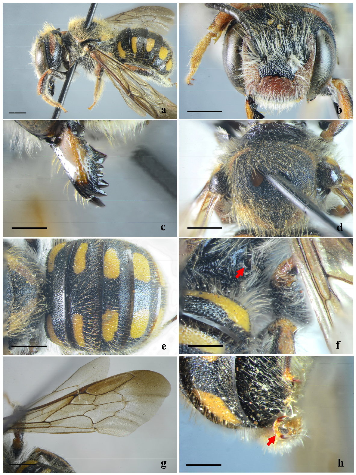
FIGURE 6. Pseudoanthidium (Pseudoanthidium) orientale (Bingham, 1897), female. a: Body in lateral view; b: Head in frontal view; c: Mandible in lateral view; d: Mesosoma in dorsal view; e: Metasoma in dorsal view; f: Propodeum in dorsolateral view, showing the propodeal spiracle; g: Fore wing in frontal view; h; Claw in frontal view. Scale bar: a, b, d, e, f, 1 mm; c, g, h, 0.5 mm.
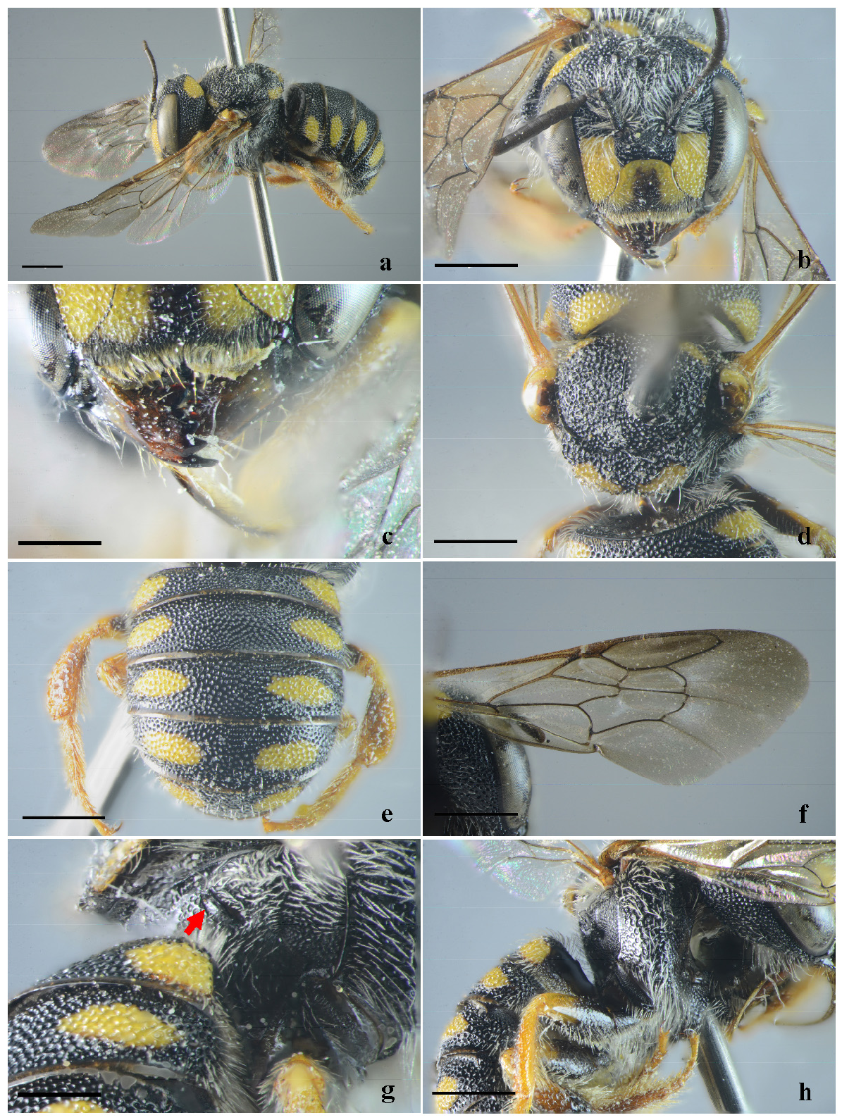
FIGURE 7. Pseudoanthidium (Pseudoanthidium) xinjiangense (Wu, 2004), female, holotype. a: Body in lateral view; b: Head in frontal view; c: Mandible in lateral view; d: Mesosoma in dorsal view; e: Metasoma in dorsal view; f: Propodeum in dorsolateral view, showing the propodeal spiracle; g; Fovea behind the propodeal spiracle; h; Omaulus in lateral view. Scale bar: a, b, d, e, f, h, 1 mm; c, g, 0.5 mm.
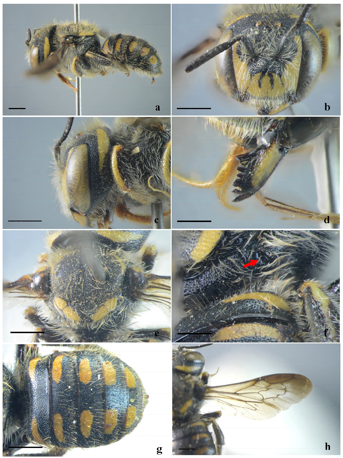
FIGURE 8. Pseudoanthidium (Pseudoanthidium) yanruae Niu & Zhu, sp. nov., female, holotype. a: Body in lateral view; b: Head in frontal view; c: Head in lateral view; d: Mandible in lateral view; e: Mesosoma in dorsal view; f: Propodeum in dorsolateral view, showing the propodeal spiracle; g: Metasoma in dorsal view; h: Wing in frontal view. Scale bar: a, b, c, e, g, h, 1 mm; d, f, 0.5 mm.

FIGURE 9. Pseudoanthidium (Pseudoanthidium) yanruae Niu & Zhu, sp. nov., male, paratype. a: Head in frontal view; b: Mandible in lateral view; c: T5-T7 in dorsal view; d: S4 in frontal view; e: S5-S6 in ventral view; f: S7 in ventral view; g: Genitalia and S8 in frontal view; h: Genitalia in dorsal view. Scale bar: a, b, c, d, e, f, 1 mm; g, h, 0.5 mm.

FIGURE 10. Pseudoanthidium (Pseudoanthidium) kunesense Niu & Zhu, sp. nov., female, holotype. a: Body in lateral view; b: Head in frontal view; c: Head in lateral view; d: Mandible in lateral view; e: Mesosoma in dorsal view; f: Metasoma in dorsal view; g: Wing in frontal view. Scale bar: a, b, c, e, f, g, 1 mm; d, 0.5 mm.

FIGURE 11. Pseudoanthidium (Pseudoanthidium) chenggongense Niu & Zhu, sp. nov., female, holotype. a: Body in lateral view; b: Head in frontal view; c: Head in lateral view; d: Mandible in lateral view; e: Mesosoma in dorsal view; f: Metasoma in dorsal view. Scale bar: a, b, c, e, f, 1 mm; d, 0.5 mm.
No known copyright restrictions apply. See Agosti, D., Egloff, W., 2009. Taxonomic information exchange and copyright: the Plazi approach. BMC Research Notes 2009, 2:53 for further explanation.
|
Kingdom |
|
|
Phylum |
|
|
Class |
|
|
Order |
|
|
Family |
|
|
Genus |