
Stillabothrium campbelli Delgado, Dedrick et Reyda, 2016
|
publication ID |
https://doi.org/10.14411/fp.2016.038 |
|
publication LSID |
lsid:zoobank.org:pub:FE2205B0-4B03-4929-8177-FEA36E9D014D |
|
DOI |
https://doi.org/10.5281/zenodo.8147821 |
|
persistent identifier |
https://treatment.plazi.org/id/03EB87AE-FFFD-FFAA-DF95-8665FD9D2ED7 |
|
treatment provided by |
Felipe |
|
scientific name |
Stillabothrium campbelli Delgado, Dedrick et Reyda |
| status |
sp. nov. |
Stillabothrium campbelli Delgado, Dedrick et Reyda View in CoL sp. n.
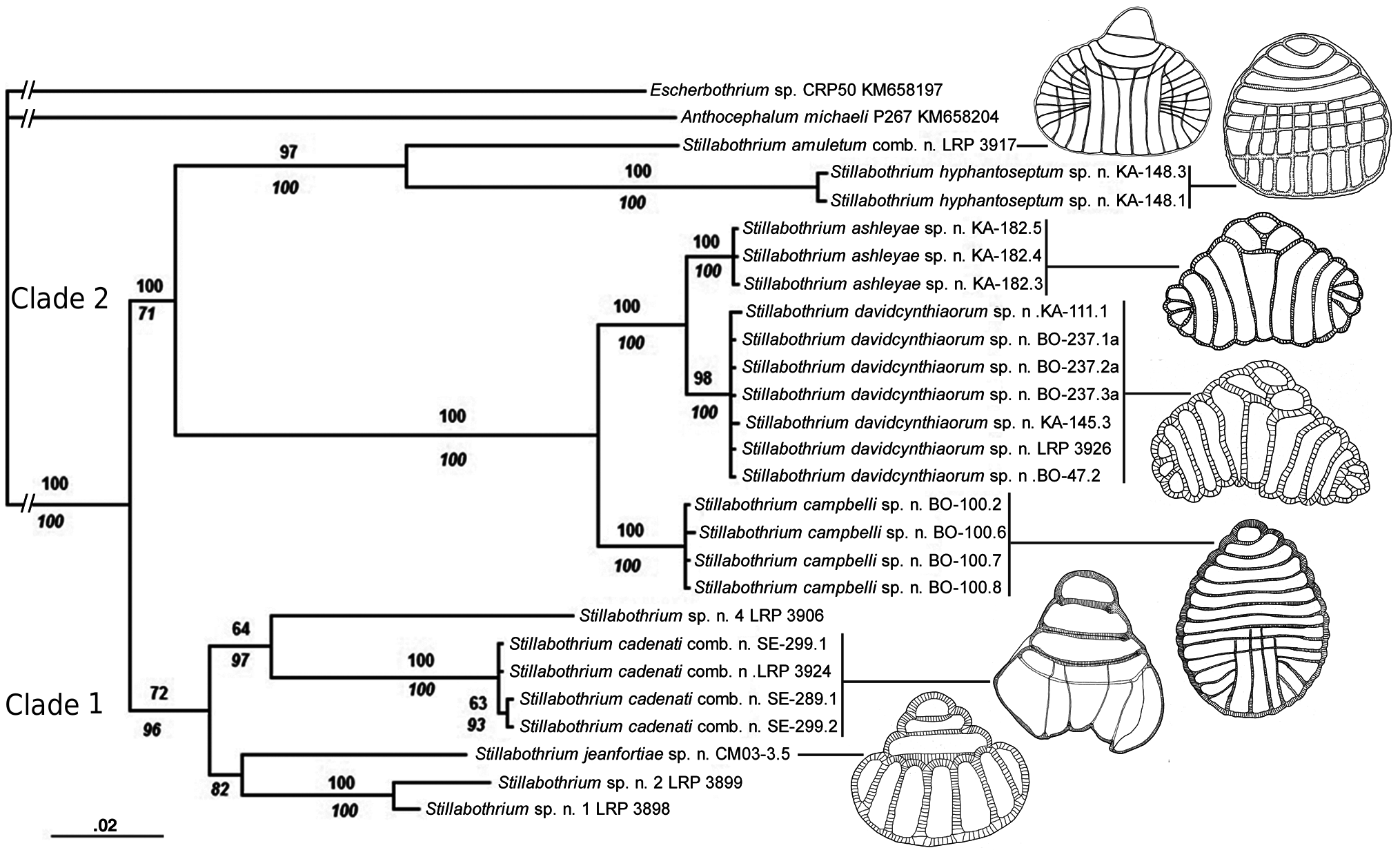
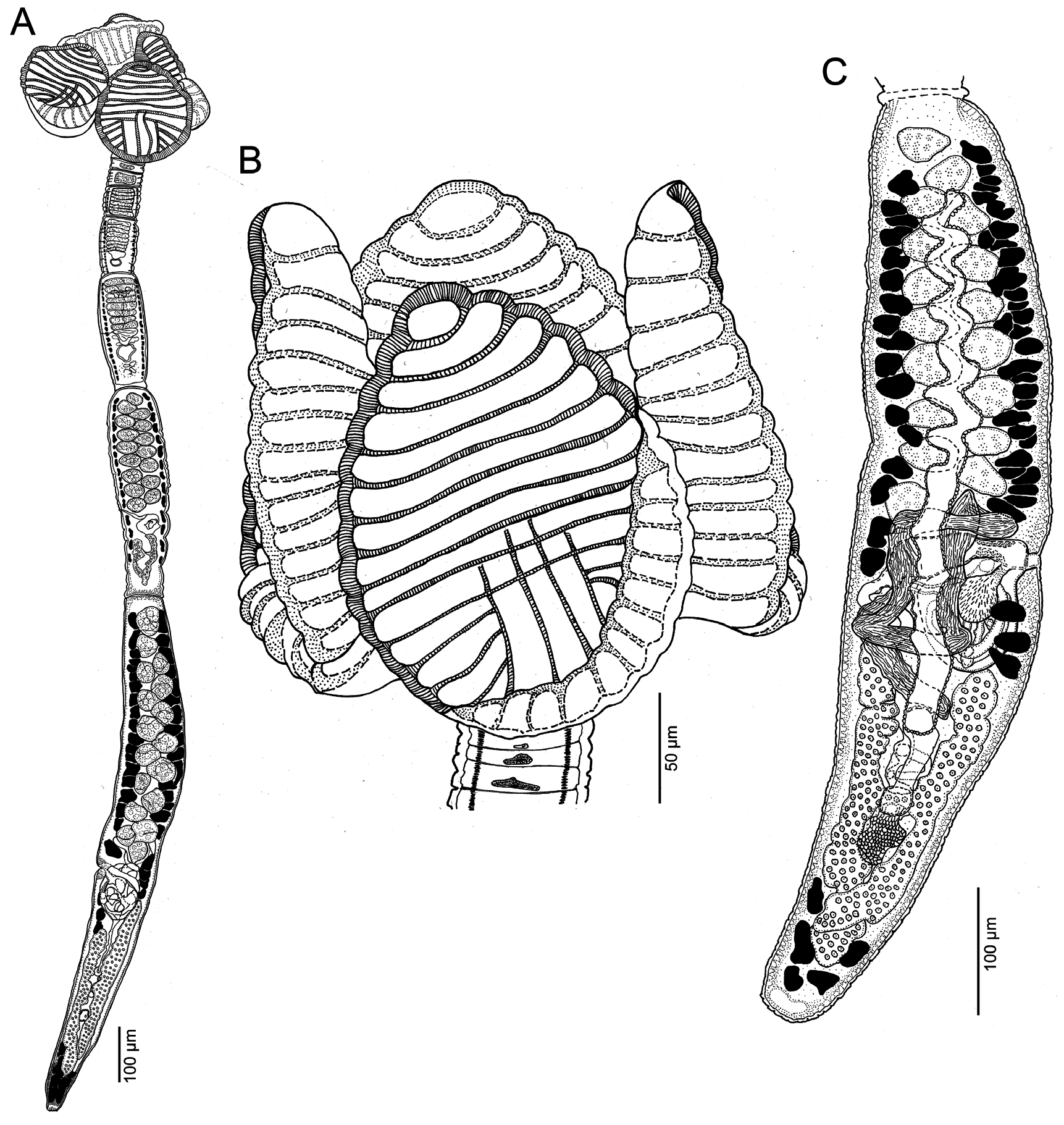
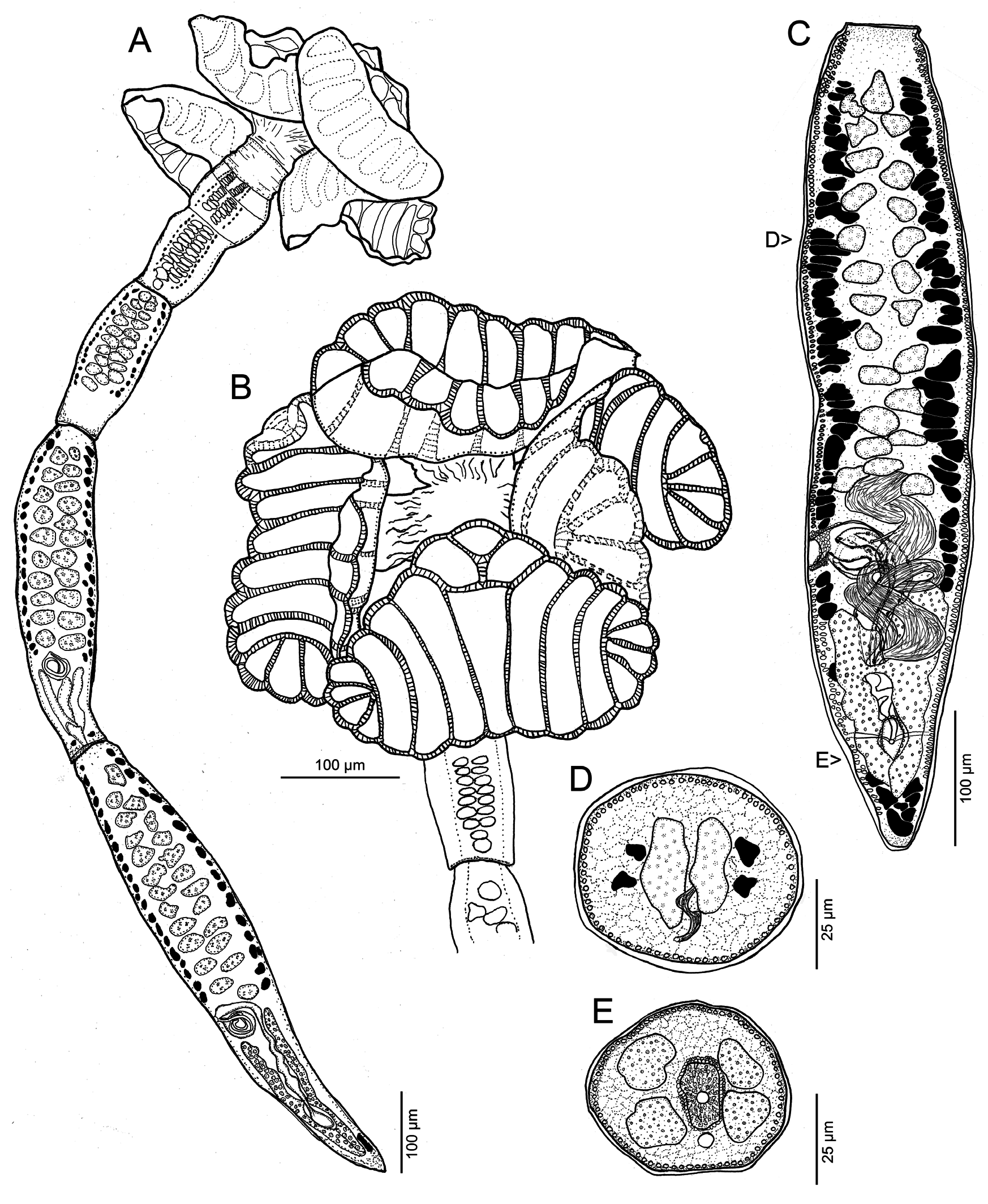
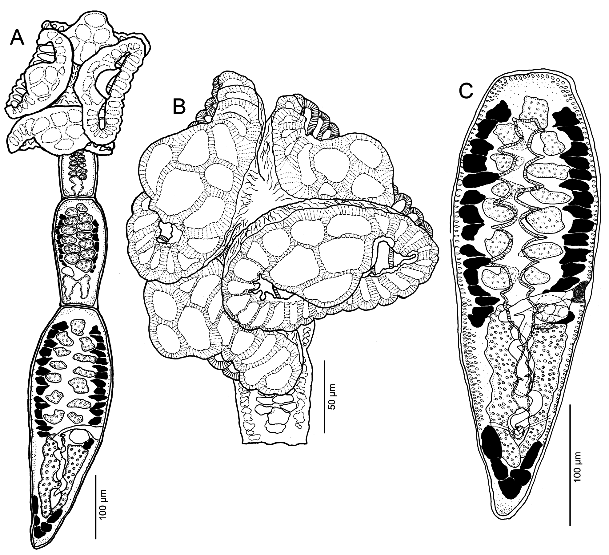
Figs. 1 View Fig , 6 View Fig , 7 View Fig , 16C View Fig
ZooBank number for species:
urn:lsid:zoobank.org:act:
Description (based on whole mounts of 14 complete mature worms, cross sections of 2 strobila and longitudinal sections of 3 scoleces and 3 scoleces prepared for SEM): Worms ( Fig. 6A View Fig ) euapolytic, acraspedote, 1.06–2.60 mm (1.68 ± 0.4; n = 14) long, greatest width 170–360 (261 ± 55; n = 14) at level of scolex; 5–8 (6 ± 1.0; n = 14) proglottids per worm. Cephalic peduncle lacking; small darkly staining germinative zone present.
Scolex ( Fig. 6B View Fig ) consisting of scolex proper bearing 4 stalked bothridia. Stalks 20–90 (49 ± 2; n = 13) long by 25–110 (68 ± 25; n = 13) wide, attached slightly posterior to middle of bothridia. Bothridia varying in shape with degree of contraction; ovoid, broadly ovoid, finely ovoid ( Fig. 6B View Fig ), deeply ovoid ( Fig. 7B View Fig ), very deeply ovoid, to finely deltoid ( Fig. 7A View Fig ), facially loculated, 166–240 (205 ± 22; n = 15) long by 110–235 (150 ± 32; n = 14) wide; bothridial margins with thin rim. Anterior region of bothridia ( Fig. 6B View Fig ) with 10–12 (10.6 ± 0.7; n = 11) horizontally oriented loculi (i.e. loculi wider than long) with 10–12 complete transverse septa. Anteriormost loculus 15–25 (20 ± 4; n = 13) long and 24–35 (31 ± 3; n = 12) wide. Posterior region of bothridia with 4 (n = 11) nonmedial longitudinal septa dividing bothridia into 5 primary loculi longer than wide; outermost primary loculi on each side subdivided by 3–4 (3.7 ± 0.5; n = 9) diagonal septa into 4–5 small subloculi; central 2 longitudinal septa of posterior region overlapping 3–4 (3.3 ± 0.5; n = 6) posteriormost transverse septa of anterior region.
Loculi of distal bothridial surfaces ( Fig. 7C View Fig ) bearing acicular and capilliform filitriches; septa on distal bothridial surfaces ( Fig. 7C View Fig ) bearing acicular and capilliform filitriches and coniform spinitriches. Proximal bothridial rim ( Fig. 7D View Fig ) bearing capilliform filitriches greater in length than those on distal bothridial surfaces ( Fig. 7C View Fig ). Proximal bothridial surfaces away from rim bearing acicular filitriches throughout ( Fig. 7D View Fig ). Posterior margin of proximal bothridial surfaces bearing patch of coniform spinitriches ( Fig. 7E View Fig ) near, but not extending to, bothridial rim. Isolated cilia observed on distal and proximal bothridial surfaces. Bothridial stalks and strobila ( Fig. 7F View Fig ) bearing capilliform filitriches only.
Strobila with 1–4 (2.3 ± 1.0; n = 14) proglottids wider than long followed by 3–5 (3.8 ± 0.6; n = 14) proglottids that are longer than wide. Strobila widest at terminal proglottid; terminal proglottid 490–1 250 (798 ± 209; n = 14) long by 78–180 (125 ± 31; n = 14) wide, genital pore located 41–55% (49 ± 4.7; n = 14) of proglottid length from proglottid posterior margin. Immature proglottids 3–6 (4.5 ± 0.9; n = 14) in number. Mature proglottids 1–2 (1.6 ± 0.5; n = 14) in number, including 0–1 (0.7 ± 0.5; n = 14) vas deferens-mature proglottid.
Testes in mature proglottids 12–19 (15 ± 2.1; n = 14) in total number, 1 layer deep in cross section, arranged in 2 columns; columns extending from anterior margin of proglottid to anterior margin of cirrus sac, 23–42 (33 ± 6; n = 13) long by 28–50 (37 ± 8; n = 13) wide. Vas deferens coiled, entering anterior margin of cirrus sac, extending from level of ovarian isthmus to overlap posteriormost testes. Cirrus sac thin-walled, oval, extending medially well past midline of proglottid; cirrus sac in terminal mature proglottid 42–80 (66 ± 17; n = 4) long by 50–70 (62 ± 9; n = 4) wide; cirrus sac in vas deferens-mature proglottids 45–90 (71 ± 13; n = 9) long by 50–100 (75 ± 18; n = 9) wide. Cirrus spinitriches present.
Vagina ( Fig. 6C View Fig ) thick-walled, sinuous, somewhat overlapping anterior margin of cirrus sac ( Fig. 16C View Fig ), extending from ootype past midline to aporal side of proglottid to anterior margin of cirrus sac, then laterally to open into genital atrium anterior to cirrus sac; vaginal sphincter absent. Seminal receptacle present. Ovary near posterior end of proglottid, H-shaped in frontal view, tetralobed in cross section, overlapping cirrus sac; ovarian lobes symmetrical; poral ovarian lobe somewhat overlapping cirrus sac; poral and aporal ovarian lobes in terminal mature proglottids 135–270 (219 ± 61; n = 5) and 140–275 (219 ± 60; n = 5) long, respectively. Poral and aporal ovarian lobes in vas deferens-mature proglottids 135–425 (258 ± 103; n = 8) and 155–450 (264 ± 108; n = 8) long, respectively. Maximum width of ovary 52–110 (78 ± 22; n = 13). Ovarian isthmus at or anterior to midpoint of ovary; poral lobe of ovary stopping 45–150 (81 ± 21; n = 13) short of genital pore. Mehlis’ gland posterior to ovarian isthmus, 30–45 (39 ± 7; n = 7) long by 25–37 (29 ± 4; n = 7) wide. Vitellarium follicular; vitelline follicles arranged in 1 dorsal and 1 ventral column on each side of proglottid; columns extending from anterior to posterior margin of proglottid, interrupted by terminal genitalia, and mostly interrupted by ovary ( Fig. 6A,C View Fig ). Uterus ventral, sacciform, extending from near isthmus of ovary to near anterior margin of proglottid.
T y p e a n d o n l y k n o w n h o s t: Himantura cf. pastinacoides ( Myliobatiformes : Dasyatidae ).
Ty p e l o c a l i t y: Sulu Sea off Kampung Tetabuan ( 06°01'10''N; 117°42'15''E), Sabah, Malaysian Borneo (BO-98, BO-100, BO-119) GoogleMaps .
A d d i t i o n a l l o c a l i t i e s: Sulu Sea off Sandakan ( 05°50'20''N; 118°07'16''E), Sabah, Malaysian Borneo (BO-79). GoogleMaps South China Sea off Sematan ( 01°48'15''N; 109°46'47''E), Sarawak, Malaysian Borneo (BO-168) GoogleMaps .
S i t e o f i n f e c t i o n: Spiral intestine.
Ty p e m a t e r i a l: Holotype MZUM (P) No. 2016.11 (H). Paratypes: IPCAS No. C-738; LRP Nos. 8995–8997; 9063– 9082; 9149 (including molecular vouchers, sections and SEM specimens); MZUM (P) No. 2016.12 (P); SBC No. P-00071; USNM Nos. 1420467–1420470 .
E t y m o l o g y: This species is named in honour of Dr. William Campbell, Nobel laureate, for his role in the development of drugs to fight parasitic infections.
Remarks. Stillabothrium campbelli sp. n. is the only Stillabothrium species treated in this study that possesses a greater number of anterior loculi than posterior loculi on the bothridia. Stillabothrium campbelli can be distinguished from both S. ashleyae and S. davidcynthiaorum in the unique configuration of the septa and loculi on its bothridia. In terms of the anterior region of the bothridia, S. campbelli possesses a total of 10–12 loculi ( Fig. 6B View Fig ) whereas both S. ashleyae and S. davidcynthiaorum each possess only 3 loculi ( Figs. 2B View Fig , 4B View Fig ). In the posterior region of the bothridia, S. campbelli possesses 5 loculi that are longer than wide whereas both S. ashleyae and S. davidcynthiaorum respectively possess 7 or 9 loculi that are longer than wide ( Figs. 2B View Fig , 4B View Fig ). In addition, the longitudinal septa in the posterior bothridia of S. campbelli extend anteriorly such that they overlap with the 3–4 posteriormost transverse septa of the anterior region of the bothridia. In S. ashleyae and S. davidcynthiaorum , the septa do not overlap.
Scolex microtriches can also be used to distinguish S. campbelli from S. ashleyae and S. davidcynthiaorum . The posterior proximal surface of bothridia in S. campbelli possess a patch of coniform spinitriches (in addition to acicular filitriches throughout) near the rim, that is restricted in distribution, whereas the proximal bothridial surface of S. ashleyae and S. davidcynthiaorum is evenly covered with coniform spinitriches and acicular filitriches. Finally, proglottid morphology can also be used to distinguish S. campbelli from S. ashleyae and S. davidcynthiaorum . The cirrus sac of S. campbelli sp. n. is larger (45–90 µm long by 50–100 µm wide) than those of S. ashleyae (26–42 µm long by 26–47 µm wide) and of S. davidcynthiaorum (18–35 µm long by 24–38 µm wide), and it extends further across the midline ( Figs. 6C View Fig , 16C View Fig ), than in the latter two species ( Figs. 2C View Fig , 4C View Fig , 16A,B View Fig ).
Stillabothrium campbelli is reported from H. cf. pastinacoides from three different localities in Malaysian Borneo. It should be noted that the stingray host specimens of S. campbelli in this study, BO-61, BO-79, BO-98, BO-100, BO-119 and BO-168, are identified as H. cf. pastinacoides ( Caira et al. 2012) , but that identification awaits expert verification. Given that Naylor et al. (2012b) showed that two species of stingray similar to H. pastinacoides (i.e. H. cf. pastinacoides 1 and H. cf. pastinacoides 2) co-occur in the region sampled, it is possible that S. campbelli sp. n. parasitises either or both. Additional specimens morphologically similar to S. campbelli were encountered in H. uarnacoides (BO-118). Further studies are needed to determine whether Stillabothrium specimens from H. uarnacoides are conspecific with S. campbelli or represent an undescribed species of Stillabothrium .
Four specimens of S. campbelli were included in the phylogenetic analysis ( Fig. 1 View Fig , Table 1 View Table 1 ).
| USNM |
Smithsonian Institution, National Museum of Natural History |
No known copyright restrictions apply. See Agosti, D., Egloff, W., 2009. Taxonomic information exchange and copyright: the Plazi approach. BMC Research Notes 2009, 2:53 for further explanation.
|
Kingdom |
|
|
Phylum |
|
|
Class |
|
|
Order |
|
|
Family |
|
|
Genus |