4.2. Diagnostic features of
E. remanei
View in CoL
Echinoderes remanei
View in CoL
belongs to the most common group of
Echinoderes
View in CoL
regarding its spine pattern. Five middorsal spines and lateroventral tubes/spines on segments 5 to 9 are shared with 46 congeners ( Yamasaki et al., 2020). However, when we combine these characters with the presence of glandular cell outlets type 2 in subdorsal, laterodorsal, midlateral/sublateral and ventrolateral positions on segment 2, we shorten the possibilities to 10 species, i.e.,
Echinoderes angustus Higgins & Kristensen, 1988
View in CoL
,
Echinoderes aquilonius Higgins & Kristensen, 1988
View in CoL
,
Echinoderes beringiensis Adrianov & Maiorova, 2022
View in CoL
,
Echinoderes cernunnos Sørensen et al., 2012
View in CoL
,
Echinoderes galadrielae Grzelak and Sørensen, 2022
View in CoL
,
Echinoderes juliae Sørensen et al., 2018
View in CoL
,
Echinoderes obtuspinosus Sørensen et al., 2012
View in CoL
,
Echinoderes pennaki Higgins, 1960
View in CoL
,
Echinoderes romanoi Landers & Sørensen, 2016
View in CoL
, and
Echinoderes xalkutaat Cepeda et al., 2019
View in CoL
( Higgins 1960; Sørensen et al., 2012; Landers & Sørensen 2016; Grzelak & Sørensen 2018; Herranz et al., 2018; Sørensen et al., 2018; Cepeda et al., 2019; Adrianov & Maiorova 2022; Grzelak & Sørensen 2022). Nevertheless,
E. beringiensis
View in CoL
,
E. cernunnos
View in CoL
,
E. galadrielae
View in CoL
,
E. romanoi
View in CoL
and
E. xalkutaat
View in CoL
can immediately be distinguished from
E. remanei
View in CoL
by the absence of glandular cell outlets type 2 on segment 4. Instead,
E. cernunnos
View in CoL
shows a pair of glands on segment 7 and very characteristic long and spiniform tergal extensions, while
E. romanoi
View in CoL
and
E. xalkutaat
View in CoL
are (in addition to their lack of gco2 on segment 4) much smaller and show different shapes of tergal extensions ( Sørensen et al., 2012; Landers & Sørensen 2016; Cepeda et al., 2019).
E. juliae
View in CoL
and
E. obtuspinosus
View in CoL
, although possessing a subdorsal pair of glandular cell outlets type 2 on segment 4, have an extra pair of glands on segment 3 (
E. juliae
View in CoL
) or can be easily differentiated from
E. remanei
View in CoL
by the presence of very short and stout lateral terminal spines (
E. obtuspinosus
View in CoL
) ( Sørensen et al., 2012; 2018).
Of the abovementioned species,
E. remanei
View in CoL
shows a much closer general resemblance with
E. aquilonius
View in CoL
,
E. angustus
View in CoL
and
E. pennaki
View in CoL
. Interestingly, the first two have been described from Disko Island, western Greenland, along with
E. tubilak ( Higgins & Kristensen 1988)
View in CoL
. The latter one, on the other hand, has been described from the San Juan Archipelago in northwest Washington, USA, and belongs to one of the first Echinoderes species described by Higgins (1960). Several years later, the species was redescribed by Higgins (1977) and recently by Herranz et al. (2018).
Echinoderes remanei
View in CoL
share identical spine patterns, distribution of glandular cell outlets type 2, and even midventral partial articulation of segment 2 ( Grzelak & Sørensen 2018; Herranz et al., 2018) with
E. aquilonius
View in CoL
,
E. angustus
View in CoL
and
E. pennaki
View in CoL
; the only exception is the absence of laterodorsal glandular cell outlets type 2 on segment 10 in
E. pennaki
View in CoL
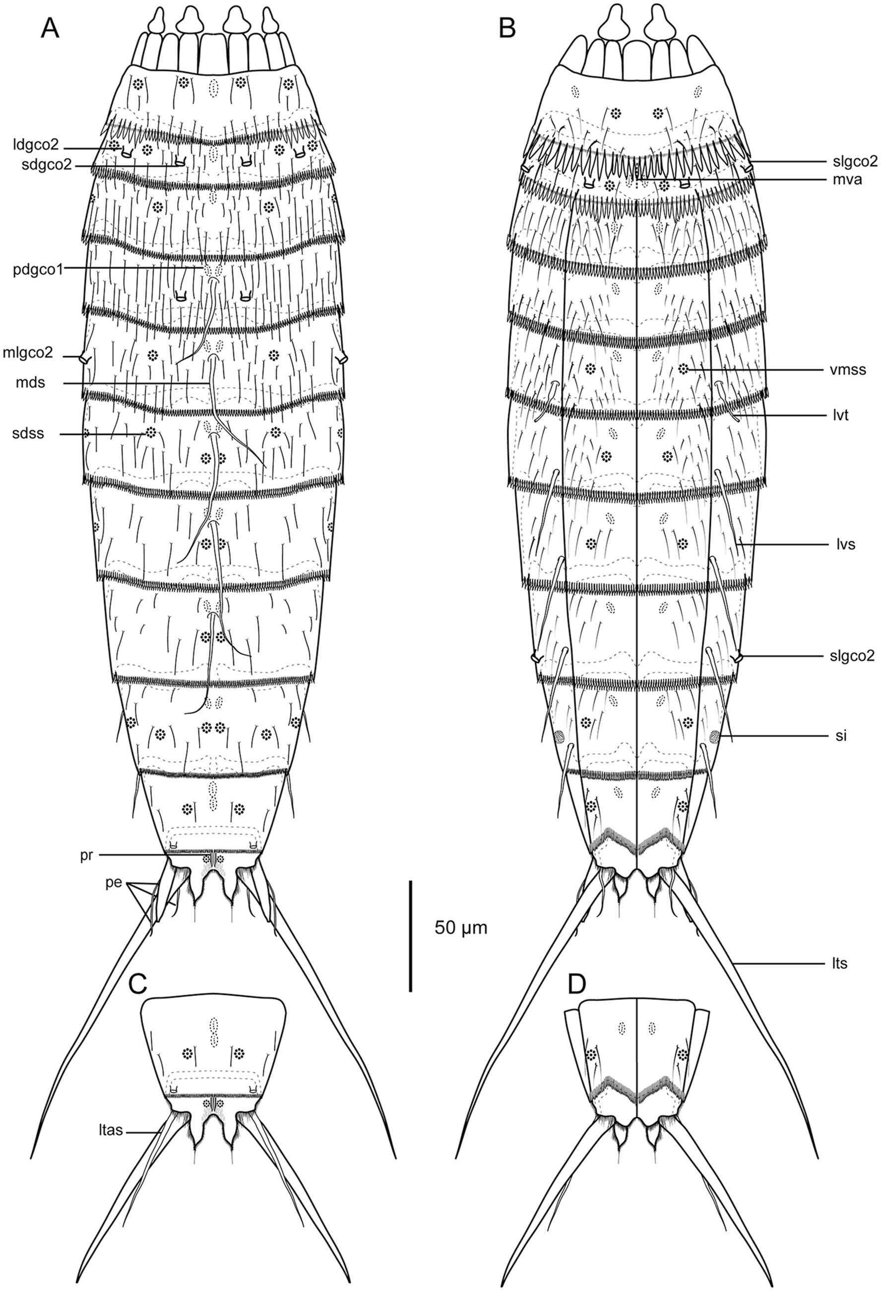
; the lack of this trait has been confirmed using SEM by Herranz et al. (2018). Therefore, the easiest and most reliable way to Abbreviation s: LA lateral accessory; LD laterodorsal; LV lateroventral; MD middorsal; ML midlateral; PD paradorsal; PV paraventral; SD subdorsal; SL sublateral; VL ventrolateral; VM ventromedial; ac acicular spine; gco1/2 glandular cell outlet type 1/2; ltas lateral terminal accessory spine; lts lateral terminal spine; pe penile spine; pr protuberance; si, sieve plate; ss sensory spot; tu tube; ♀ female condition of sexually dimorphic characters; 6 male condition of sexually dimorphic characters.
distinguish
E. remanei
View in CoL
from its three congeners is by using morphometric differences in combination with a comparison of the pectinate fringe on segments 1 and 2. Unlike
E. remanei
View in CoL
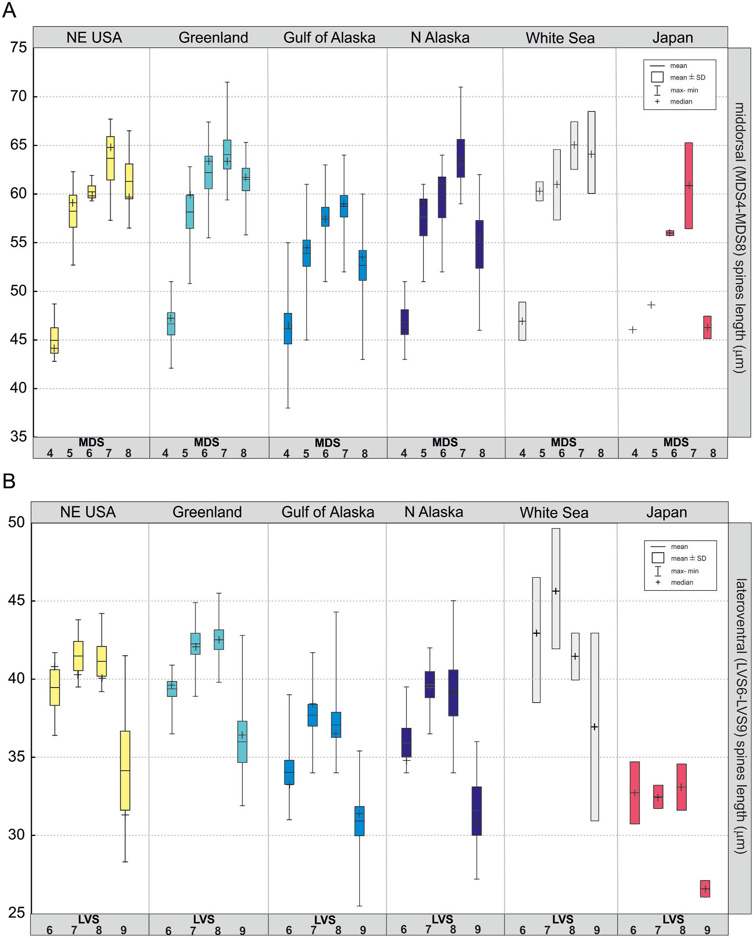
, none of these species is characterized by the middorsal spine on segment 8 being significantly shorter than the one on segment 7 ( Table 2; Fig. 8
View Fig
) ( Higgins & Kristensen 1988; Herranz et al., 2018). This feature, which is unusual among
Echinoderes
View in CoL
, is consistently found in all individuals in
E. remanei
View in CoL
, irrespective of sampling location. Moreover,
E. angustus
View in CoL
and
E. pennaki
View in CoL
, in contrast to
E. remanei
View in CoL
, do not show significant differences in lateral terminal spine lengths between males and females ( Higgins & Kristensen 1988; Herranz et al., 2018). Even if only the length of spines for males would be compared, both species have longer spines in comparison to those of
E. remanei
View in CoL
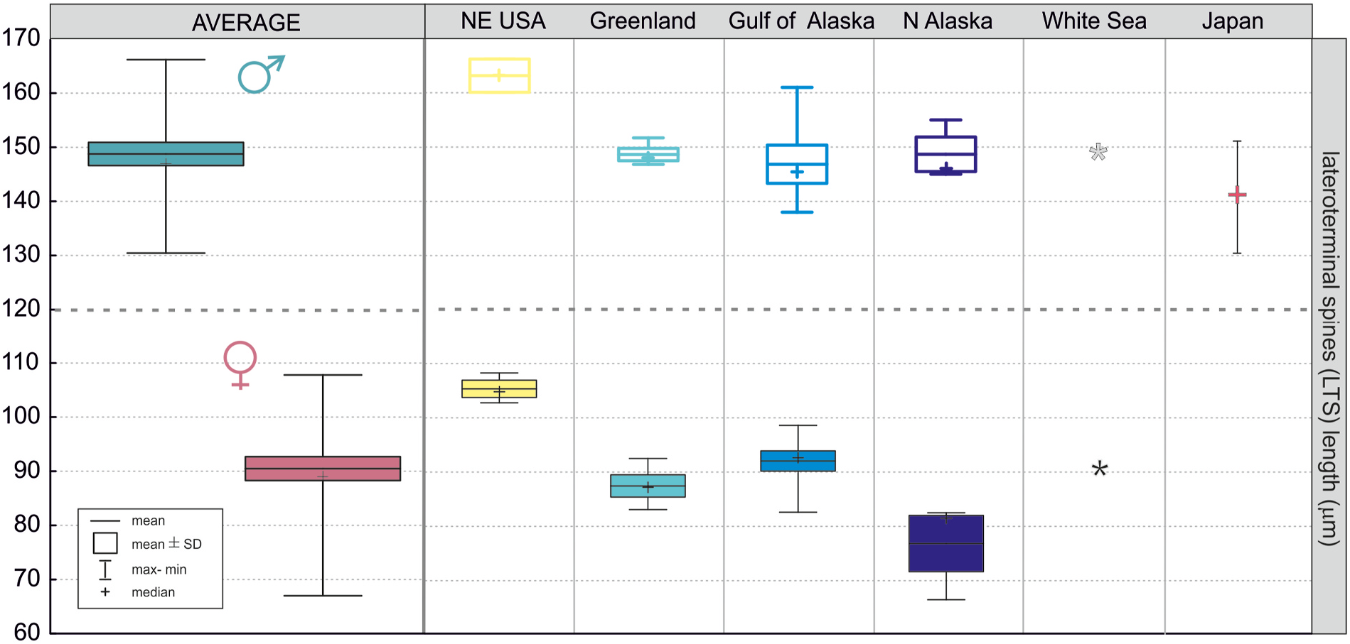
(avg. LTSm: > 170 μm vs 149 μm, respectively).
Echinoderes aquilonius
View in CoL
is, however, similar to
E. remanei
View in CoL
in sexual dimorphism expressed in longer lateral terminal spines in males than in females ( Higgins & Kristensen, 1988; own data), but still both species can be distinguished from one another since the lateral terminal spines in
E. aquilonius
View in CoL
are shorter than those in
E. remanei
View in CoL
(avg. LTS ♀: 70 μm vs 90 μm, respectively; avg. LTS 6: 135 μm vs. 149 μm, respectively). In addition, all three species differ from
E. remanei
View in CoL
by being larger (avg.TL: 405–460 μm vs. 369 μm), and having markedly less conspicuous and more uniform midventral pectinate fringe on segment 1 and 2.
It seems that the morphometric pattern for middorsal and terminal spines itself represents a rather unique feature for
E. remanei
View in CoL
. In species of
Echinoderes
View in CoL
segment 8 usually has the longest middorsal spine, or is at least similar in length to the spine on segment 7. Therefore, a significantly shorter spine on segment 8 certainly stands out and distinguishes
E. remanei
View in CoL
among other
Echinoderes species.
Also, the sexual dimorphism expressed in lateral terminal spine lengths is not a very common trait. According to our knowledge, this trait has only been observed for five other species so far, i.e.,
E. aquilonius
View in CoL
,
Echinoderes bengalensis ( Timm, 1958)
View in CoL
,
Echinoderes blazeji Grzelak & Sørensen, 2022
View in CoL
,
Echinoderes coulli Higgins, 1977
View in CoL
, and
Echinoderes lusitanicus Neves et al., 2016
View in CoL
( Higgins 1977; Higgins & Kristensen 1988; Neves et al., 2016; Grzelak & Sørensen 2022; Sørensen, unpubl. obs). The difference in length due to sexual dimorphism observed in
E. remanei
View in CoL
is very conspicuous ( Table 2; Figs. 2
View Fig
and 9
View Fig
) and the size ranges between the sexes never overlapped ( Fig. 9
View Fig
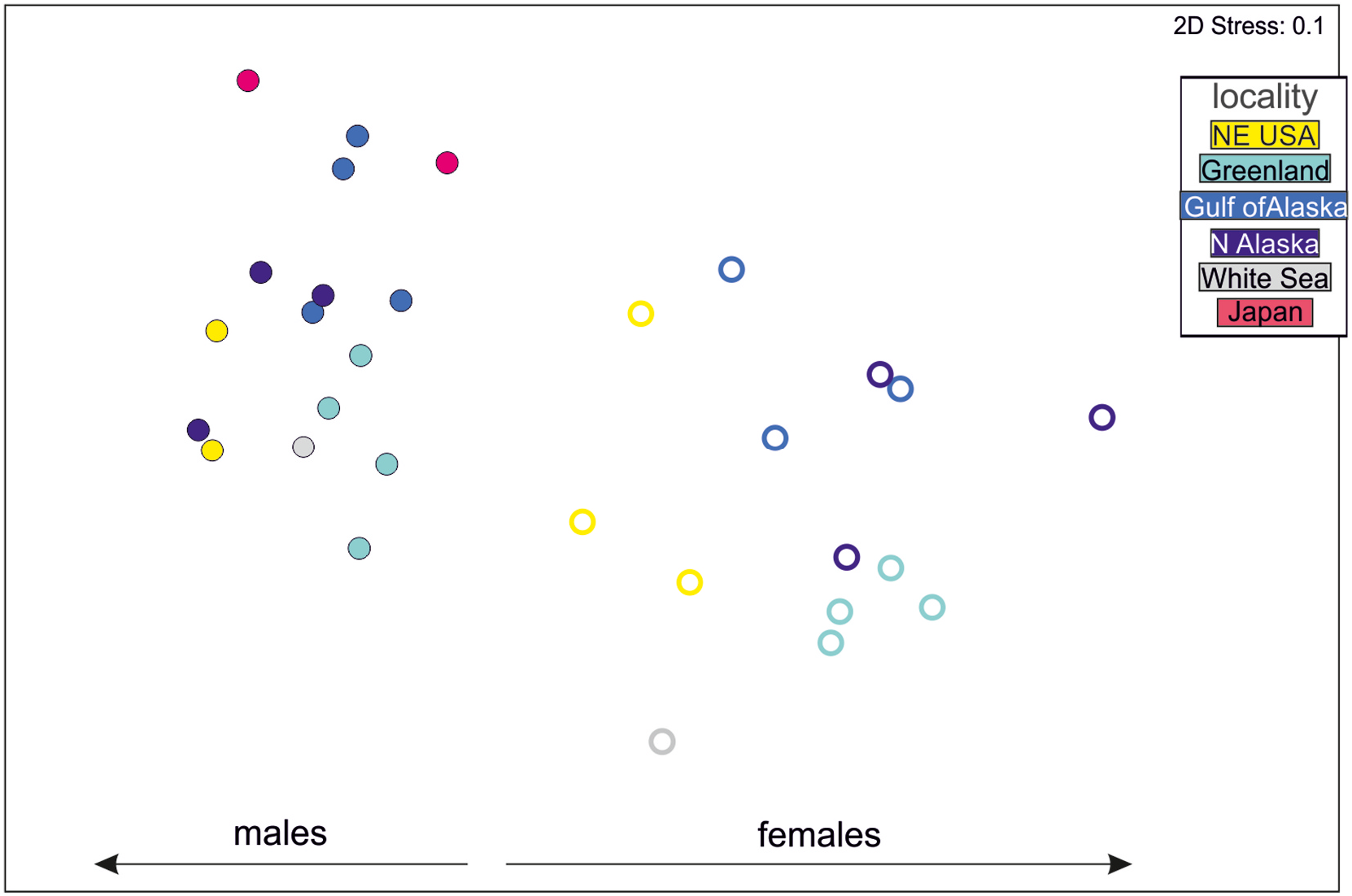
); males were characterized by considerably longer lateral terminal spines than those in females, and this pattern was very consistent for all localities. Consequently, when all morphometric data were taken into account, regardless of sampling location males were plotted closer together and form a distinct group ( Fig. 10
View Fig
, left side of the plot) in comparison to females ( Fig. 10
View Fig
, right side of the plot). This result only emphasizes that the specimens, despite the large geographic distance between their locations, are indeed morphologically highly similar, and that differences between the sexes are consistent and driven by the length of lateral terminal spines.