
Maemonstrilla longipes, (A. SCOTT, 1909), 2008
|
publication ID |
https://doi.org/ 10.1111/j.1096-3642.2007.00381.x |
|
publication LSID |
lsid:zoobank.org:pub:942A51C7-EE79-4C60-8B80-9EA8835889CC |
|
persistent identifier |
https://treatment.plazi.org/id/03EBB12D-F773-0C01-FF37-EFB6E85B7C87 |
|
treatment provided by |
Felipe |
|
scientific name |
Maemonstrilla longipes |
| status |
comb. nov. |
MAEMONSTRILLA LONGIPES (A. SCOTT, 1909)
COMB. NOV.
Monstrilla longipes A. Scott, 1909: 238 , pl. LVIII, figs 3–4. Davis 1949: 248 (footnote), 249; Sewell 1949: 141; Wickstead 1961: 61, 62; Zheng, Li & Xu 1984, 1989: both fig. 141H.
Monostrilla longipes – Al-Kholy 1963: 130, fig. I, 1–4.
Remarks: Scott (1909) described and illustrated (habitus dorsal view, detail of reticulation and legs 5) this species based on a single, 1.85-mm-long female captured during the Siboga Expedition at sta. 142, off Laiwui, coast of Obi Major, 0°24 ′ S, 127°36 ′ E, northern part of Ceram Sea, Indonesia. In 1994 this holotype was not found together with the other Siboga monstrilloids that are deposited in the Institute for Systematics and Population Biology, University of Amsterdam, and it is presumed lost. The description is detailed enough to recognize Monstrilla longipes as a member of Maemonstrilla ( M. hyottoko species group) on the basis of its body form, the spinose-reticulate cuticle of the cephalothorax and free metasomal pedigers, and the form and setation of leg 5. A suture crosses the dorsum of the genital segment, as in most species of Maemonstrilla . The ovigerous spines were not described. It is possible that one of the newly described species of the M. hyottoko species group described herein (but not M. simplex ) is synonymous with M. longipes , but this cannot be demonstrated conclusively.
More recent, non-illustrated records of supposed female Monstrilla longipes from Nankauri Harbour, Nicobar Islands ( Sewell, 1949; 2.25 mm long with a groove subdividing the genital segment) and the Singapore Strait ( Wickstead, 1961), and a poorly illustrated record from Ghardaqa, Egypt, on the Red Sea ( Al-Kholy, 1963), must be considered unsubstantiated at the specific level, although the specimens involved presumably do belong to the Maemonstrilla hyottoko species group. The 1.55-mm-long Egyptian specimen has a minute, non-setose, endopodal lobe on leg 5 ( Al-Kholy, 1963: fig. I, 4), and so it very likely represents an undescribed species. Perhaps the ‘short hairs’ along the outer surface of the antennule are optically sectioned, high cuticular ridges such as those present in M. hyottoko . The ovigerous spines are 0.63 mm long but their direction was not mentioned. We have not tried to borrow this specimen for redescription.
MAEMONSTRILLA TURGIDA SPECIES GROUP (MONOTYPIC) MAEMONSTRILLA TURGIDA (A. SCOTT, 1909) COMB. NOV.
FIGURES 1B View Figure 1 , 13B View Figure 13 , 24–29 View Figure 24 View Figure 25 View Figure 26 View Figure 27 View Figure 28 View Figure 29
Monstrilla turgida A. Scott, 1909: 239 , pl. LVIII, figs 5–6. Davis, 1949: 248 (footnote), 250; Wickstead, 1961: 61, 62; Martin Thompson & Meiyappan, 1980: 207, fig. 1.
[?] Monstrilla sp. – Krishnaswamy 1953: 75, textfigs 17–18.
Diagnosis: Cephalothorax non-reticulated, but with faint encircling striations. Cuticle appearing smooth by light microscopy, but patches of minute spinules revealed by SEM dorsally on metasomal pedigers and all urosomal segments except telson, and on outer face of coxa of legs 1–4. Pair of spinulose, spine-like scales posteriorly near dorsal midline on both first and second free pedigers. Outer basis seta of leg three shorter than exopod. Inner seta present on first segment of each ramus of legs 1–4. Leg 5 bilobed, its exopodal lobe with three setae and endopodal lobe with one seta (perhaps
KEY TO THE RYUKYU SPECIES OF THE MAEMONSTRILLA HYOTTOKO SPECIES GROUP
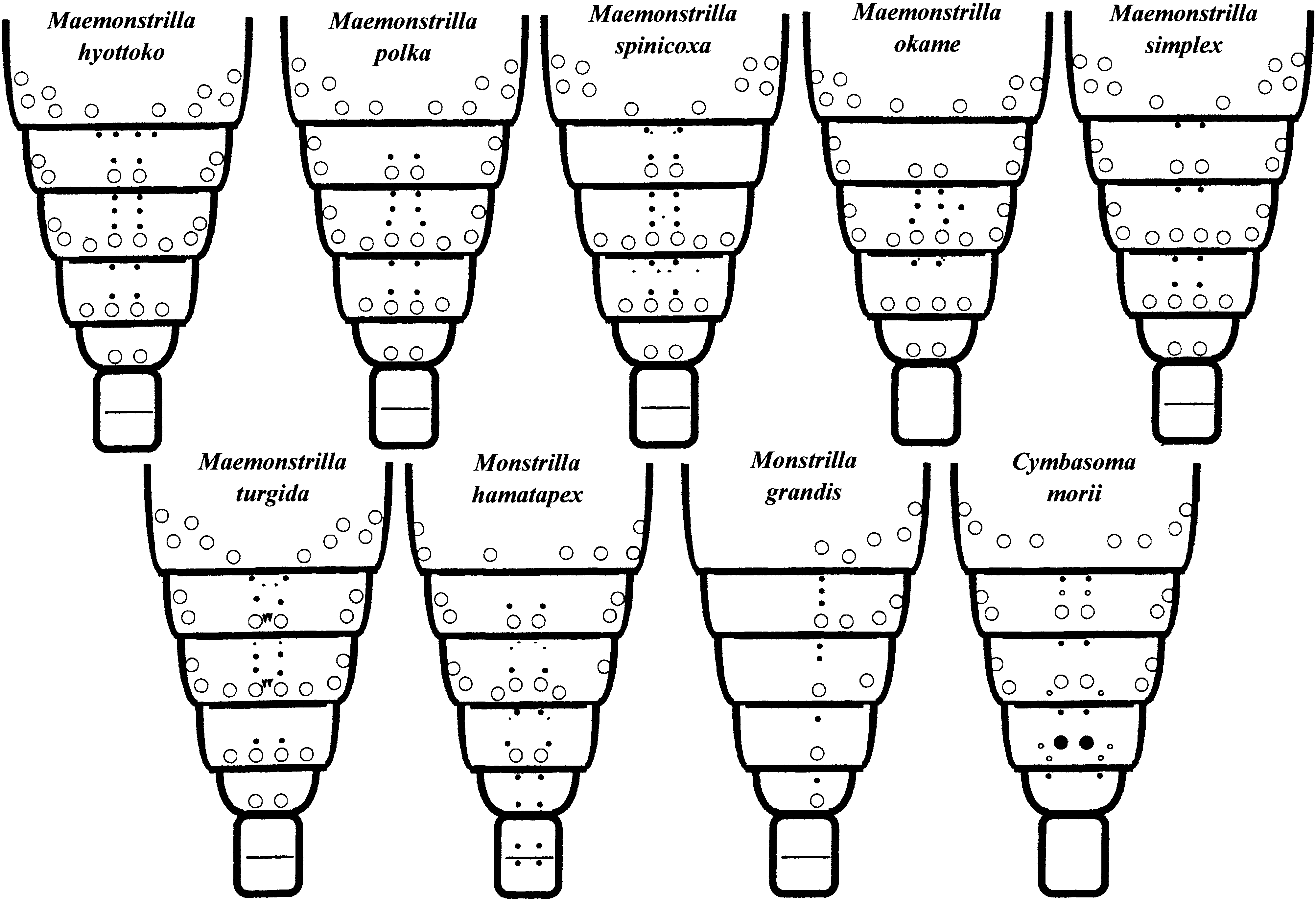
1. (a) Cuticle of cephalothorax, antenules, lateral sides of trunk, dorsum of telson, and caudal rami reticulated; outer faces of legs 1–4 and dorsum of free pedigers, genital compound somite, and penultimate somite spinulose. Cephalothoracic reticulations comprising ridges with abundant or sparse spinules; simple or complex cuticular ornamentation within at least some meshes...........................................................................................2 (b) Only cephalothorax reticulated, and no obviously spinulose regions present. Ridges of cuticular meshwork lacking spinules, and cuticular surface within meshes plain......................................................... M. simplex
2. (a) Oral papilla large and conical, directed ventrally. Genital compound somite distinctly divided by transverse ridge ...............................................................................................................................................3 (b) Oral papilla small, directed more anteriorly than ventral and flanked by ‘puffed cheeks’. Genital compound somite lacking dorsal transverse ridge ....................................................................................... M. okame
3. (a) Legs 1–4 each with two low, rounded lobes on outer proximal part of coxa; rest of outer face of coxa with fields of minute spinules separated by bare lanes. Cephalothoracic reticulatations lacking side branches but bearing sporadic or sparse spinules; meshes uniformly polygonal over most of surface..............................................4 (b) Legs 1–4 lacking such coxal lobes; outer face of coxa evenly covered with larger spines. Cephalothoracic reticulatations with many side branches; besides polygonal meshes anteriorly and posteriorly, wide zone of narrow meshes present behind level of oral papilla .............................................................................. M. hyottoko
4. (a) Proximal coxal lobes of legs 1–4 bearing spinules only slightly larger than those on rest of coxa. Body length (as defined herein)> 2 mm. Spherical pigmented spots (red when fresh) found throughout body. Posteroventral part of genital compound somite produced into apron-like flap; posterodorsal margins of this somite and penultimate somite not denticulate .............................................................................................................. M. polka (b) Proximal coxal lobes of legs 1–4 bearing large, bluntly rounded denticles. Body length <2 mm, and pigment spots absent. Posteroventral part of genital compound somite produced into spur; posterodorsal margins of this somite and penultimate somite distinctly denticulate.......................................................................... M. spinicoxa up to two setae). No ventral protrusion of posterior part of genital compound segment.
Distribution: ‘Siboga’ Expedition sta. 142, northern Ceram Sea, Indonesia; Kundugal Channel and Minicoy Island lagoon, India; east of Linggau Archipelago, South China Sea; Ryukyu Islands.
Material examined: Syntype female, ‘ Siboga’ Expedition sta. 142, off Laiwui , coast of Obi Major , 0°24 ′ S, 127°36 ′ E, northern part of Ceram Sea , Indonesia, on loan from Institute for Systematics and Population Biology (Zoölogisch Museum), University of Amsterdam (Cat. no. ZMA Co. 201478) GoogleMaps .
Twelve females collected at Sesoko Island by M. J. Grygier (on first occasion, with H. Ueda), intact unless otherwise noted: one on 12.vii.1988 ( USNM 1093750 View Materials ), one on 26.xii.1988 ( LBM Reg. No. 1430000932); two on 7.vii.1989 (SO lab; both used for SEM), three on 13.viii.1989 ( ZMA Co. 205910), two on 31.viii.1989 ( KMNH IvR 700 224, 700 225; latter including slide with legs 1–4, used for microscopic drawings), one on 28.ix.1992 ( USNM 1093751 View Materials ), one on 2.x.1992 ( NHM Reg. No. 2006. 1912), one on 9.vii.1996 ( LBM Reg. No. 1430000933) .
Twelve females collected at Ishigaki Island, all intact: one at Kabira Bay, 9.iii.1993, by M. J. Grygier ( NHM Reg. no. 2006. 1913); two on south coast, 30.iv.1994, by S. Ohtsuka ( KMNH IvR 700 226, 700 227; former used for photography); 9 off Kabira, 14.iv.1996, by A. Murase (intended for LBM, but now misplaced) .
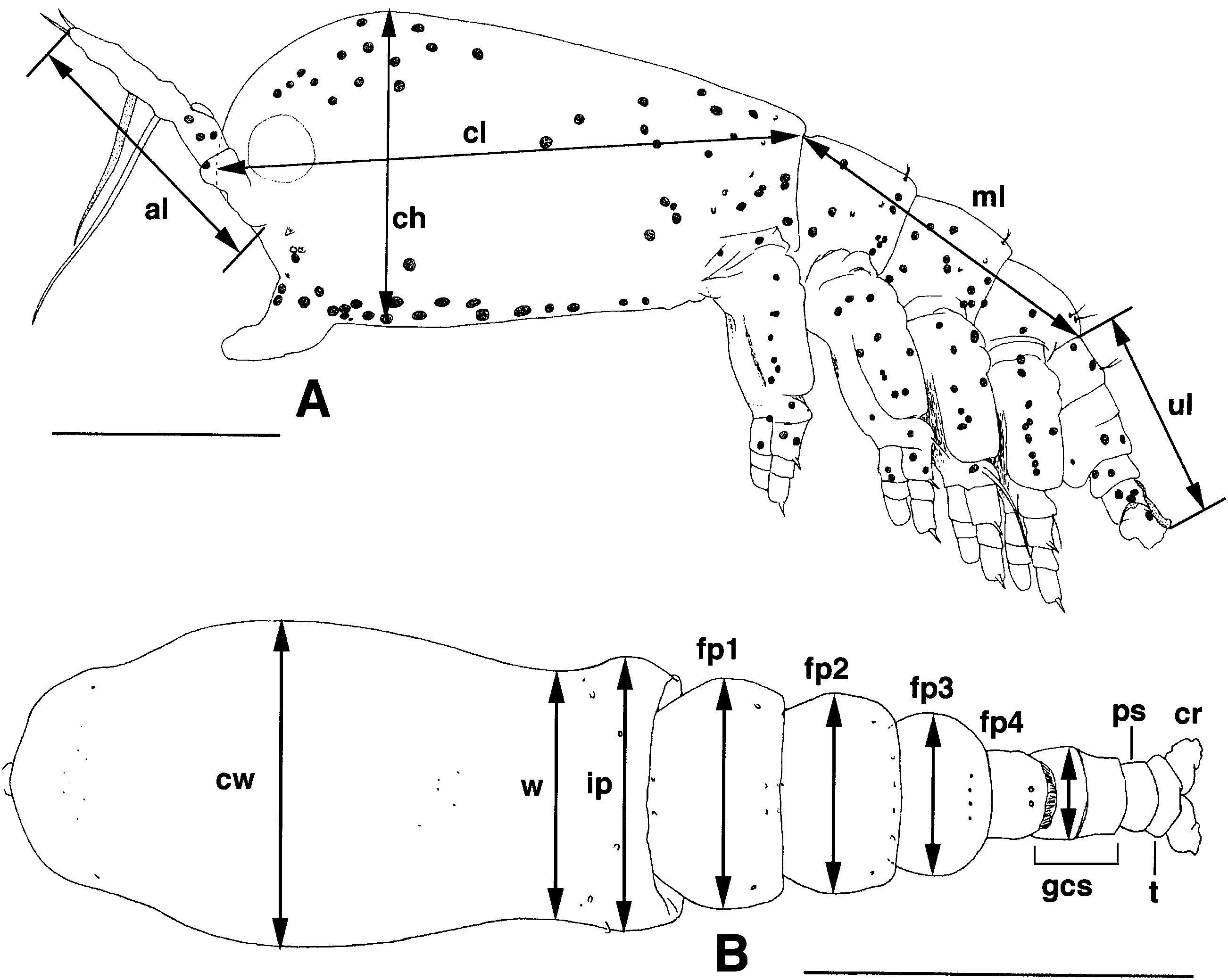
Description of Ryukyuan specimens: Overall appearance with bulbous cephalothorax much the same as in M. hyottoko , but considerably larger and with relatively longer ovigerous spines ( Figs 1B View Figure 1 , 13B View Figure 13 , 24A View Figure 24 , 25A View Figure 25 ). Measurements taken from ten or (for some) 11 specimens, excluding specimens from Kabira in 1996. Length in lateral view (sum of lengths of cephalothorax, metasome and urosome as defined in Fig. 1A View Figure 1 ) 1.38–2.21 mm, with these body regions contributing 49.4–54.8, 26.1–30.4 and 17.2–20.8%, respectively (this sum exceeding body length as seen in dorsal view by 2.4–8.7%; largest individual 2.11 mm long in dorsal view). Height and greatest width of cephalothorax, respectively, 40.9–57.9 and 41.9–57.9% of cephalothorax length; in several specimens height and width equal. Antennule length 41.6–51.3% that of cephalothorax. Width of incorporated pediger 68.4–88.5% of greatest width. Widths of succeeding three free pedigers and genital compound somite relative to that of incorporated pediger 76.1–89.1, 64.8–77.2, 53.8–63.5 and 30.7–40.3%, respectively. Ovigerous spines (seven pairs measured) 45.1–56.1% as long as body in dorsal view.
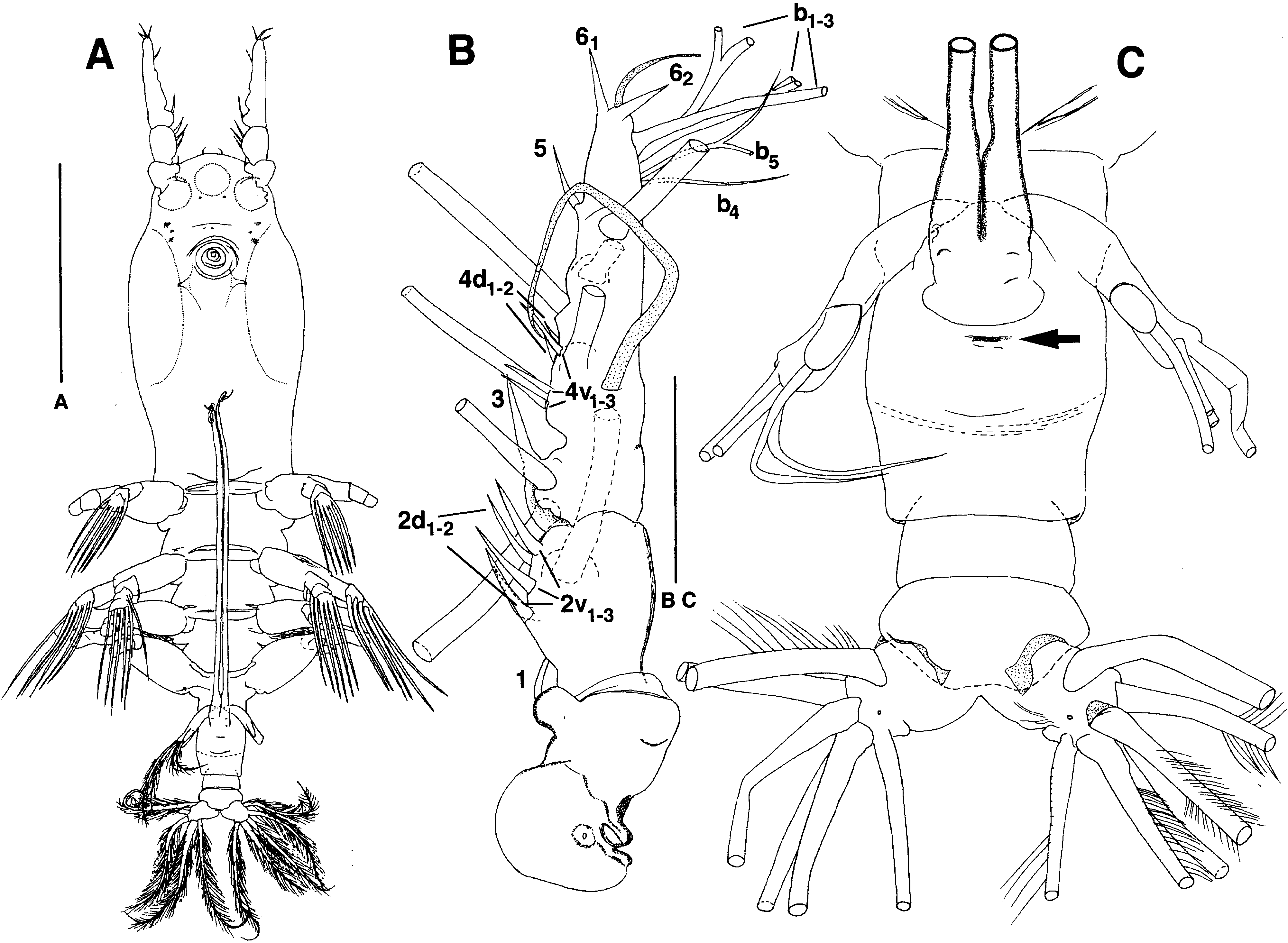
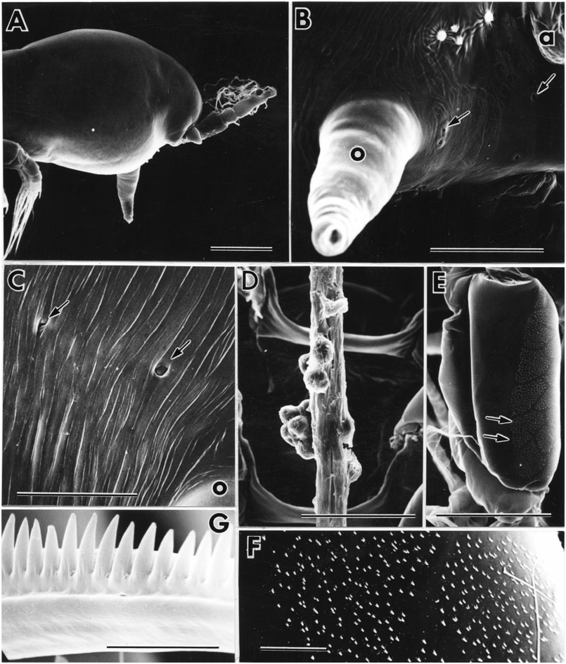
Cuticular reticulation completely absent, but cephalothorax with faint but dense circular striae ( Fig. 25C View Figure 25 ); scattered minute dots also seen by phase contrast. Oral papilla long, straight, conical, directed ventrally ( Fig. 25A, B View Figure 25 ). Pair of closely spaced pores just in front of it; another pair, further apart, at level of posterior bases of antennules ( Fig. 25B View Figure 25 ). Two more pairs of pores, even further apart, behind oral papilla ( Fig. 25C View Figure 25 ). Three or four small scars behind base of each antennule, middle one(s) smallest, anterior and posterior ones surrounded by radiating ridges ( Fig. 25B View Figure 25 ). Pair of hair-like sensilla on forehead; dorsal to them, symmetical pattern of about nine small pores and dorsolateral pair of larger pores (not illustrated); apparently no wrinkled pit like that of M. hyottoko . Three cups of naupliar eye large, equal in size ( Fig. 24A View Figure 24 ).
Antennules with non-articulating boundary between third and fourth segments suggested by constriction; first and second segments distinct ( Fig. 24B View Figure 24 ). Full setation probably present in life ( Grygier & Ohtsuka, 1995), but one or two setae missing on each antennule observed. Among spiniform setae, 2-setae all rather short, less than twice as long as longest 4-seta; 3-seta a little longer than 2-setae; near tip, b-setae all bifurcating twice with first split rather far out; apical 6-setae short and equal.
Patches of minute, bluntly conical teeth separated by smooth lanes present dorsally beginning with first free pediger ( Fig. 26A, C–E View Figure 26 ). Patches arrayed in six rows, with numbers per row as follows: 2-3-6-6-3-2 on first free pediger, 4-4-7-7-4-4 on second, 3-3-4-4-3-3 on third, fourth unknown due to wrinkling,?(3-3-3-3-3- 3) + (3-3-3-3-3-3) on genital compound somite before and after transverse dorsal suture (suture: Fig. 26E View Figure 26 ) and 2-2-2-2-2-2 on penultimate segment. Patches generally square to oblong, but all patches on second free pediger and outer ones on third free pediger irregular in shape ( Fig. 26D View Figure 26 ). Rear-most patches of middle two rows of first and second free pedigers extending onto pair of posteriorly directed, spine-like scales ( Fig. 26B, C View Figure 26 ).
Several pores near dorsal midline at two-thirds length of cephalothorax. Distribution of more posterior dorsal pores and dorsal and lateral pit-setae shown in Figure 29 View Figure 29 ; essentially as in M. hyottoko , but anterior dorsal pores of first free pediger in two rows with medial pair small ( Fig. 26A View Figure 26 ), and anterior dorsal pores of third free pediger not confirmed. Larger pores of first three free pedigers apparently provided with valves ( Fig. 26A, C, D View Figure 26 ).
Intercoxal sclerites and segmentation of legs 1–4 as in M. hyotokko ( Figs 25D View Figure 25 , 27A–F View Figure 27 ). About 12 patches of minute, rather widely spaced denticles along outer face of coxa ( Fig. 25E, F View Figure 25 ); these patches in two interdigitating rows and separated by bare lanes. Similar spinules present on outer face of at least least first exopodal segment. Full plesiomorphic setation of legs 1–4 present as outlined by Huys & Boxshall (1991), including inner seta of first endopodal segment and weak inner seta of first exopodal segment ( Fig. 27A, C, D, F View Figure 27 ). Outer basis seta short and hair-like in legs 1, 2 and 4, longer and plumose in leg three but shorter than exopod; pore anterior to this seta in each leg. Tiny pore on anterior face of third segment of each ramus (illustrations all in posterior view, so pore not shown). Outer spiniform setae of exopod simple, that on third segment a little longer than that on first. Outer side of outer apical exopodal seta lined with closely adjacent, bluntly conical denticles ( Figs 25G View Figure 25 , 27E View Figure 27 ).
Leg 5 with wide exopodal lobe bearing two apical setae and one subapical outer seta; narrower but longer endopodal lobe with one apical seta; all setae biserially plumose, outer two exopodal setae longer than others ( Figs 24C View Figure 24 , 27G View Figure 27 ).
Round copulatory pore on midline within crescentshaped slot at posterior base of conical process bearing ovigerous spines ( Fig. 24C View Figure 24 ). Spines extending to halfway between legs 1 and oral papilla, slender and cylindrical with tapered tips ( Figs 13B View Figure 13 , 24A View Figure 24 ). Caudal rami with ventral pore and six setae distributed as in M. hyottoko ( Fig. 24A View Figure 24 ); short dorsal seta usually simple, but sometimes with distal setules; other setae biserially plumose and medial one longest.
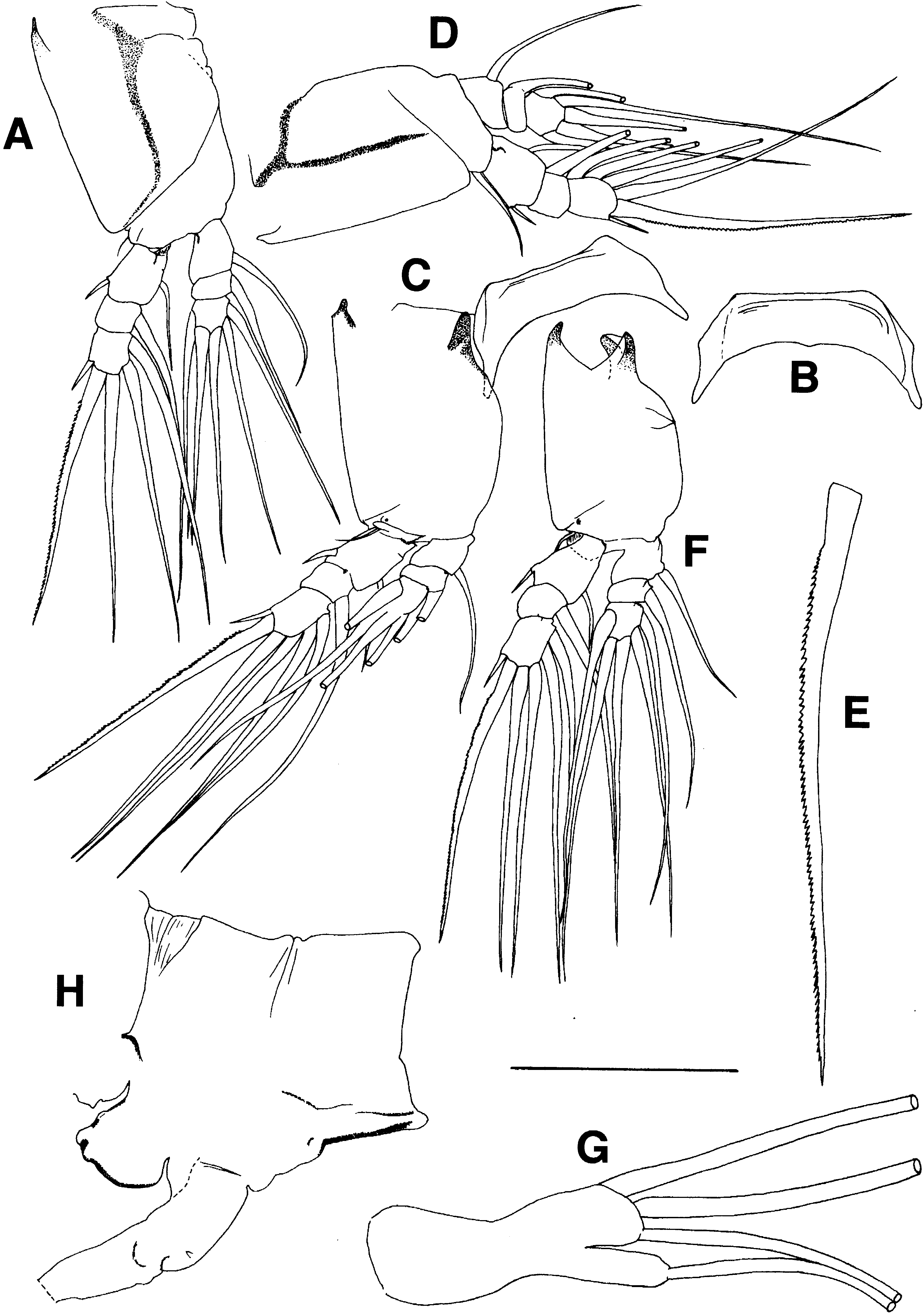
Description of syntype: Damaged ( Fig. 28A View Figure 28 ), lacking most leg setae besides those of endopods of legs 3 and 4 ( Fig. 28E–G View Figure 28 ), lacking many antennular setae, especially distal to 4-setae and Vm ( Fig. 28B View Figure 28 ), and lacking all except one pair of furcal setae ( Fig. 28I View Figure 28 ). In addition, all rami of legs 1 and left leg 2 torn away, along with exopod of right leg 2 (a few loose pieces of these legs found; Fig. 28C, D View Figure 28 ). Dimensions in lateral view as follows: length (sum of lengths of cephalothorax, metasome and urosome as defined in Fig. 1A View Figure 1 ) 2.39 mm, with these body regions contributing 53.3, 28.1 and 18.6%, respectively; height of cephalothorax 42.1% of cephalothorax length; antennule length 37.0% that of cephalothorax; unbroken part of longer ovigerous spine 34.1% as long as body. Cephalothorax full of unlaid brood. Three pairs of scars behind bases of antennules. Inner seta of first exopod segment not retained on any leg; that of first endopodal segment retained on right leg 2 and left leg 4 ( Fig. 28C, E View Figure 28 ), represented by socket on some other legs. Leg 5 biramous, at least left one originally with three setae on exopod (1 remaining) and one setae on endopod ( Fig. 24H View Figure 24 ). Ovigerous spines arising from process with large, rounded, anterior protrusion ( Fig. 24H View Figure 24 ).
Remarks: The full setation of legs 1–4 and the biramous condition of leg 5 are plesiomorphic features compared with the M. hyottoko species group. The spine-like scales of the first two free pedigers are similar to but much smaller and closer together than the pairs of spines present on the same segments in the eponymous Monstrilla spinosa Park, 1967 (see Park, 1967; Suárez-Morales & Vásquez-Yeomans, 1996). Monstrilla spinosa is an odd species with an assortment of other unique specializations, none of which indicates any relationship with Maemonstrilla turgida .
Scott (1909) reported two syntypes of his Monstrilla turgida from the same sample, but only one was in the vial obtained on loan from Amsterdam; the other must be assumed lost. The remaining syntype (original description and present observations) shows enough of the distictive features of the present Ryukyuan specimens so as to allow assignment of the latter to M. turgida for the time being. It was not possible to confrm in the syntype the inner seta of the first exopodal segment of legs 1–4, nor the presence of the pair of spine-like scales dorsally on the first two free pedigers. Although the first antennae of the syntype are relatively shorter than in the measured Ryukyuan specimens, other measurements and proportions fall within the range of the latter.
The unidentified Monstrilla figured by Krishnaswamy (1953) from Kundugal Channel, India, seems to be a female of Maemonstrilla turgida or a similar species, to judge from the bulbous cephalothorax and bilobed leg 5. The ovigerous spines were neither mentioned nor drawn; possible differences from the Ryukyuan specimens are the supposed presence of two setae, not one, on the endopod of leg 5 and five setae, not six, on the caudal rami.
Suárez-Morales & Dias (2001b) compared two new or redescibed species of Monstrilla , both with ordinary ovigerous spines and swimming legs, with what was then called Monstrilla turgida , while citing A. Scott (1909) and Grygier & Ohtsuka (1996) in regard to the latter. In particular they judged that M. pustulata Suárez & Dias, 2001 from Brazil shows ‘some affinities’ to M. turgida , i.e. a similar cephalothoracic shape and a bilobed female leg 5 with 3 + 1 setation. But they noted several differences as well, especially the opposed direction of the ovigerous spines, and did not press a case for a phyletic connection.


No known copyright restrictions apply. See Agosti, D., Egloff, W., 2009. Taxonomic information exchange and copyright: the Plazi approach. BMC Research Notes 2009, 2:53 for further explanation.
|
Kingdom |
|
|
Phylum |
|
|
Class |
|
|
Order |
|
|
Family |
|
|
Genus |
Maemonstrilla longipes
| Grygier, Mark J. & Ohtsuka, Susumu 2008 |
Monostrilla longipes
| Al-Kholy AA 1963: 130 |
Monstrilla longipes A. Scott, 1909: 238
| Wickstead JH 1961: 61 |
| Davis CC 1949: 248 |
| Sewell RBS 1949: 141 |
| Scott A 1909: 238 |
Monstrilla turgida A. Scott, 1909: 239
| Martin Thompson PK & Meiyappan MM 1980: 207 |
| Wickstead JH 1961: 61 |
| Davis CC 1949: 248 |
| Scott A 1909: 239 |