
Victoria cruziana, Orbign.
|
publication ID |
https://doi.org/ 10.1016/j.phytochem.2021.112899 |
|
DOI |
https://doi.org/10.5281/zenodo.8269001 |
|
persistent identifier |
https://treatment.plazi.org/id/03EBF734-A030-1624-0125-F9B0FD84F8AD |
|
treatment provided by |
Felipe |
|
scientific name |
Victoria cruziana |
| status |
|
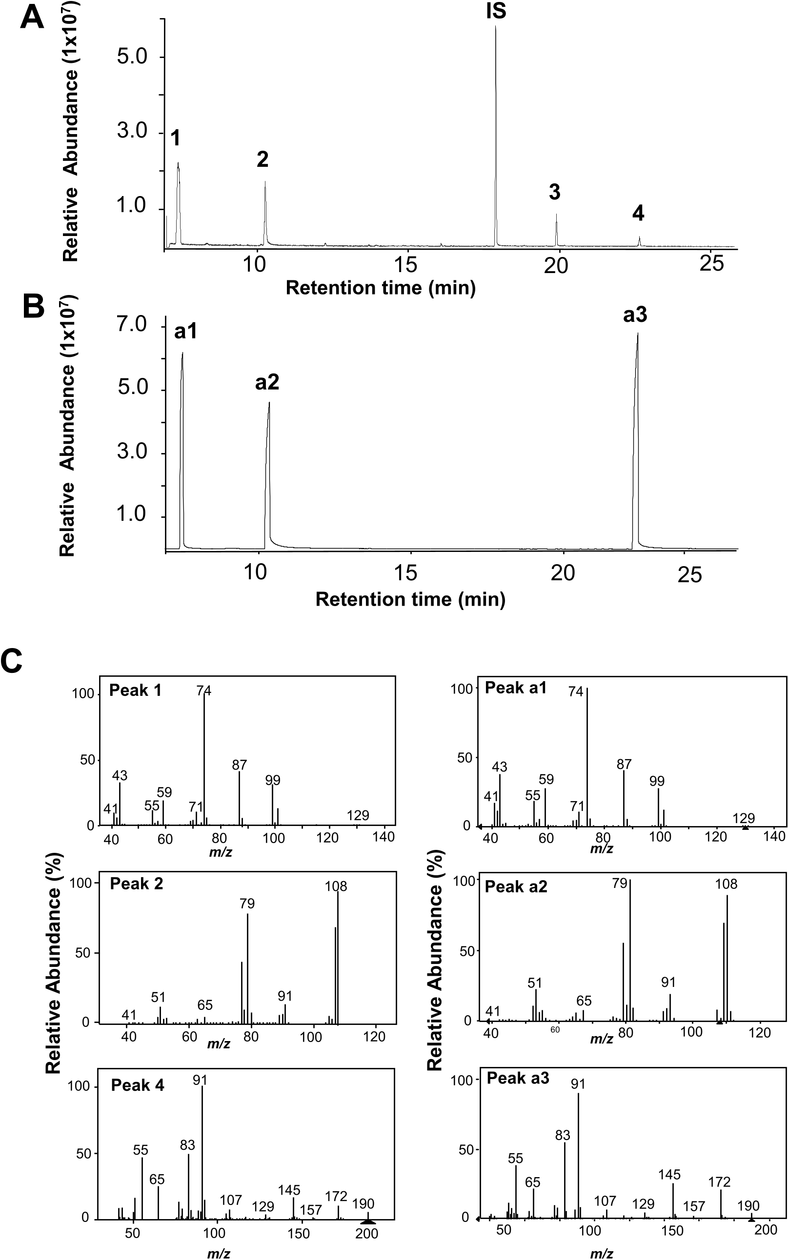
2.1. Chemical profiling of floral scent from V. cruziana View in CoL during first bloom
Combining headspace collection with gas chromatography–mass spectrometry (GC-MS) analysis, four volatile compounds were detected from the fully-opened flowers of V. cruziana during their first bloom ( Fig. 1A View Fig ). These four compounds were identified to be methyl hexanoate, benzyl alcohol, benzyl 2-methylbutanoate and benzyl tiglate. Except benzyl 2-methylbutanoate, the chemical identities of the other three compounds were verified by comparing their chromatograms ( Fig. 1B View Fig ) and mass spectra ( Fig. 1C View Fig ) with those of authentic standards. Among these compounds, methyl hexanoate was the most abundant constituent accounting for 45.5 % of the total emission. Benzyl alcohol, benzyl 2-methylbutanoate and benzyl tiglate accounted for 37.8 %, 11.3 %, and 5.4 % of the total emission, respectively.
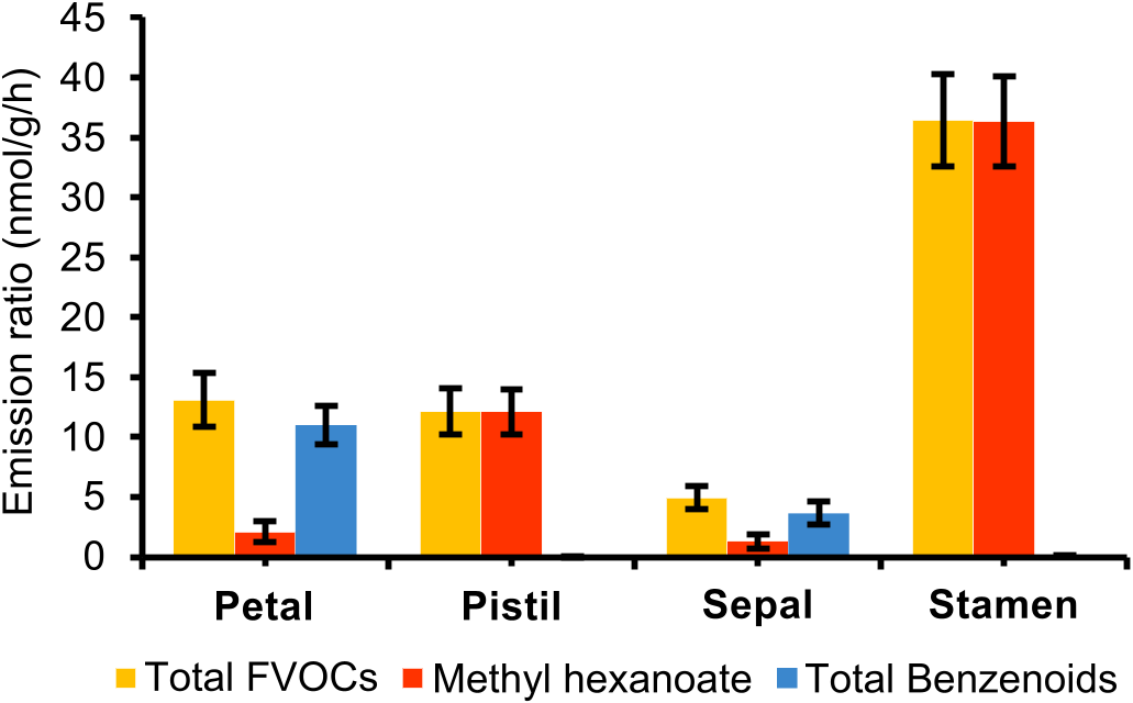
2.2. Emission of floral volatiles from different parts of V. cruziana flowers
To determine the contribution of different parts of V. cruziana flowers to floral scent profile, the flowers of V. cruziana were divided into sepals, petals, stamen and pistils. These four parts were subjected to headspace collection, separately. The four floral volatiles, i.e. methyl hexanoate, benzyl alcohol, benzyl 2-methylbutanoate and benzyl tiglate, were detected from each of the four parts. ( Fig. 2 View Fig ). Of the four parts, stamen showed the highest rates of total emission (36.42 ± 7.6 nmol/g / h). Methyl hexanoate exhibited the highest rates of emission from stamens at 36.31 ± 7.5 nmol/g/h ( Fig. 2 View Fig ), followed by pistils (12.11 ± 3.8 nmol/g / h), petals (2.09 ±1.6 nmol/g/h) and sepals (1.31 ± 1.24 nmol/g/h). In contrast, the three benzenoids exhibited the highest rates of emission from the petals with the emission rate of 10.99 ±3.3 nmol/g/h ( Fig. 2 View Fig ). Their emission rates from sepals, stamens and pistils were 3.64 ± 1.9 nmol/g/h, 0.11 ± 0.01 nmol/g/h and pistils (0.01 ± 0.001 nmol/g/h), respectively.
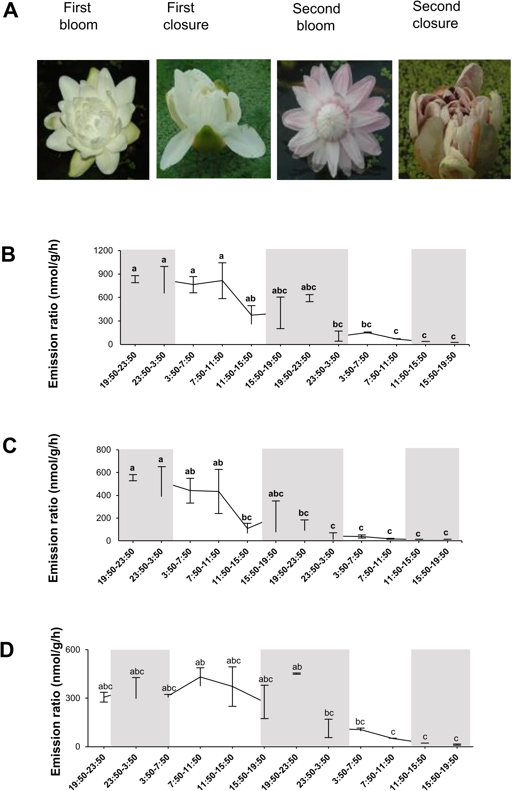
2.3. Emission dynamics of floral volatiles from V. cruziana View in CoL
In our observation, the flowers of V. cruziana opened fully at 19:00 on the first day, began to close in the following morning and became completely closed between 12:00 and 13:00. The flowers started to reopen between 16: 00 and 17: 00 on the same day. The representative flowers at the four stages were shown in Fig. 3A View Fig . To examine the emission dynamics, floral volatiles of V. cruziana were continuously collected (1-h collection followed by a 3-h interval) for two days (12/12 h light/dark) and analyzed by GC-MS. The emission rates of total volatiles remained constant before a significant drop towards the end of second bloom ( Fig. 3B View Fig ). The emission rates of benzenoids and methyl hexanoate were also analyzed separately. The emission patterns of benzenoids were very similar to those of total volatiles ( Fig. 3C View Fig ). In contrast, there was a reduction in mission rates of methyl hexanoate towards the end of first closure before increasing during the second bloom ( Fig. 3D View Fig ). Like total volatiles, the emission of both benzenoids ( Fig. 3C View Fig ) and methyl hexanoate ( Fig. 3D View Fig ) dropped significantly at the of the end of second bloom and after that.
2.4. Identification of candidate genes for the biosynthesis of methyl hexanoate
To understand the molecular basis of floral volatile biosynthesis in V. cruziana , an RNA-Seq library using the RNA samples of whole flowers of V. cruziana at full blooming stage was constructed and sequenced. High quality sequencing data was obtained, and the clean bases of flower and leaf were 6.11 G (Table S1). The de novo assembly yielded 64,472 unigenes (Table S1). The unigenes longer than 1k bp were 34483 that accounted for 53.49 % of the total unigenes and the N50 was 2476 bp (Table S1). More than 63.51 % unigenes were annotated in at least one database, and 16 % unigenes were annotated in all seven bioinformatics databases (Table S1). The annotated unigenes were clustered in three GO function classification (biological process, cellular component, molecular function, Fig. S1 View Fig ).
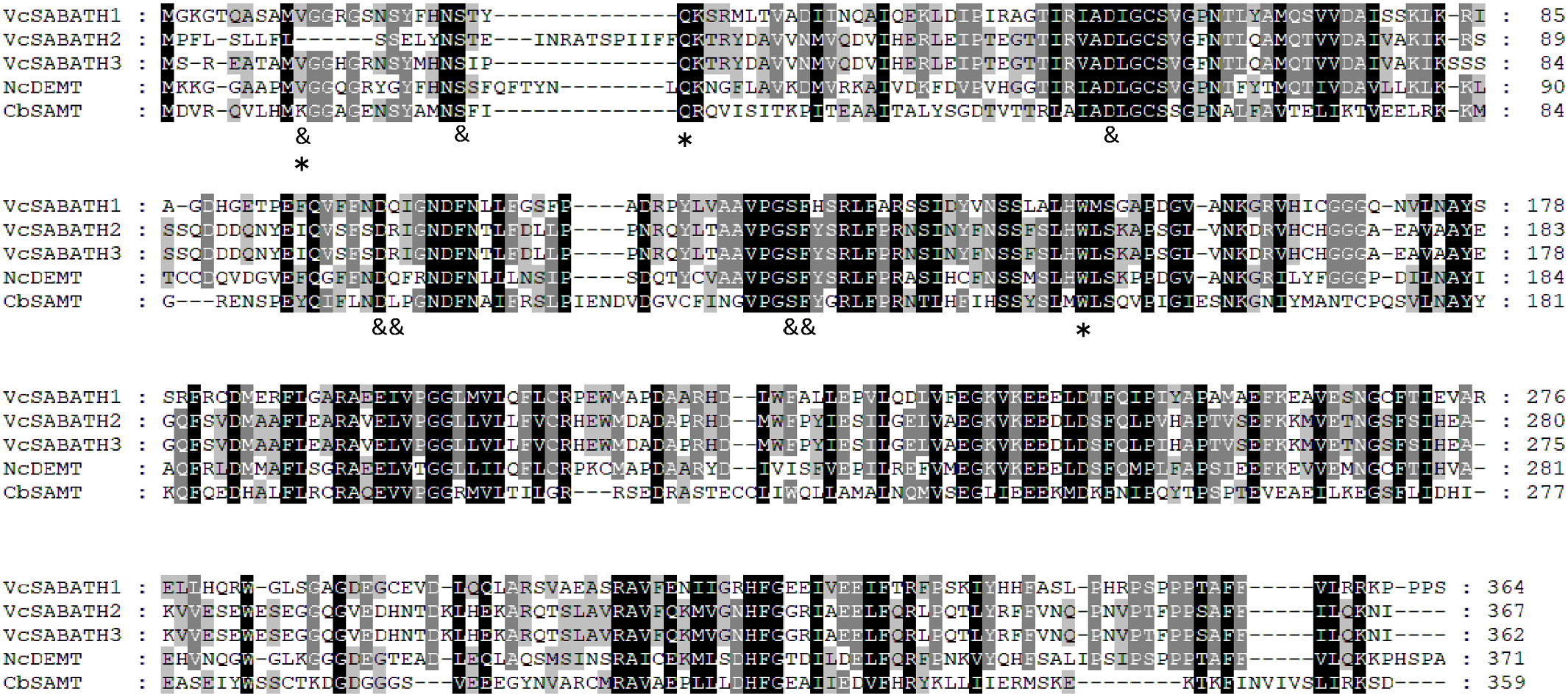
Next, the transcriptome was specifically searched for candidate biosynthetic genes. This study focused on the biosynthesis of methyl hexanoate. Prior to this study, the biosynthesis of fatty acid methyl esters has been studied in another water lily N. colorata , in which methyl decanoate and methyl octanoate are biosynthesized by methyltransferases that belong to the SABATH family ( Zhang et al., 2019). It was our hypothesis that the formation of methyl hexanoate in V. cruziana is also catalyzed by SABATH enzymes. As such, the flower transcriptome of V. cruziana was created ( Fig. S1 View Fig ) and searched for SABATH genes. Three putative full-length SABATH genes were identified, which were designated as VcSABATH1, VcSABATH2 and VcSABATH3 with GenBank accession number of MZ541994 View Materials , MZ541995 View Materials and MZ541996 View Materials , respectively. The proteins they encode are 364 (VcSABATH1), 367 (VcSABATH2), and 362 (VcSABATH3) amino acids in length. Multiple sequence alignment revealed that all three VcSABATH proteins contain conserved residues for interacting with the carboxyl moiety of the substrate and the methyl donor S -adenosyl-L-methionine ( Fig. 4 View Fig ).
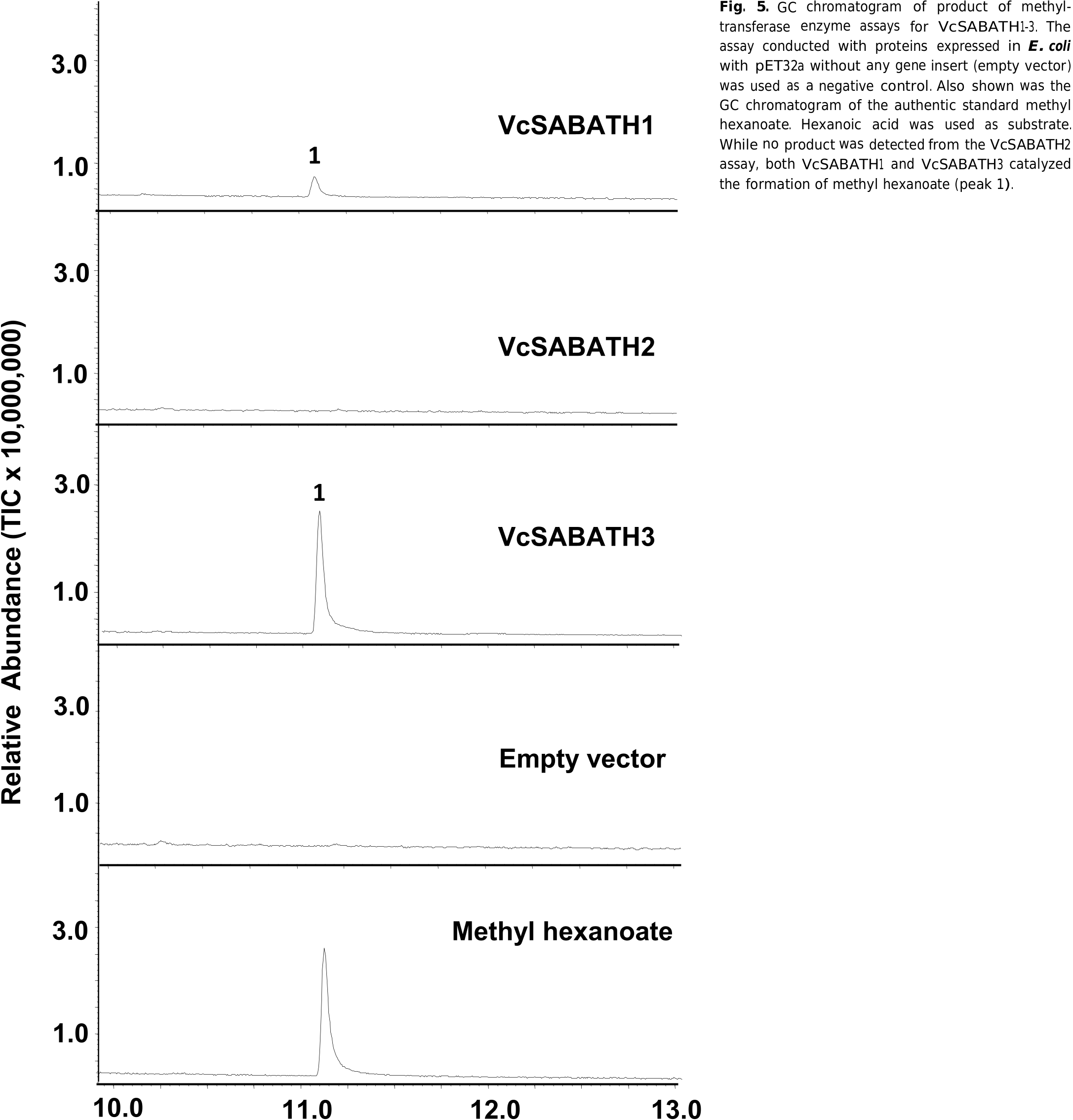
2.5. Biochemical assay of VcSABATHs
Next, in vitro methyltransferase enzyme assays were performed to determine which of the three VcSABATH genes is responsible for the biosynthesis of methyl hexanoate in V. cruziana flowers. A full-length cDNA for each of the three VcSABATH genes was synthesized and cloned into a protein expression vector (pET32a) and expressed in E. coil to produce recombinant enzymes. Individual recombinant VcSABATH proteins were tested in methyltransferase enzyme assays using hexanoic acid as substrate. While VcSABATH2 was inactive, both VcSABATH1 and VcSABATH3 catalyzed the formation of methyl hexanoate ( Fig. 5 View Fig ). Next, E. coli -expressed recombinant VcSABATH1 and VcSABATH3 were purified ( Fig. S2 View Fig ) and the purified proteins used to measure their respective specific activities. The specific activity of VcSABATH1 and VcSABATH3 using hexanoic acid as substrate was determined to be 23.2 ±2.41 pkat/mg protein and 150.4 ±3.6 pkat/mg protein, respectively. To determine substrate specificity of VcSABATH1 and VcSABATH3, they were also tested using three other fatty acids as substrate. The relative activity of VcSABATH1 using octanoic acid, decanoic acid or dodecanoic acid as substrate was 39.4 %, 33.6 % and 16.9 % of that with hexanoic acid respectively. Similarly, the relative activity of VcSABATH3 using octanoic acid, decanoic acid or dodecanoic acid as substrate was 33.5 %, 38.8 % and 17.3 % of that with hexanoic acid respectively.
2.6. Expression of VcSABATH1 and VcSABATH 3 in different flower organs
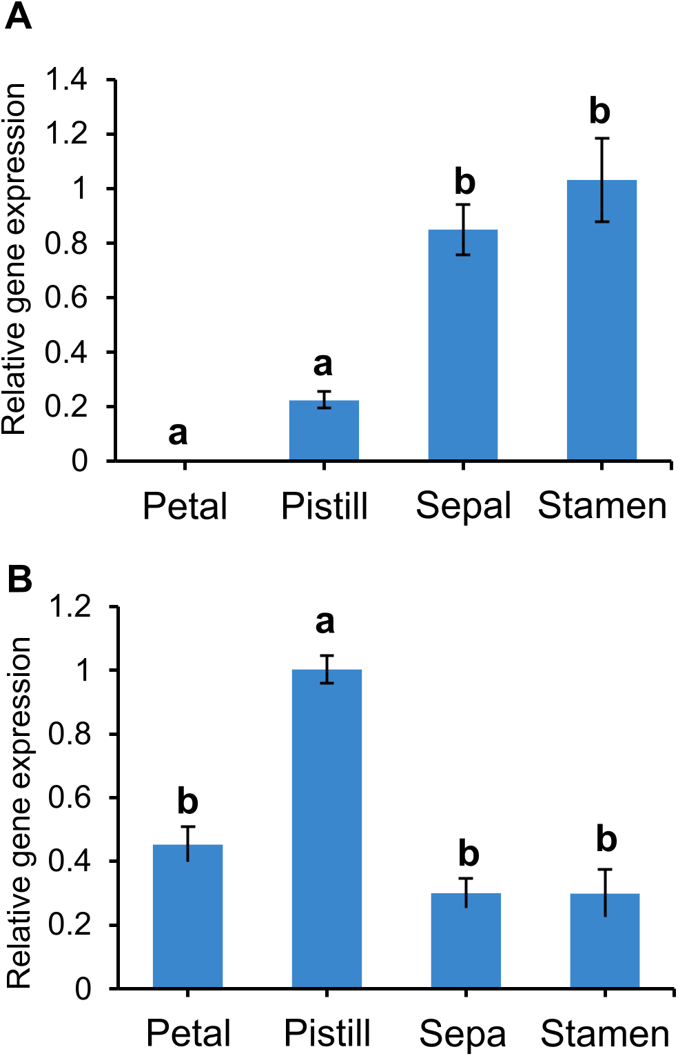
With VcSABATH3 and VcSABATH1 demonstrated to encode hexanoic acid methyltransferase, their expression in different parts of V. cruziana flowers was measured using reverse transcriptionquantitative PCR (RT-qPCR). The highest level of expression of VcSABATH1 was detected in stamens ( Fig. 6A View Fig ), whereas VcSABATH3 showed the highest level of expression in pistils ( Fig. 6B View Fig ).
2.7. Phylogenetic analysis
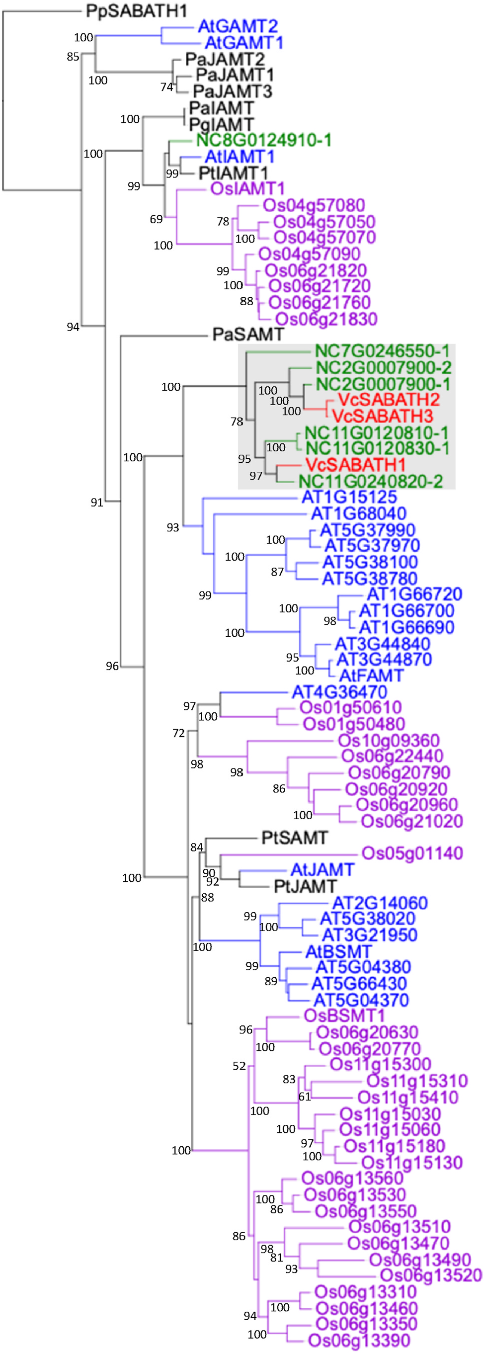
Prior to this study, the SABATH family of methyltransferases in plants has been relatively well studied with a number of members characterized ( D’ Auria et al., 2003). These include salicylic acid MT ( Ross et al., 1999; Chen et al., 2003, Zhao et al., 2010), jasmonic acid MT ( Seo et al., 2001; Zhao et al., 2013), indole-3 acetic acid MT ( Qin et al., 2005; Zhao et al., 2007, 2008), gibberellic acid MT ( Varbanova et al., 2007; Zhang et al., 2020). To understand the evolutionary relatedness of the two fatty acid methyltransferases from V. cruziana (VcSABATH1 and VcSABATH3) with known SABATH proteins, phylogenetic analyses were performed. Three VcSABATHs form a water lily-specific cluster with 6 full-length SABATH members from N. colorata except the putative IAMT (Nc8G0124910-1) ( Fig. 7 View Fig ). VcSABATH1 was most closely related to three NcSABATHs including Nc11G0120830-1 which was characterized to be decanoic acid methyltransferase ( Zhang et al., 2019). VcSABATH2 and VcSABATH3 were most closely related to each other and together with two NcSABATHs of unknown functions they form a subcluster in water lily-specific cluster ( Fig. 7 View Fig ).
No known copyright restrictions apply. See Agosti, D., Egloff, W., 2009. Taxonomic information exchange and copyright: the Plazi approach. BMC Research Notes 2009, 2:53 for further explanation.
|
Kingdom |
|
|
Phylum |
|
|
Class |
|
|
Order |
|
|
Family |
|
|
Genus |