
Pseudoraphes Franz, 1980
|
publication ID |
https://doi.org/ 10.11646/zootaxa.4612.2.4 |
|
publication LSID |
lsid:zoobank.org:pub:7525A835-B189-4F33-B17B-59D722E50951 |
|
persistent identifier |
https://treatment.plazi.org/id/03EC5172-7449-2456-BBEF-F880B433FA85 |
|
treatment provided by |
Plazi |
|
scientific name |
Pseudoraphes Franz |
| status |
|
Pseudoraphes Franz View in CoL
Pseudoraphes Franz, 1980b: 209 View in CoL . Type species: Pseudoraphes huallagae Franz, 1980b View in CoL (orig. des.).
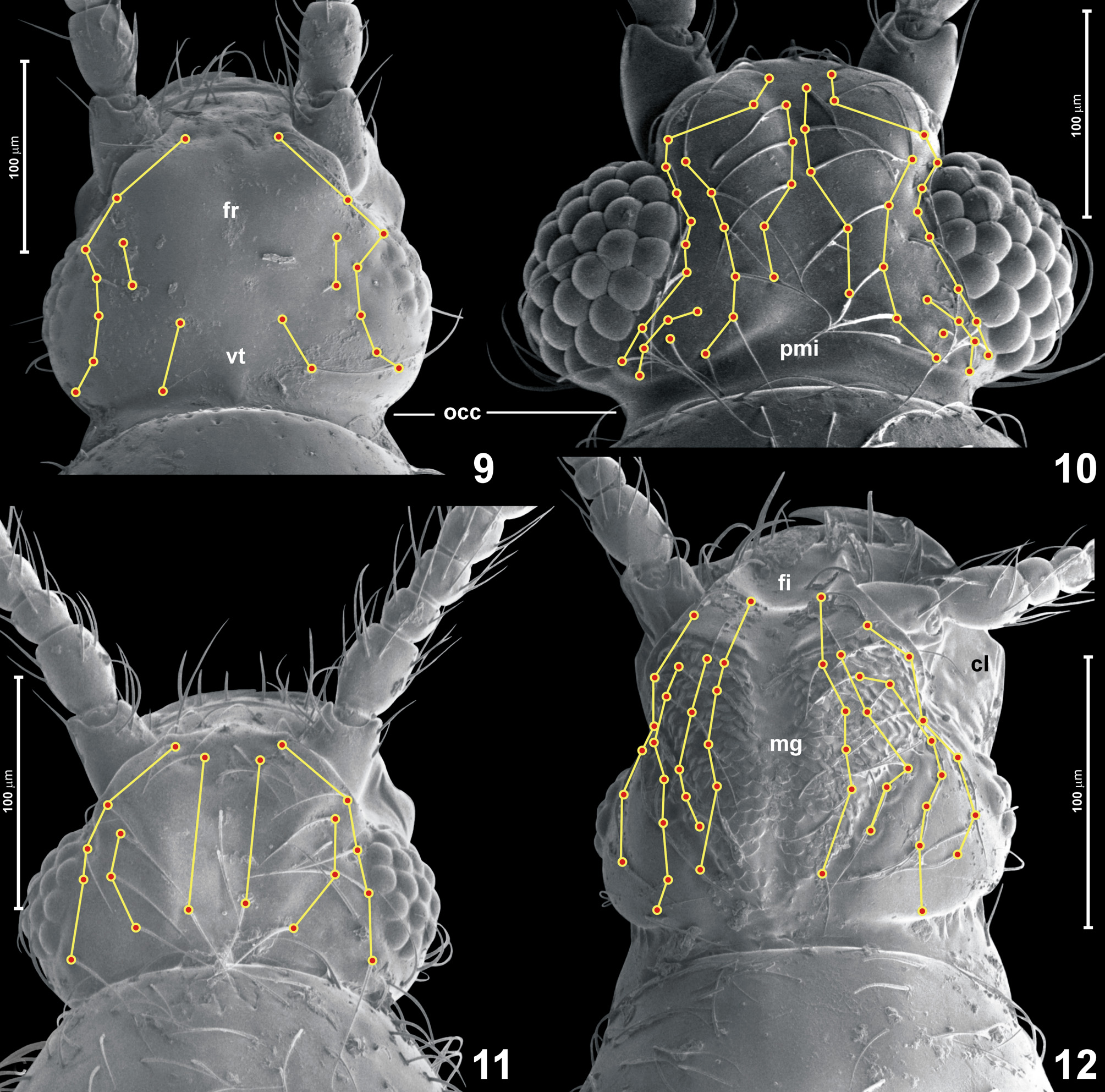
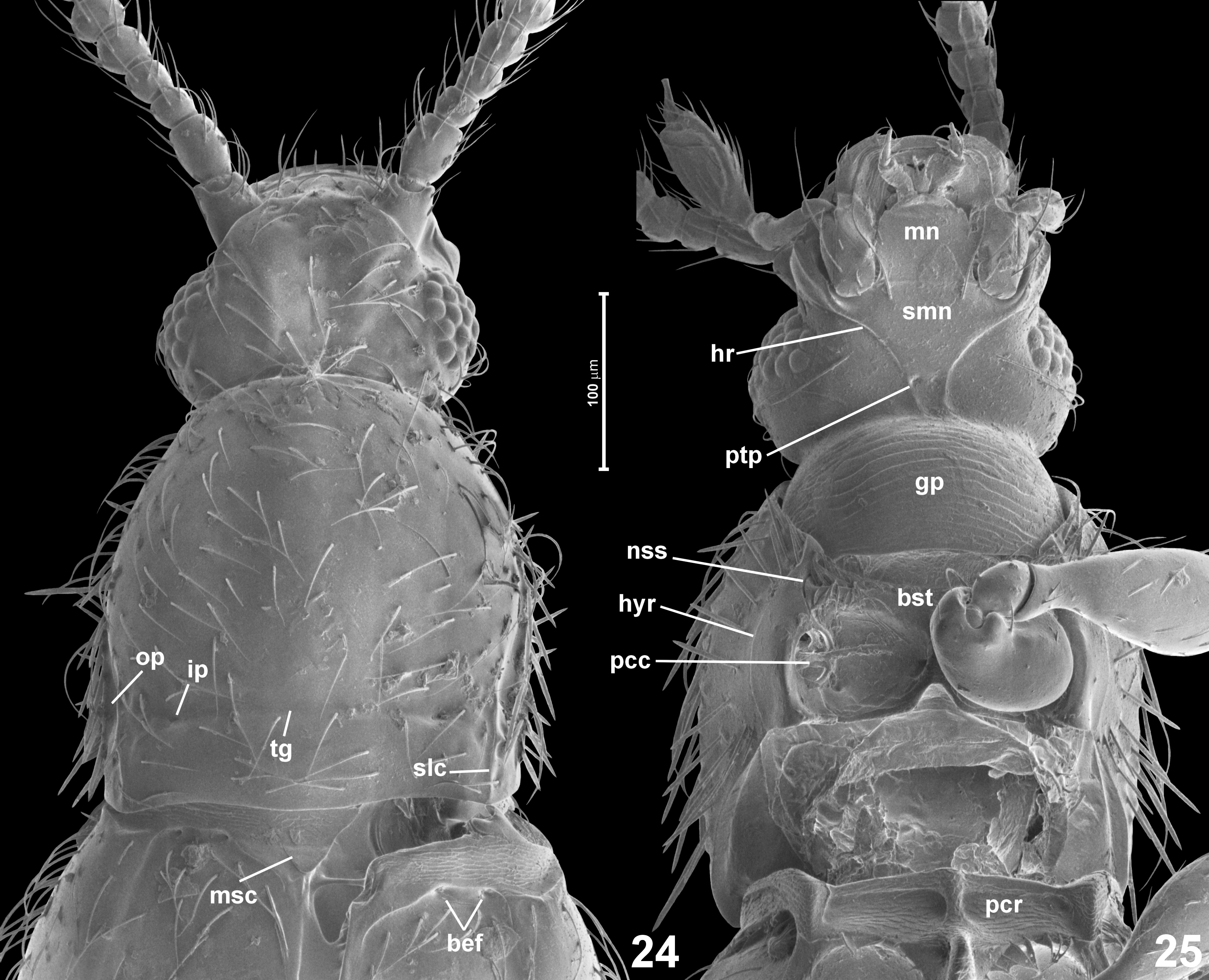
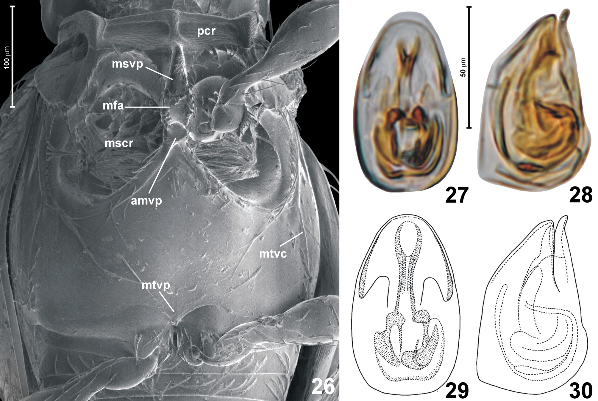
Revised diagnosis. Body (Fig. 3) moderately stout, distinctly constricted between head and prothorax and between prothorax and elytra. Head ( Fig. 11 View FIGURES 9–12 ) about as long as broad with tempora shorter than eyes; tempora, vertex, genae and postgenae lacking thick bristles; occipital constriction distinctly, but not much, narrower than vertex; submen- tum lacking lateral sutures; hypostomal ridges present and complete, obliquely running toward posterior tentorial pits where they are not connected; antennae gradually thickened; head lacking frontal impression, posteromedian impression and median subtriangular 'platform', but bearing a diffuse longitudinal groove and largely symmetrical setae ( Fig. 11 View FIGURES 9–12 ); maxillary palpomere IV slightly constricted before apex, so that its apical portion is broadened and truncate; pronotum ( Fig. 24 View FIGURES 24–25 ) broadest in front of middle, sides with lateral edges or carinae developed in posterior half and with distinct sublateral carinae; transverse antebasal groove present and connecting small but distinct inner lateral pits, also with a pair of indistinct outer lateral pits; sides of pronotum with dense bristles; prosternum ( Fig. 25 View FIGURES 24–25 ) with basisternal part shorter than half length of coxal part; prosternal intercoxal process developed as a diffuse ridge; procoxal cavities closed; notosternal sutures and hypomeral ridges complete; mesoscutellum ( Fig. 24 View FIGURES 24–25 ) largely exposed between elytral bases; mesocoxal rests with diffuse marginal carina and a faint postmarginal carina; mesoventral intercoxal process ( Fig. 26 View FIGURES 26–30 ) carinate and strongly elevated, narrowly separating mesocoxae, with a broadened, flattened and smooth median area, with a well-defined posterior tip and a subtriangular posteromedian emargination; metaventral carinae present; anterior metaventral process present, well-defined, conspicuously large, with subrectangular anterior tip and broadly triangular base; metaventral intercoxal process ( Fig. 26 View FIGURES 26–30 ) not separating metacoxae, subtriangular, lacking median notch and lacking lateral spines; each elytron ( Fig. 24 View FIGURES 24–25 ) with two vestigial asetose foveae; and aedeagus ( Figs 27–30 View FIGURES 26–30 ) symmetrical with free, slender parameres (broken off in the studied specimen, but Franz (1989b) illustrated parameres for P. huallagae , which has a very similar aedeagus).
Redescription. Body (Fig. 3) elongate but not very slender, distinctly constricted between head and prothorax and less so between prothorax and elytra, strongly convex, brown.
Head ( Figs 11 View FIGURES 9–12 , 24–25 View FIGURES 24–25 ) with moderately large eyes; tempora short but distinct, rounded; frons lacking frontal impression, with diffuse longitudinal median impression; vertex anteriorly confluent with frons, lacking posteromedian impression; numerous setae on frons and vertex largely symmetrical ( Fig. 11 View FIGURES 9–12 ); gular plate ( Fig. 25 View FIGURES 24–25 ; gp) subtrapezoidal; posterior tentorial pits ( Fig. 25 View FIGURES 24–25 ; ptp) in front of transverse impression demarcating 'neck' region ventrally, minute; hypostomal ridges ( Fig. 25 View FIGURES 24–25 ; hr) complete, reaching level of posterior tentorial pits, not connected; submentum ( Fig. 25 View FIGURES 24–25 ; smn) lacking lateral sutures. Antennae (Fig. 3) slender, gradually thickened distad.
Pronotum ( Fig. 24 View FIGURES 24–25 ) bell-shaped, broadest in front of middle; with blunt lateral carinae developed in posterior half, and with short sublateral carinae ( Fig. 24 View FIGURES 24–25 ; slc), with distinct antebasal transverse groove ( Fig. 24 View FIGURES 24–25 ; tg) and two pairs of antebasal lateral pits: inner ( Fig. 24 View FIGURES 24–25 ; ip) and outer ( Fig. 24 View FIGURES 24–25 ; op). Prosternum with vestigial basisternal region ( Fig. 25 View FIGURES 24–25 ; bst), prosternal process carinate but weakly elevated; notosternal sutures ( Fig. 25 View FIGURES 24–25 ; nss) complete; hypomeral ridges ( Fig. 25 View FIGURES 24–25 ; hyr) complete; inner (adcoxal) portion of hypomeron broad and glabrous, outer portion (confluent with side of pronotum) covered with thick bristles. Procoxal cavities ( Fig. 25 View FIGURES 24–25 ; pcc) closed.
Mesoventrite with a pair of transverse impressions behind its anterior ridge that function as procoxal rests ( Fig. 26 View FIGURES 26–30 ; pcr); mesoventral intercoxal process ( Fig. 26 View FIGURES 26–30 ; msvp) carinate, long, strongly elevated, with distinctly broadened posterior portion bearing a median flattened area ( Fig. 26 View FIGURES 26–30 ; mfa) that is smooth and devoid of setae, posterior tip of process with subtriangular emargination.
Mesoscutellum ( Fig. 24 View FIGURES 24–25 ; msc) exposed between elytral bases in intact beetles, subtriangular, about as long as broad.
Metaventrite with massive anterior metaventral process ( Fig. 26A View FIGURES 26–30 ; amtp), its anterior portion subrectangular, posterior portion broadly subtriangular; each side with metaventral carina ( Fig. 26 View FIGURES 26–30 ; mtvc) running posterad and slightly toward lateral margin of ventrite; mesocoxal rests ( Fig. 26A View FIGURES 26–30 ; mscr) very deep and filled with setae, their posterior marginal carina diffuse and incomplete; metaventral intercoxal process ( Fig. 26 View FIGURES 26–30 ; mtvp) lacking lateral spines, not separating metacoxae.
Elytra (Figs 3, 24) oval, each elytron with one pair of vestigial asetose basal elytral foveae ( Fig. 24 View FIGURES 24–25 ; bef), lacking basal impression, and with distinct humeral callus. Hind wings developed.
Legs do not differ from other genera of Glandulariini, unmodified in males.
Abdomen unmodified, with sternites III and IV subequal in length.
Aedeagus ( Figs 27–30 View FIGURES 26–30 ) stout, weakly sclerotized, with symmetrical median lobe, asymmetrical endophallic structures with indistinct flagellum, and with free parameres bearing apical setae (parameres broken off in the studied specimen, but Franz (1989b) illustrated parameres for P. huallagae , which has a very similar aedeagus).
Remarks. Pseudoraphes is most similar to Meridaphes and Stenichnoconnus ; differences were discussed at each of the latter genera.
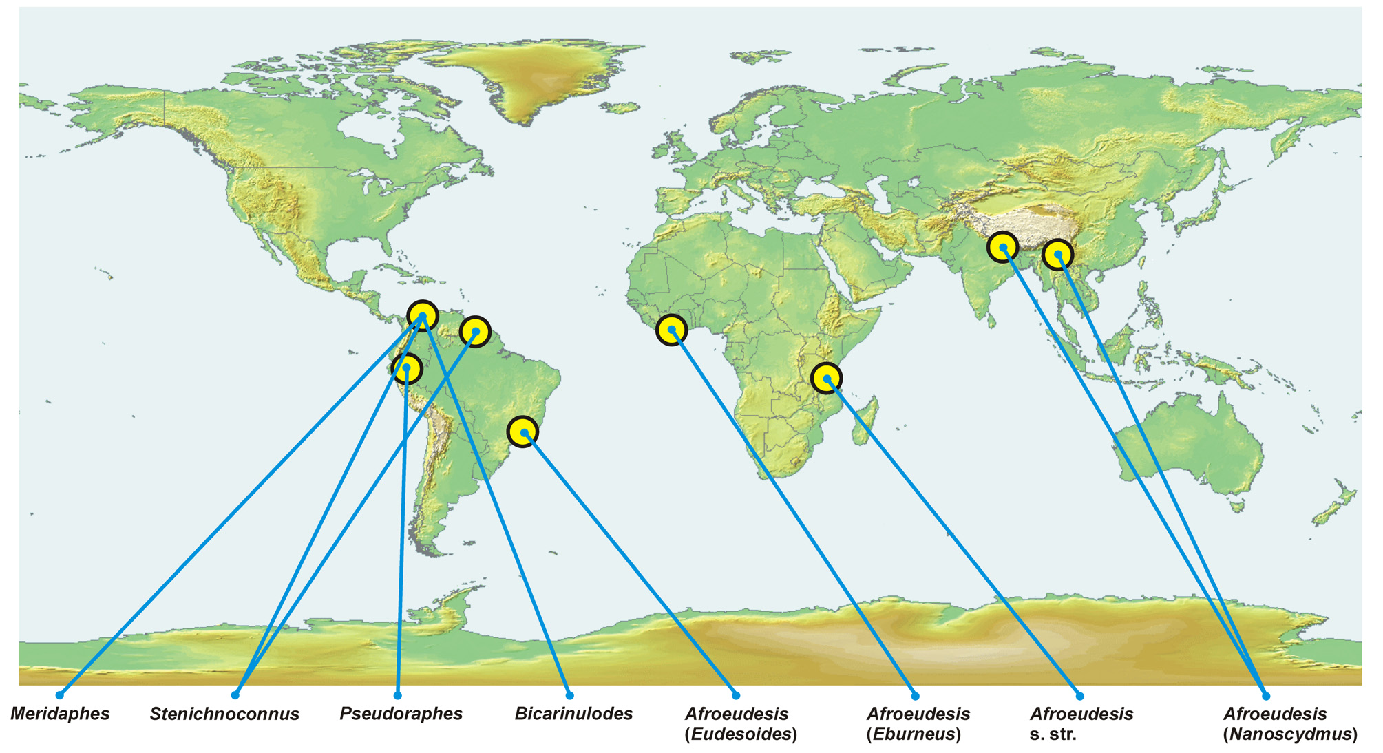
Composition and distribution. Pseudoraphes comprises two species: P. huallagae known to occur in Peru, and a sympatric P. minor ( Fig. 41 View FIGURE 41 ).
No known copyright restrictions apply. See Agosti, D., Egloff, W., 2009. Taxonomic information exchange and copyright: the Plazi approach. BMC Research Notes 2009, 2:53 for further explanation.
|
Kingdom |
|
|
Phylum |
|
|
Class |
|
|
Order |
|
|
Family |
Pseudoraphes Franz
| Jałoszyński, Paweł 2019 |
Pseudoraphes
| Franz, H. 1980: 209 |