
Bruchophagus ribesi Askew, 2019
|
publication ID |
https://doi.org/10.11646/zootaxa.4597.1.1 |
|
publication LSID |
lsid:zoobank.org:pub:F8FD30CA-1B84-4134-91BC-B69736DB0EA8 |
|
persistent identifier |
https://treatment.plazi.org/id/03ED8793-FFE3-3B1C-D9F0-A3FAE38FF8C0 |
|
treatment provided by |
Plazi |
|
scientific name |
Bruchophagus ribesi Askew |
| status |
sp. nov. |
Bruchophagus ribesi Askew sp. n.
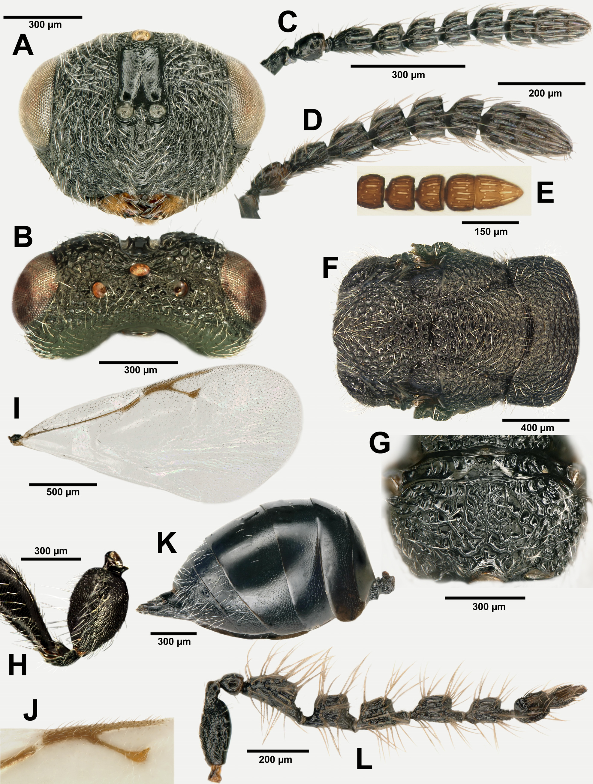
( Figs 11 View FIGURE 11 , 12 View FIGURES 12 A–L; Tab. 4 View TABLE 4 , 5 View TABLE 5 )
Type material. Holotype ♀: SPAIN, Lleida, Tunel de Viella north end, 1390 m, 42.67647°N 0.77150°E, ex fruits of A. albus delphinensis , collected 18.viii.2008, emerged 27.iv.2010 ( A. Ribes) (in BMNH) GoogleMaps . Allotype ♂, same data as holotype (in BMNH) GoogleMaps . Paratypes. 20 ♀ 21 ♂, same data as holotype except some with different emergence dates in 2010 ( 4 ♀ 5 ♂ in BMNH, 2 ♀ 2 ♂ in NMS, 2 ♀ 2 ♂ in MNCN, 12 ♀ 12 ♂ in RAPC) GoogleMaps .
Other material. FRANCE: Alpes - Maritimes, Saint-Dalmas-le-Selvage, GR 5-56 Bousieyas-point 44a, N Combe Male , 1930–2000 m, 44.31833°N 6.85833°E, ex seeds of A. albus delphinensis , 20.vi.2012, adults emerged on 01–15.v.2013 ( G. Delvare) ( 1 ♀ 3 ♂, in GDPC) GoogleMaps ; Aveyron, Lapanouse-de-Cernon, Causse Viel , 810 m, 43.98222°N 3.57417°E, collected on flowers of A. cerasiferus , 01.vi.2009 ( G. Delvare) (vouchers GDEL1636 ♀, GDEL1637 ♀ & GDEL1638 ♀, in CIRAD; 1 ♀, in GDPC); same data but collected on 23.v.2011 (vouchers GDEL1584 ♀, GDEL1585 ♀, GDEL1586 ♀ & GDEL1587 ♀, in CIRAD; 1 ♀, in GDPC) GoogleMaps ; Dordogne, La Douze, Forêt Domaniale de la Barade , 235 m, 44.87777 °N 0.86028°E, 14 & 19.vi.2011 ex seeds of A. albus albus , 14.xi.2009 ( R.R. Askew) ( 60 ♀ 51 ♂, in RAPC) GoogleMaps , Hautes-Pyrénées, Gavarnie-Gèdre, Plateau de Saugué , 1615 m, 42.78555°N 0.00888°W, ex seeds of A. albus delphinensis , 10.vii.2009 ( R.R. Askew) ( 2 ♀ 2 ♂, in RAPC) GoogleMaps ; Gavarnie-Gèdre, Vallée d'Ossoue , 1655 m, 42.75111°N 0.12166°W, 9.vii.2009, same asphodel and collector ( 10 ♀ 10 ♂, in RAPC; 2 ♀ 1 ♂, in GDPC) GoogleMaps ; Hérault, Grabels , 07–13.iv.1989, sweeping herbaceous layer ( H. Tussac) ( 1 ♀, in GDPC) ; Pyrénées-Orientales, Banyuls-sur-Mer, E Col de la Créu , 240 m, 42.46278°N 3.14222°E, on flowers of A. ramosus , 27.iv.2014 ( J. Lecomte) ( 1 ♀, in GDPC) GoogleMaps ; Vienne, Châtellerault, Aire de Chagnats on A 10, 131 m, 48.81778°N 0.54611°E 7, ex seeds of A. a. albus , 14.xi.2009 ( R.R. Askew) ( 1 ♀ 2 ♂, in RAPC) GoogleMaps ; SPAIN: Huesca, Fraga , 125 m, 41.41892°N 0.10911°E, ex seed of A. cerasiferus , 29.iii.2011, adult emerged 15.iv. 2011 ( A. Ribes) ( 1 ♀, in GDPC) GoogleMaps ; Bielsa, Col de Larri, Val de Pineta , 1200 m, 42.64507°N 0.212893°E, ex seeds of A. a. delphinensis, 06.vii.2007 ( R.R. Askew) ( 15 ♀ 6 ♂, in RAPC) GoogleMaps . Lleida, Tunel de Viella north end, 18.viii.2008 ex fruits A. a delphinensis, additional specimens reared with the type series ( A. Ribes) (vouchers GDEL1582 ♀ & GDEL1583 ♂, in CIRAD; 2 ♀ 5 ♂, in GDPC; 79 ♀ 94 ♂ in RAPC) .
Etymology. This species is named for Antoni Ribes who made a very substantial contribution to this study before his death in December 2014.
Condition of female holotype. Specimen complete, glued by right side to rectangular card.
Description of female holotype. Body length 3.6 mm (range in type series 3.2–3.6 mm). Body black, two poorly defined pale spots on anterior face of pronotum, pilosity whitish; legs black with only knees and a narrow stripe on anterior face of protibia brownish, tarsi uniformly infuscate; wings clear, venation brown, setation white ( Fig. 12I View FIGURES 12 ).
Head in dorsal view rather more than 2× as broad as long, eyes separated by 1.8× their own height; POL 1.8× OOL and OOL 2.2× long diameter of posterior ocellus; temples about 0.4× eye length ( Fig. 12B View FIGURES 12 ); head in front view ( Fig. 12A View FIGURES 12 ) almost 1.4× as wide as high; gena weakly curved malar space 0.7× as long as height of eye and 0.6× as long as width of oral fossa. Toruli about equidistant from anterior margin of clypeus and anterior ocellus. Clypeus almost smooth and weakly protuberant, anterior tentorial pits visible; lower face with a median ridge from clypeus to between toruli and with rather weak radiating striae from sides of clypeus to about two-thirds distance to eye, but mostly with reticulate microsculpture in large areoles with poorly-defined margins and central setae; setation of lower face oriented downwards and mesally. Frons with large punctures, bottoms shining but with reticulate microsculpture and central setae directed laterally; malar sulcus traceable for only a short distance below the small suborbital fovea. Genal carina weak, visible for only a short distance above oral fossa. Antennal scrobe with outer margin slightly raised.
Antenna ( Figs 12C, D View FIGURES 12 ) with scape slightly stouter in proximal two-thirds, about 3.7× as long as broad. Pedicel plus flagellum as long as width of head. Pedicel 1.4× as long as wide. Flagellum less stout than in B. abscedus with F1 hardly broader than pedicel, about 1.6× as long as broad; F2 as long as broad; F3 to F5 increasingly transverse; seventh flagellar segment (C1) separated from the eighth (C2) by a deep constriction (in some prepared specimens, as in the holotype, a dried white exudate has erupted from this suture), but C2 and C3 are fused so that the funicle is considered here to be 5-segmented and the clava effectively 2-segmented (composed of C1 + C2+3) ( Fig. 12E View FIGURES 12 ), C1 about 2× as broad as long, C2+3 about 1.6× as long as broad, not quite as long as F5 plus C1.
Mesosoma ( Fig. 12F View FIGURES 12 ) 1.54× as long as wide, 1.4× as long as high. Pronotum 3.35× and mesoscutum 1.65× as wide as long; mesoscutellum 1.1× as long as wide. Dorsal surface of mesosoma in profile with pronotum and mesoscutum evenly arched, mesoscutellum more strongly arched, propodeum declived at an angle of about 85° to plane of mesoscutellum and mesoscutum. Pro- and mesonotum with closely-packed umbilicate punctures, the areolar walls contiguous and at most with only an extremely few very small, microreticulate interspaces, mesonotum anteriorly with a few transverse wrinkles between rows of punctures. Notauli impressed except for extreme posterior part and with inner and outer margins step-like. Scutello-axillar grooves deep, terminating anteriorly just mesad of posterior ends of notauli. Mesoscutellum with umbilicate punctures larger than on mesoscutum and interspersed by larger patches of microsculpture; axilla dorsally with rather few and small umbilicate punctures, these separated by microreticulation which becomes finely striate ventrally. Mesosoma viewed laterally with prepectus about as long as tegula, delimited dorsally by a broad, smooth ridge and ventrally by a finer ridge, its disc dull with irregular sculpturation. Lateral panel of pronotum squamous reticulate. Mesepisternum with anterior margin of epicnemium almost straight, some convexity only in front of mesocoxae, epicnemium finely reticulate, epicnemial carina complete and fronted by a row of large punctures, distance between pro- and mesocoxae relatively short. Lower mesepimeron with reticulate sculpture coarser than on epicnemium, upper mesepimeron with reticulation irregular and strigose on lower part but finer and more even dorsally. Metapleuron with moderately coarse punctures but appearing longitudinally striate when viewed from certain angles. Propodeum ( Fig. 12G View FIGURES 12 ) rather less than 2× as broad as long, its dorsal surface relatively flat with large punctures except on poorly defined median groove which is finely reticulate. Procoxa without depression on frontal surface. Metacoxa with dorsal surface finely reticulate and bare.
Fore wing ( Fig. 12J View FIGURES 12 ) 2.1× as long as wide; lengths of costal cell: marginal vein: stigmal vein: postmarginal vein as 60:13:14.5:17. Stigmal vein straight, stigma not quite half as wide as its distance from anterior margin of wing, uncus with 2 pairs of placodea. Basal cell with about 12 upper surface setae; costal cell on upper surface with a complete anterior row of setae plus some additional setae at apex, anterior one-third of lower surface setose; speculum small, reaching only base of parastigma and partially closed below.
Metasoma ( Fig. 12K View FIGURES 12 ). Petiole about 0.7× as long as broad, dorsally with strong reticulate sculpture. Gaster about as long as head plus mesosoma, in lateral view 1.5× as long (excluding ovipositor) as deep; GT4 dorsally 1.3× as long as GT3 with a band of sparse setae dorsolaterally; syntergum in dorsal view about 1.6× as broad as long; exposed part of ovipositor sheath about 0.7× length of syntergum.
Male. Length 2.4–3.2 mm. Wing pilosity darker than in female but colouration otherwise similar. Antenna ( Fig. 12L View FIGURES 12 ) with pedicel plus flagellum about 1.3× as long as head breadth; funicle of 5 segments and clava 2- segmented although a deep constriction may separate flagellar segments 7 and 8; F 1 in profile not quite 2× as deep as pedicel, its body 1.5× as long as broad; F6 rather less than 2× as long as broad; setae on funicle segments longer and finer than in B. abscedus , those on F1–F5 longer than their segments, arranged in 3 irregular whorls on F1 and in 2 whorls on the other funicle segments; claval segment slightly shorter than F6, about 2× as long as broad.
Metasoma. Gaster including petiole about as long as mesosoma; body of gaster ovate, its dorsal surface rounded and not sharply peaked at apex of GT2; petiole 1.6× as long as broad, its dorsal surface punctured and flat, about one-third as long as body of gaster with its apex slightly beyond apex of hind coxa.
Variation. The size varies according to the associated asphodel. There is a strong contrast between females reared respectively from A. albus and A. ramosus on the one hand and A. cerasiferus on the other. In the former phenotype the gaster is relatively shorter, about as long as head plus mesosoma ( Fig. 12F View FIGURES 12 ), but in the latter it is distinctly longer and the ovipositor sheaths are strongly upturned ( Fig. 11 View FIGURE 11 ). For some time it was hypothesized that the two phenotypes may belong to separate species but the molecular data clearly indicate that only one species is involved ( Figs 8–9 View FIGURE 8 View FIGURE 9 ). Another variation, this apparently independent of the associated asphodel and appearing within populations at the same site, concerns the segmentation of the flagellum. In some females a strong constriction, in addition to a suture, may be present between the 7 th and 8 th flagellomeres ( Fig. 12D View FIGURES 12 ); the funicle hence appearing 6-segmented whereas it is more distinctly 5-segmented in most females ( Fig. 12C View FIGURES 12 ).
Diagnosis. Both sexes. Species of medium size, 3.2–3.6 mm in length; mesonotum with dense puncturation and narrow interspaces showing no coriaceous sculpture; notauli clearly impressed; anterior outline of mesepisternum mostly straight, somewhat convex only ventrally.
Female. OOL 1.7× as long as posterior ocellus diameter; POL 1.9× as long as OOL; clava bisegmented (the penultimate segment being fused with the terminal one), occasionally with a constriction between the 7 th and 8 th flagellomeres, hence the funicle, although usually 5-segmented, sometimes appears 6-segmented; setation of fore wing entirely white; stigmal and postmarginal veins respectively 1.25× and 1.55× as long as marginal vein.
Male. Scape with moderately protruding apicoventral swelling; flagellum 5-segmented and clava 2-segmented, C1 and C2 separated only by a suture.
Recognition. Both sexes of B. ribesi may be distinguished from B. gijswijti and B. lecomtei by their larger size and the straight anterior outline of the mesepisternum ( Fig. 11 View FIGURE 11 ). The species may be separated from B. abscedus by the very dense punctuate sculpture of the mesoscutellum without coriaceous interspaces ( Fig. 12F View FIGURES 12 ). The white setation of the fore wing in the female separates B. ribesi from both B. insulare , to which it is extremely close but which has bicoloured setation, and from B. abscedus in which the setation is entirely dark. The female of B. ribesi may also be separated from that of B. abscedus by having a 5-segmented funicle and 2-segmented clava, although in some specimens of B. ribesi a deeper constriction may separate the 6 th from the 7 th flagellomere, giving the funicle a 6-segmented appearance. The two terminal flagellomeres are fused in B. ribesi but separated in B. abscedus . Bruchophagus ribesi is distinguished from B. asphodelinae by the relative proportions of OOL and POL and by the venation of the fore wing. The form of the gaster is also useful in distinguishing B. ribesi , although it varies somewhat according to the associated asphodel. In specimens emerging from A. albus the gaster is short with the ovipositor sheaths hardly upturned ( Fig. 12K View FIGURES 12 ), but in those associated with A. cerasiferus the gaster is longer with the sheaths clearly upturned ( Fig. 11 View FIGURE 11 ). The gaster of female B. asphodelinae is also relatively long but the ovipositor sheaths are only slightly upturned ( Fig. 15 View FIGURE 15 ).
Specimens of B. ribesi and B. abscedus from their respective type localities are quite distinct, but in France individuals approaching B. ribesi in morphology, but reared from A. albus albus , are not uncommon. All Bruchophagus associated with A. albus delphinensis in our study can be confidently attributed to B. ribesi , but those obtained from A. albus albus may comprise both species with B. abscedus usually dominant.
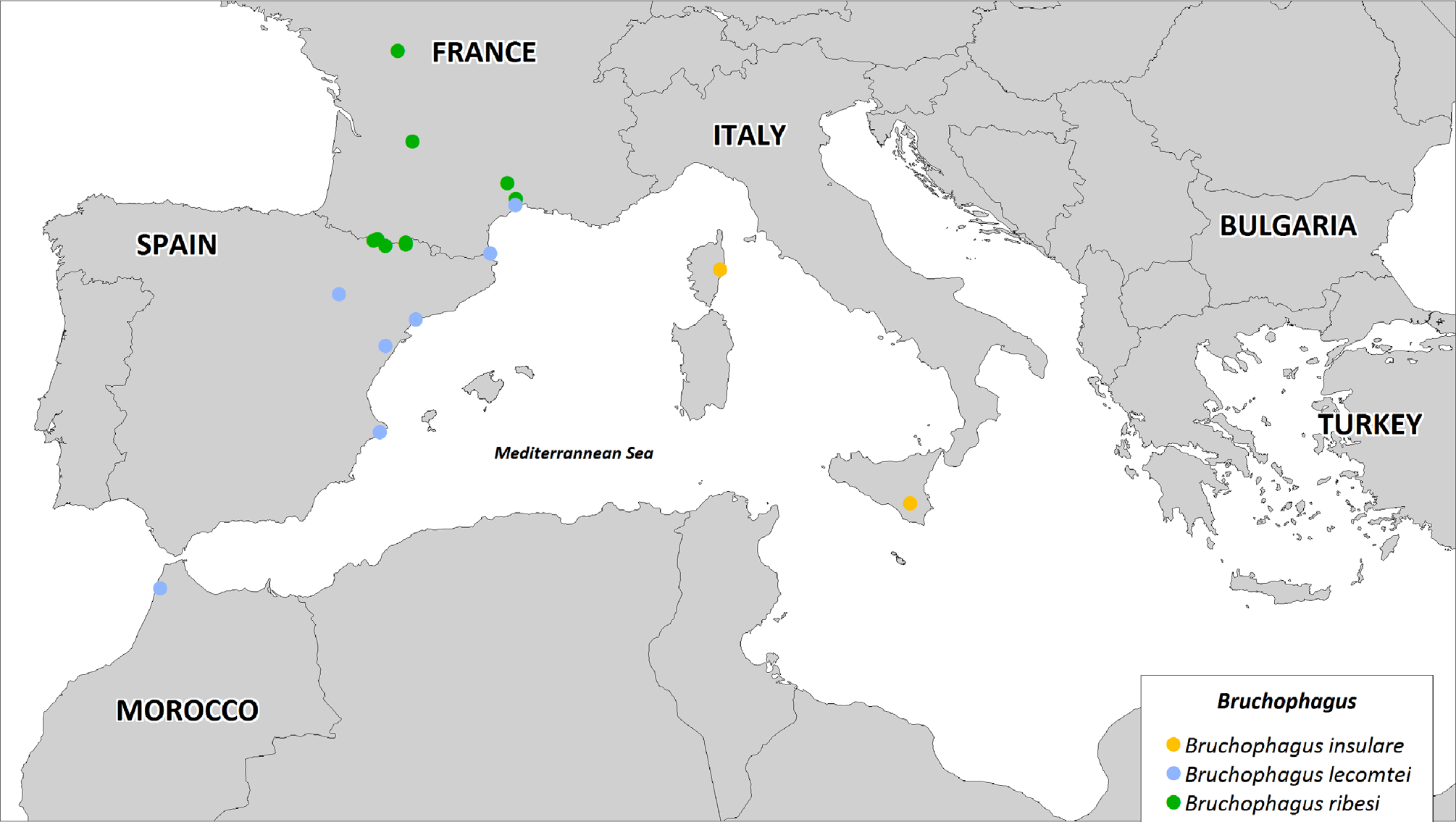
Distribution ( Fig. 35 View FIGURE 35 ). Spain and France.
Biology. Host plants. Bruchophagus ribesi is most usually associated with A. albus delphinensis ( Fig. 40 View FIGURE 40 ) and it is the only Bruchophagus species reared from seeds of this plant. It was observed in large numbers on flowering spikes with green fruits in both the French and Spanish Pyrénées. In the Val de Pineta near Bielsa (Huesca), Spanish Pyrénées, a sample ( 7 ♀ 4 ♂) was collected in the field on 6.vii 2007 from flowering stems of A. albus delphinensis (together with larger numbers of Eurytoma asphodeli ), and 6 ♀ were found drilling fully formed green fruits of the same asphodel on 9.vii. 2009 in Val d’Ossoue, French Pyrénées. Bruchophagus ribesi , however, is not confined to high altitude and apparently also occurs in A. a. albus seeds, together with B. abscedus , in more northerly parts of this plant’s range, being identified in rearings from seed capsules of A. albus albus collected in lowland localities in France (Vienne, Dordogne). On the Causse du Larzac, in southern France, at an elevation around 800 m, it was the single Bruchophagus species associated with A. cerasiferus ( Fig. 41 View FIGURE 41 ) and a single female was reared from A. ramosus at Banyuls-sur-Mer near the coast ( Fig. 42 View FIGURE 42 ). Another female was collected by sweeping near Montpellier by Hubert Tussac before its association with asphodels was known. Thus B. ribesi is primarily a montane species but it may also, on occasion, be found on mediterranean or submediterranean asphodels. Its larval life is passed within a single seed in which it is believed to be phytophagous.
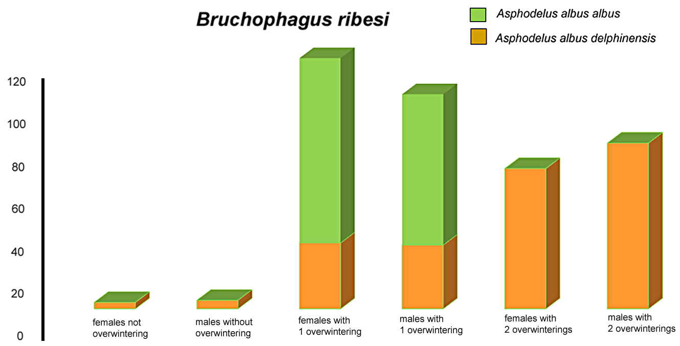
Phenology. A sample of fruits collected in the Val de Pineta in July 2007 produced 2 ♀ B. ribesi in August of the first year, 6 ♀ 4 ♂ overwintered once to emerge during April and May 2008 and 1 ♀ 2 ♂ overwintered twice to emerge in 2009 ( Fig. 46 View FIGURE 46 ).


No known copyright restrictions apply. See Agosti, D., Egloff, W., 2009. Taxonomic information exchange and copyright: the Plazi approach. BMC Research Notes 2009, 2:53 for further explanation.
|
Kingdom |
|
|
Phylum |
|
|
Class |
|
|
Order |
|
|
SuperFamily |
Chalcidoidea |
|
Family |
|
|
Genus |