
Bruchophagus insulare Delvare, 2019
|
publication ID |
https://doi.org/10.11646/zootaxa.4597.1.1 |
|
publication LSID |
lsid:zoobank.org:pub:F8FD30CA-1B84-4134-91BC-B69736DB0EA8 |
|
persistent identifier |
https://treatment.plazi.org/id/03ED8793-FFE6-3B23-D9F0-A1D9E653F94A |
|
treatment provided by |
Plazi |
|
scientific name |
Bruchophagus insulare Delvare |
| status |
sp. nov. |
Bruchophagus insulare Delvare sp. n.
( Figs 13 View FIGURE 13 , 14 View FIGURES 14 A–D; Tab. 4 View TABLE 4 , 5 View TABLE 5 )
Type material. Holotype ♀: ITALY: Sicilia, Vizzini, Monte Iblei, SE Lago Dirillo , contrado Rubalé , 420 m, 37.10778°N 14.72578°E, ex seed of A. ramosus , 19.vi.2014, adult emerged 10.iv.2015 ( G. Delvare) (in BMNH) GoogleMaps . Paratypes. Same data as holotype (vouchers GDEL1715 ♀, GDEL1716 ♀ & GDEL1717 ♀, in CIRAD; 3 ♀ 2 ♂, in MNHN) GoogleMaps ; same locality and host plant, 350 m, 7.12103°N 14.72166°E, 21.vi.2014, adults emerged 09–17.iv.2015 (G. Delvare ) ( 7 ♀ 6 ♂, in GDPC) GoogleMaps ; same data except host plant is Asphodeline lutea ( 1 ♀, in GDPC) GoogleMaps . FRANCE: Corse, Aléria, Vaccaja , 20 m, 42.12861°N 9.46556°E, ex seed of A. ramosus , 22.ix.2011, adult emerged on 20–30.iv 2012 ( J. Balajas) (voucher GDEL1617 ♂, in CIRAD; 1 ♂, in GDPC) GoogleMaps
.
Etymology. The species name refers to the known distribution of the species which has been collected only on the islands of Corse and Sicilia.
Condition of holotype. Specimen complete, glued on rectangular card, with left wings removed and glued on the same card.
Description of female holotype. Body size 2.8 mm. Body colour and setation as in B. ribesi but setation of fore wing dark on basal and costal cells, also dark in front of the median fold beyond the speculum, otherwise white ( Fig. 13 View FIGURE 13 ). Mandible brown on disc, galea and labium whitish; exserted part of ovipositor sheaths entirely dark.
Head 1.91× as wide as long in dorsal view; eyes separated by 1.62× their own height; POL 2.04× OOL and OOL 1.52× posterior ocellus diameter; temple 0.29× eye length; head 1.33× as wide as high in frontal view. Lateral margin of gena forming blunt angle above edge of oral fossa. Malar space 0.67× as long as height of eye and 0.7× as long as width of oral fossa. Lower edge of antennal toruli closer to ventral margin of clypeus than to anterior ocellus. Labrum with 5 ventral setae. Clypeus smooth, somewhat protruding ventrally; anterior tentorial pits visible; lower face with radiating strigose wrinkles originating from sides of clypeus and alternating with punctures, with coriaceous supraclypeal stripe reaching edge of antennal toruli and punctured laterally; setation oriented downwards on face, outwards on the punctured frons; malar sulcus faintly impressed but complete. Gena very faintly carinate just above oral fossa. Postgena with vestigial ventral depression. Surface of scrobal depression with superficial squamose sculpture.
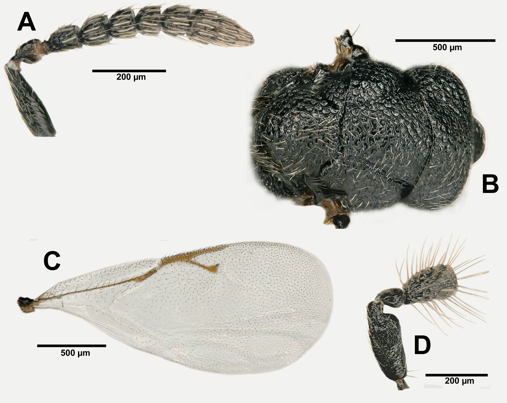
Antenna ( Fig. 14A View FIGURES 14 ). Scape linear, 3.63× as long as wide. Pedicel plus flagellum taken together 0.78× as long as width of head. Pedicel 1.42× as long as wide. Funicle 5-segmented; F1 about as long as pedicel; F1 and F5 respectively 1.48× and 0.84× as long as wide; each funicular with a single row of MPS; F2–F5 and C1 with 2 whorls of long setae. Clava effectively 2-segmented with the vestige of the suture visible between the second row of MPS and the tapering apex of the clava.
Mesosoma 1.76× as long as wide and 1.47× as long as high. Pronotum, and mesoscutum respectively 3.28× and 1.61× as wide as long; mesoscutellum 0.97× as long as wide. Dorsal surface of mesosoma nearly regularly arched, only mesoscutellum somewhat more so; propodeum strongly sloping. Epicnemium flat, the anterior outline of mesepisternum straight, somewhat convex ventrally. Pro- and mesonotum densely areolate-punctured with narrow interspaces; transverse wrinkles alternating with punctures on anterior third of mid lobe of mesoscutum ( Fig. 14B View FIGURES 14 ). Lateral panel of pronotum reticulate squamose. Notauli impressed throughout but partly obliterated by sculpture of mesoscutum, their inner and outer margins step-like. Axillar grooves facing notauli on transscutal line deeply and step-like impressed, with a pit at anterior third; hind margin of mesoscutellum regularly arched. Postscutellum areolate rugulose. Propodeum with incompletely delimited, areolate rugose, median groove; surface irregularly and coarsely areolate between that groove and spiracle, areolate laterally. Lateral panel of prepectus ventrally delimited by a ridge. Mesepisternum with incomplete epicnemial carina, exhibiting a row of punctures in front of femoral depression; epicnemium reticulate, femoral depression and lower mesepimeron more coarsely so; upper mesepimeron strigose dorsally. Metepimeron subtriangular and areolate. Procoxa without depression on frontal surface. Metacoxa bare basidorsally.
Fore wing ( Fig. 14C View FIGURES 14 ) 2.24× as long as wide, costal cell virtually as long as wing width; stigmal and postmarginal veins respectively 1.35× and 1.79× as long as marginal vein. Stigmal vein straight; stigma about half as wide as its own distance from anterior margin of wing; parastigma with 2 sensilla placodea, uncus with 2 narrowly separated pairs of placodea. Basal cell and cubital fold bearing respectively 10–12 and 3–5 setae; costal cell on the upper side with one complete setal row and a few setae behind it, completely setose ventrally; speculum bare dorsally and with a few setae on the underside.
Metasoma ( Fig. 13 View FIGURE 13 ). Petiole with 2 laterobasal and 1 mediodorsal small teeth, bare ventrally. Gaster ovate, about as wide as and 1.08× as long as mesosoma, 1.68× as long as high. GT4 somewhat longer than GT3 on the median line. GT3 and GT4 with few setae dorsally, GT5 and GT6 entirely but sparsely setose. Syntergum very short and not upturned. Dorsal outline of ovipositor sheaths sloping; apex of sheaths narrowly blunt, not sharp.
Male. Differs from female mostly as follows: scrobal depression short, hardly higher than wide; cells on mid lobe of mesoscutum isodiametric, not transverse; postscutellum and propodeum with sculpture less coarse; setation of fore wing entirely dark. Scape with protruding apicoventral swelling ( Fig. 14D View FIGURES 14 ); funicle 5-segmented, the funiculars with short apical bottle-necks; clava 2-segmented with distinct constriction between segments.
Variation. Very little in specimens examined, all being of identical size and most having developed in the same species of asphodel.
Diagnosis. Both sexes. Mesonotum with dense puncturation; notauli clearly impressed; anterior outline of mesepisternum mostly straight, somewhat convex only ventrally.
Female. OOL 1.5× as long as posterior ocellus diameter, POL× as long as OOL. Funicle 5-segmented and clava effectively bisegmented the penultimate segment being fused with the terminal one (C1+C2+3). Setation of fore wing bicolored, with special pattern of white and dark setae; stigmal and postmarginal veins respectively 1.35× and 1.8× as long as marginal vein. Gaster short and oval with ovipositor sheaths hardly upturned.
Male. Scape with protruding apicoventral swelling; flagellum 5-segmented and clava 2-segmented, C1 and C2 separated by suture; setation of fore wing entirely dark.
Recognition. The female of B. insulare can be distinguished from that of B. abscedus by the segmentation of the clava, the penultimate segment being fused with the terminal segment in B. insulare but separated in B. abscedus . The male of B. insulare may also be separated from that of B. abscedus by the segmentation of the clava, the segments being separated by a constriction only in B. insulare but by a short peduncle in B. abscedus . The male of B. insulare differs from that of B. ribesi in having a more protruding apicoventral swelling of the scape.
Distribution. The species is presently known only from two islands: Corse and Sicilia.
Biology. Host plant. Mostly reared from A. ramosus ( Fig. 42 View FIGURE 42 ), but a single female emerged from a seed of A. lutea collected at the same Sicilian locality where the A. ramosus grew that yielded the type series.
Phenology. All specimens hibernated and emerged in the year following seed collection. Hence the species is apparently univoltine as are the other Bruchophagus species associated with asphodels.

No known copyright restrictions apply. See Agosti, D., Egloff, W., 2009. Taxonomic information exchange and copyright: the Plazi approach. BMC Research Notes 2009, 2:53 for further explanation.
|
Kingdom |
|
|
Phylum |
|
|
Class |
|
|
Order |
|
|
SuperFamily |
Chalcidoidea |
|
Family |
|
|
Genus |