
Pyura gangelion (Savigny, 1816)
|
publication ID |
https://doi.org/10.11646/zootaxa.4545.2.6 |
|
publication LSID |
lsid:zoobank.org:pub:276F1D88-3186-4D14-B0BF-F345D043382B |
|
DOI |
https://doi.org/10.5281/zenodo.3510418 |
|
persistent identifier |
https://treatment.plazi.org/id/03ED87D1-A252-B344-FF24-FE5EE182FEB1 |
|
treatment provided by |
Plazi |
|
scientific name |
Pyura gangelion (Savigny, 1816) |
| status |
|
Pyura gangelion (Savigny, 1816) View in CoL
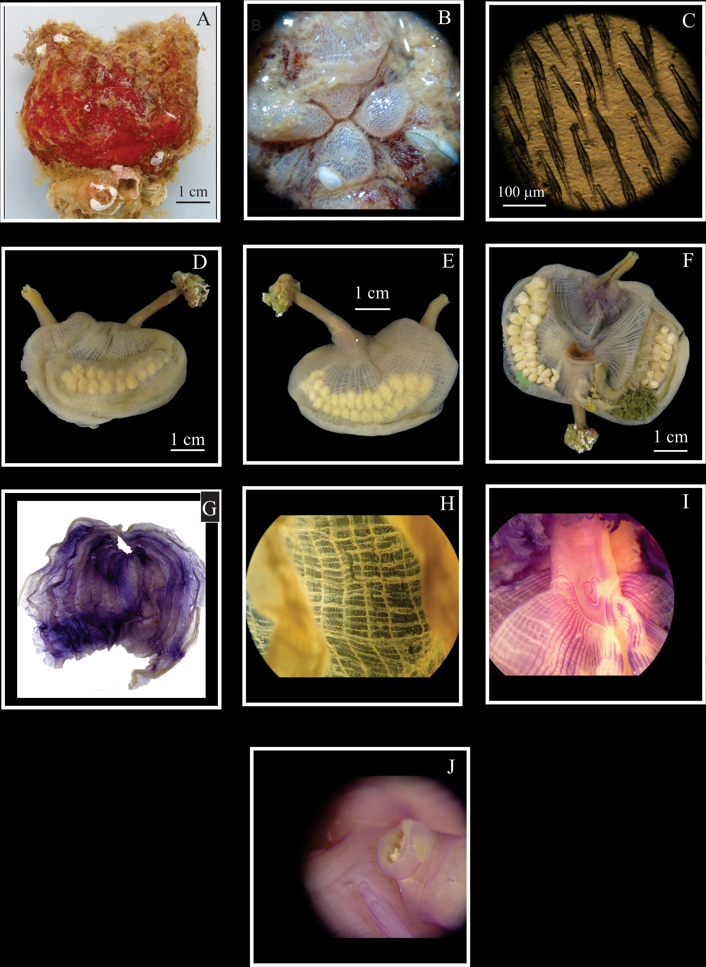
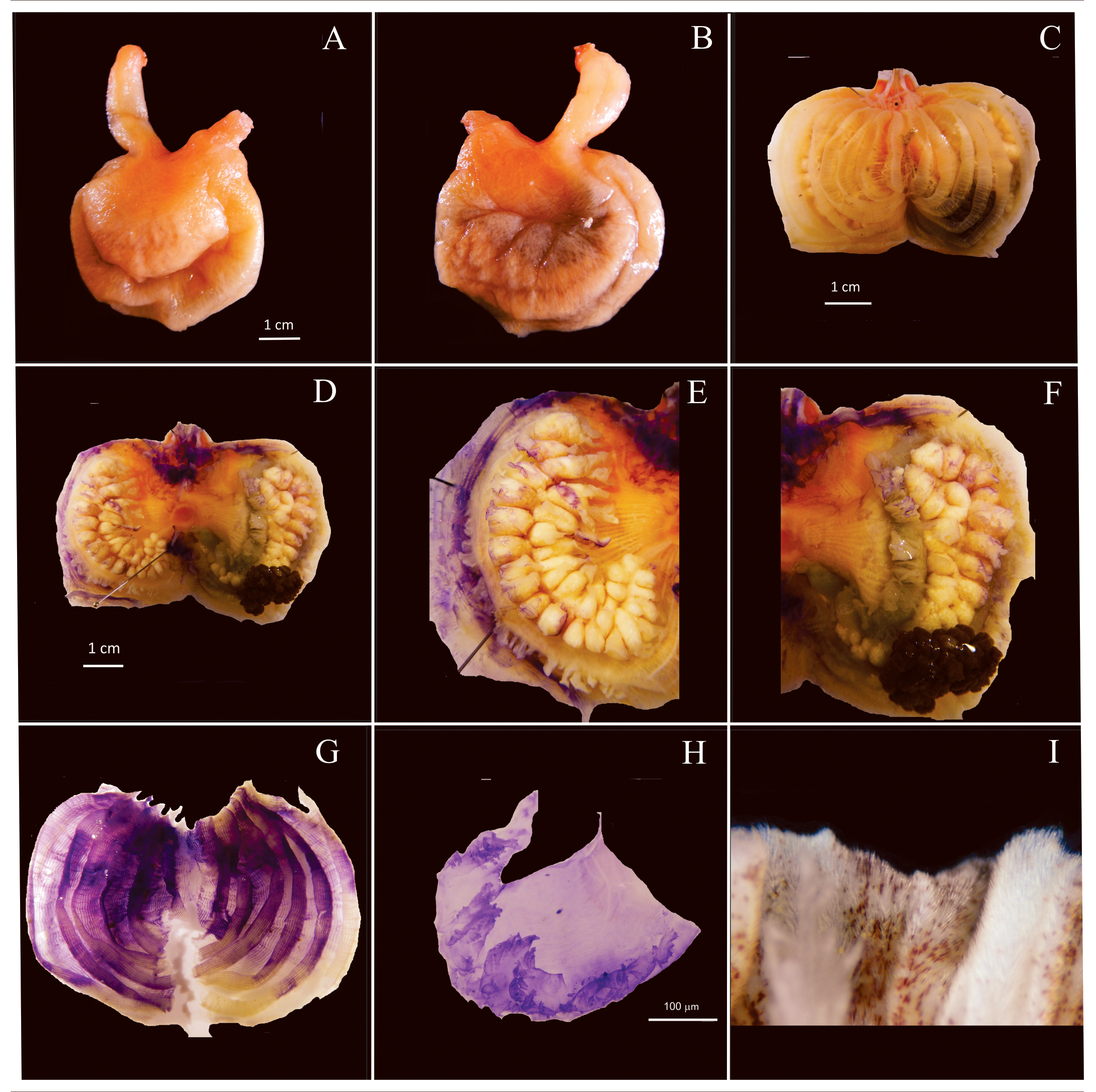
Figs. 5–6 View FIGURE 5 View FIGURE 6
Cynthia gangelion Savigny, 1816 .
Pyura gangelion: Monniot 2018: 13 View in CoL View Cited Treatment , Fig. 12–15 and other synonyms
Material examined. DZUP PYU 74 View Materials , Escalvada Island , Guarapari, ES, 26/I/2012, 1 ind., R.M. Rocha ; DZUP PYU-160 Escalvada Island , Guarapari, ES, 27/I/2012, 2 ind. R.M. Rocha ; DZUP PYU-153 , PYU-154 , PYU-155 Rasa de Terra Island , Guarapari, ES, 23/III/2017, 1 ind. each, R.M. Rocha .
Description. Animals are rounded with very long siphons, 4 to 7 cm in total length including the tunic. They attach to the substrate by the posterior region of the tunic. Tunic is leathery, 2 mm thick around the body but up to 5 mm thick when contracted around the siphons, and covered by many fouling organisms like algae, sponges, hydroids, polychaetes, colonial ascidians and others. The animal is reddish to wine color, with some orange areas and the tunic matrix is wine or pale wine with a mucous yellow membrane in contact with the animal epidermis ( Fig. 5A View FIGURE 5 ). The siphons are located anteriorly. The oral siphon is shorter ( 1–2 cm) than the atrial (1.5–3.5 cm). Both siphons have four lobes covered by a layer of dense spinules measuring up to 200 µm ( Fig. 5B, C View FIGURE 5 , 6I View FIGURE 6 ) that extend from inside the siphons. The body wall is almost opaque because of the well developed muscles forming thin bands underneath a mat of fibers crossing each other in different directions and extend to the endostyle ( Fig. 5D, E View FIGURE 5 , 6A, B View FIGURE 6 ). In freshly preserved animals the anterior region of the body, including the siphons, is orange.
The oral tentacles are laminar and curved, with dense ramifications up to second order along the posterior margin ( Fig. 6H View FIGURE 6 ); the number ranges from 18 to 23, in three different sizes. The largest measure 7 mm. The prepharyngeal region is very narrow and smooth, and the prepharyngeal groove margins do not have projections. The peritubercular region is a deep V with the dorsal tubercle aperture heart-shaped or as a Y ( Fig. 5I View FIGURE 5 ).
The dorsal lamina is divided into many filamentous languets (73–112) and extends to the esophagus. The ends of the longitudinal vessels project towards the esophagus at both sides of its aperture, forming long languets. There are six high pharyngeal folds that overlap a little at each side of the body ( Figs 5G View FIGURE 5 , 6G View FIGURE 6 ). The number of longitudinal vessels ranges from 177–209 on the right side and 172–216 on the left. Transverse vessels 70 (only counted in one individual). Parastigmatic vessels are present. The number of stigmata ranges from four to six per mesh ( Fig. 5H View FIGURE 5 ). The longitudinal vessel formula, from the right to left side in two different animals is:
E 11 (23) 7 (24) 7 (24) 7 (24) 8 (24) 7 (27) 4 DL 4 (27) 5 (27) 5 (30) 5 (26) 5 (23) 4 (26) 8 E
E 4 (25) 4 (24) 5 (30) 6 (32) 4 (26) 5 (26) 6 DL 5 (26) 5 (33) 5 (32) 7 (31) 6 (28) 4 (26) 8 E
The gut occupies about 2/3 of the left side of the body. The digestive gland is one large mass formed by many tubular projections, brownish to light/dark green ( Figs. 5F View FIGURE 5 , 6F View FIGURE 6 ). There are three connections between the digestive gland and the intestine and small groups of projections along the esophagus. The intestine is isodiametric, with a first loop that is long and wide containing the left gonad and a short second loop. Fleshy and irregular-shaped, very dense endocarps are present from the anterior region close to the digestive gland, following the outer intestinal curvature to the anus ( Figs 5F View FIGURE 5 , 6D, E View FIGURE 6 ). Sometimes a slight constriction of the pre-anal region could be observed. The anus is sometimes bilobed, with smooth margins and opening very close to the atrial siphon ( Fig. 5J View FIGURE 5 ). The atrial siphon velum forms four lobes projecting internally.
On the right side, the gonad is attached to the center of the body wall; it is curved and when fully developed it can occupy most of the space. It is formed by 24–33 spherical or elongated lobes disposed irregularly along the oviduct ( Figs 5E, F View FIGURE 5 , 6D, E View FIGURE 6 ). The left gonad has 22–30 lobes organized in pairs in the wide portion of the loop ( Fig. 5D, F View FIGURE 5 , 6D, F View FIGURE 6 ). The oviduct and sperm duct run together for a short distance and open close to the atrial siphon on both sides. Endocarps on the gonadal lobes are irregular and smaller than the ones on the intestine, but there is a group of elongated endocarps around the anterior end of the right gonad and a line of endocarps along the heart, between the right gonad and the endostyle, especially noticeable in Fig. 6D, E View FIGURE 6 .
Remarks: Pyura gangelion is known from the Red Sea ( Monniot 1973), Tanzania, Mozambique, Madagascar, Maldivas and many countries in the Indo-Pacific ( Monniot & Monniot 2001). Monniot (2018) re-examined Pyura torpida from Martinique which has been previously studied by C. Monniot (1983) and decided that the correct identification should be P. gangelion , showing that the species has been introduced in the Atlantic for at least 35 years. The strong armature of spinules in the siphons, four lobed atrial velum, lack of endocarps on the body wall and the line of endocarps present along the heart are diagnostic features of this species (compare our Fig. 6D, E View FIGURE 6 with Monniot (2018) Figs. 13A and 14C). The spinules have a similar shape as in P. vittata but are more iridescent in the latter. It also differs from P. vittata mainly by the shape of the body (more round), stronger body wall musculature, rectum not dilated and the line of endocarps along the heart ( Monniot 1983). The very long siphons of the specimens found in Brazil make it similar to Microcosmus exasperatus Heller, 1878 in the field.
| DZUP |
Universidade Federal do Parana, Colecao de Entomologia Pe. Jesus Santiago Moure |
No known copyright restrictions apply. See Agosti, D., Egloff, W., 2009. Taxonomic information exchange and copyright: the Plazi approach. BMC Research Notes 2009, 2:53 for further explanation.
|
Kingdom |
|
|
Phylum |
|
|
Class |
|
|
Order |
|
|
Family |
|
|
Genus |
Pyura gangelion (Savigny, 1816)
| Skinner, Luís Felipe, Rocha, Rosana M. & Counts, Bailey K. 2019 |