
Chilodontopsis simplex Ozaki & Yagiu, 1941
|
publication ID |
https://doi.org/ 10.5281/zenodo.279782 |
|
DOI |
https://doi.org/10.5281/zenodo.5691541 |
|
persistent identifier |
https://treatment.plazi.org/id/03EDC957-E25A-AD5A-FF23-9096FE83FC66 |
|
treatment provided by |
Plazi |
|
scientific name |
Chilodontopsis simplex Ozaki & Yagiu, 1941 |
| status |
|
Chilodontopsis simplex Ozaki & Yagiu, 1941
( Figs. 1–4 View FIGURES 1 – 10 , 11–18 View FIGURES 11 – 18 ; Table 1 View TABLE 1 , 2)
Improved diagnosis. Cell size 90–160 × 45–95 µm in vivo, cell flattened, oval to long elliptical in outline; 59–78 somatic kineties, 11–13 nematodesmal rods; synhymenium constricted to ventral side, composed of 45–90 dikinetids, running across cell width; single contractile vacuole in posterior cell end; marine habitat.
Description of the Qingdao population. Cell size 90–160 × 45–95 µm in vivo. Cell flexible and non-contractile; shape oval to long elliptical in outline; both ends rounded, with posteriorly slightly tapering in some individuals. Both margins convex with anterior portion slightly projected to left ( Figs. 1, 2 View FIGURES 1 – 10 , 11, 12 View FIGURES 11 – 18 ). Cell distinctly dorsoventrally flattened about 3–4:1. Cyrtos composed of 11–13 nematodesmal rods, each about 50 µm long in vivo ( Fig. 1 View FIGURES 1 – 10 ). Cytostome at anterior 1/4–1/5 of cell length. Cytoplasm colourless, floc-like, with many granules (3– 5 µm across) and up to 50 ingested diatoms ( Figs. 1 View FIGURES 1 – 10 , 17, 18 View FIGURES 11 – 18 ). Macronucleus ovoid, positioned near cell centre, about 40 × 30 µm in size after protargol impregnation ( Figs. 1, 4 View FIGURES 1 – 10 , 13, 16 View FIGURES 11 – 18 ). Micronuclei not observed. Single large contractile vacuole, terminally located, usually formed by aggregating of several small vacuoles around ( Figs. 1, 2 View FIGURES 1 – 10 , 12 View FIGURES 11 – 18 ). Cilia about 5–7 µm long in vivo, uniformly distributed on ventral and dorsal surfaces. Movement moderately rapid, swimming or gliding on substrate.
Synhymenium mainly constricted to ventral side, composed of 45–90 dikinetids, running obliquely from anterior 1/8 to 1/3 of cell ( Figs. 3 View FIGURES 1 – 10 , 13, 15, 16 View FIGURES 11 – 18 ). In total 59–78 somatic kineties, with slightly fewer rows on dorsal than ventral side ( Figs. 3, 4 View FIGURES 1 – 10 , 13, 14 View FIGURES 11 – 18 , Table 1 View TABLE 1 ). About 10 ventral kineties curving around and anterior to cytostome ( Figs. 3 View FIGURES 1 – 10 , 15, 16 View FIGURES 11 – 18 ).
Remarks. To our knowledge, since the establishment of the genus Chilodontopsis Blochmann, 1895 , about 16 nominal species had been assigned to the taxon, and several have been either recognized as a junior synonym (e.g. C. planicaudata Song & Wilbert, 1989 ), or transferred into other genera (e.g. Nassula oblonga (Maupas, 1883) Kahl, 1931 , Chilodonella bengalensis (Ghosh, 1921) , Zosterodasys transverses ( Kahl, 1928) Foissner et al., 1994 , and Nassulopsis elongata ( Kahl, 1931) Foissner et al., 1994 ; for details see Kahl 1931 and Foissner et al. 1994). We therefore accept 11 species of Chilodontopsis (see Figs. 27–37 View FIGURES 27 – 39 ), including: C. depressa (Perty, 1852) Blochmann, 1895 , C. vermiformis Deroux, 1978 , C. kurensis Alekperov, 1985 , C. muscorum Kahl, 1931 , C. acuta Ozaki & Yagiu, 1941 , C. caudata Kahl, 1933 , C. hisioensis Ozaki & Yagiu, 1941 , C. nemerosa Ozaki & Yagiu, 1941 , C. ovalis Biernacka, 1963 , C. pseudonassula ( Penard, 1922) and C. simplex Ozaki & Yagiu, 1941 (for references see Alekperov 1985; Biernacka 1963; Blochmann 1895; Deroux 1978; Foissner 1984; Foissner et al. 1994; Kahl 1931, 1933; Ozaki & Yagiu 1941; Penard 1922).
Character Min Max Mean SD SE CV n Abbreviations: CV = coefficient of variation in %, Max = maximum, Mean = arithmetic mean, Min = minimum, n = number of individuals examined, SD = standard deviation, SE = standard error of mean.
In 1941, Ozaki and Yagiu established the species Chilodontopsis simplex , with describing some important living features, for example, cell size on average 110 × 45 µm, single macronucleus measuring 29 × 11 µm, with one large, irregularly shaped contractile vacuole which is always located near the posterior region of the left side of the cell ( Fig. 34 View FIGURES 27 – 39 ; Ozaki & Yagui 1941). The authors also pointed out that the species had no “postoral ciliary line”, which most probably referred to the synhymenium. We argue that the absence of synhymenium is either a character of a genus other than Chilodontopsis , or a mis-observation, and we assume the latter is the case. Since no redescription with infraciliature features has been available for this species so far, our population of C. simplex is identified based on the living features (e.g. cell shape and size, with one contractile vacuole) and marine habitat ( Ozaki & Yagui 1941).
Compared with the four species (i.e. Chilodontopsis depressa , C. vermiformis , C. kurensis and C. muscorum ) for which the infraciliature is known, C. simplex most resembles the freshwater species C. kurensis in terms of cell size and shape ( Fig. 29 View FIGURES 27 – 39 ; Table 2; Alekperov 1985). Nevertheless, C. simplex has more somatic kineties (59–78 vs. 50–60), fewer nematodesmal rods (11–13 vs. 20), and higher ratio of synhymenium length to cell length (1:1 vs. 1:3), hence can be recognized ( Alekperov 1985). Chilodontopsis depressa and C. vermiformis can be distinguished from C. simplex by their smaller cell size (50–80 vs. 90–160 μm in length), and having fewer somatic kineties (27– 42 vs. 59–78) ( Figs. 27, 28 View FIGURES 27 – 39 ; Table 2; Foissner et al. 1994; Deroux 1978). Chilodontopsis muscorum differs from C.
simplex in cell size (50–70 vs. 90–160 μm in length), numbers of somatic kineties (ca. 21 vs. 59–78) and dikentids in the synhymenium (ca. 32 vs. 45–90), and the position of contractile vacuoles (middle-left vs. posterior) ( Figs. 33, 36, 37 View FIGURES 27 – 39 ; Table 2; Kahl 1931; Foissner 1984).
Chilodontopsis simplex is comparable with C. acuta in terms of cell size ( Fig. 30 View FIGURES 27 – 39 ; Ozaki & Yagiu 1941). However, the former differs from the latter in having fewer nematodesmal rods (11–13 vs. 16) and three contractile vacuoles on right of cell (vs. one, located in posterior end of cell). Chilodontopsis caudata , C. pseudonassula and C.
ovalis are smaller than C. simplex in cell size (35–80 vs. 90–160 µm in length). In addition, C. simplex is posteriorly rounded (vs. posteriorly tailed in C. caudata ), inhabiting marine environments (freshwater for C. pseudonassula ) ( Figs. 31, 32, 35 View FIGURES 27 – 39 ; Kahl 1931, 1933; Penard 1922).
Chilodontopsis simplex can be distinguished from C. hisioensis and C. nemerosa in cell size (90–160 vs. 150– 250 µm in length), and the number of contractile vacuoles (single vs. many) ( Figs. 38, 39 View FIGURES 27 – 39 ; Ozaki & Yagiu 1941).
C. simplex C. depressa C. vermiformis C. kurensis C. muscorum
Cell size in vivo in μm 90–160 × 45–95 50–80 × 20–35 50–65 × 17–25 80–100 ×? 50–75 × 20–30 Cell shape in outline Oval Ellipsoid Oval Oval Oval Size of macronucleus in μm 25–55 × 20–40 15 × 15?? 10 × 5 Number of somatic kineties 59–78 27–42 40 50–60 ca. 21 Number of nematodesmata 11–13 12–15? 20 13–14 Ratio of length of ca. 1:1 ca. 1:1 ca. 1:2 ca. 1:3 ca. 1:1.2 synhymenium to cell width
Number of dikinetids 45–90 20–32?? ca. 32 in synhymenium
Contractile vacuoles One, terminal One, terminal?? One, middle-left Habitat Marine Freshwater & Marine Marine Freshwater Soil Data source Present work Foissner et al. 1994 Deroux 1978 Alekperov 1985 Foissner 1984
? data not available.
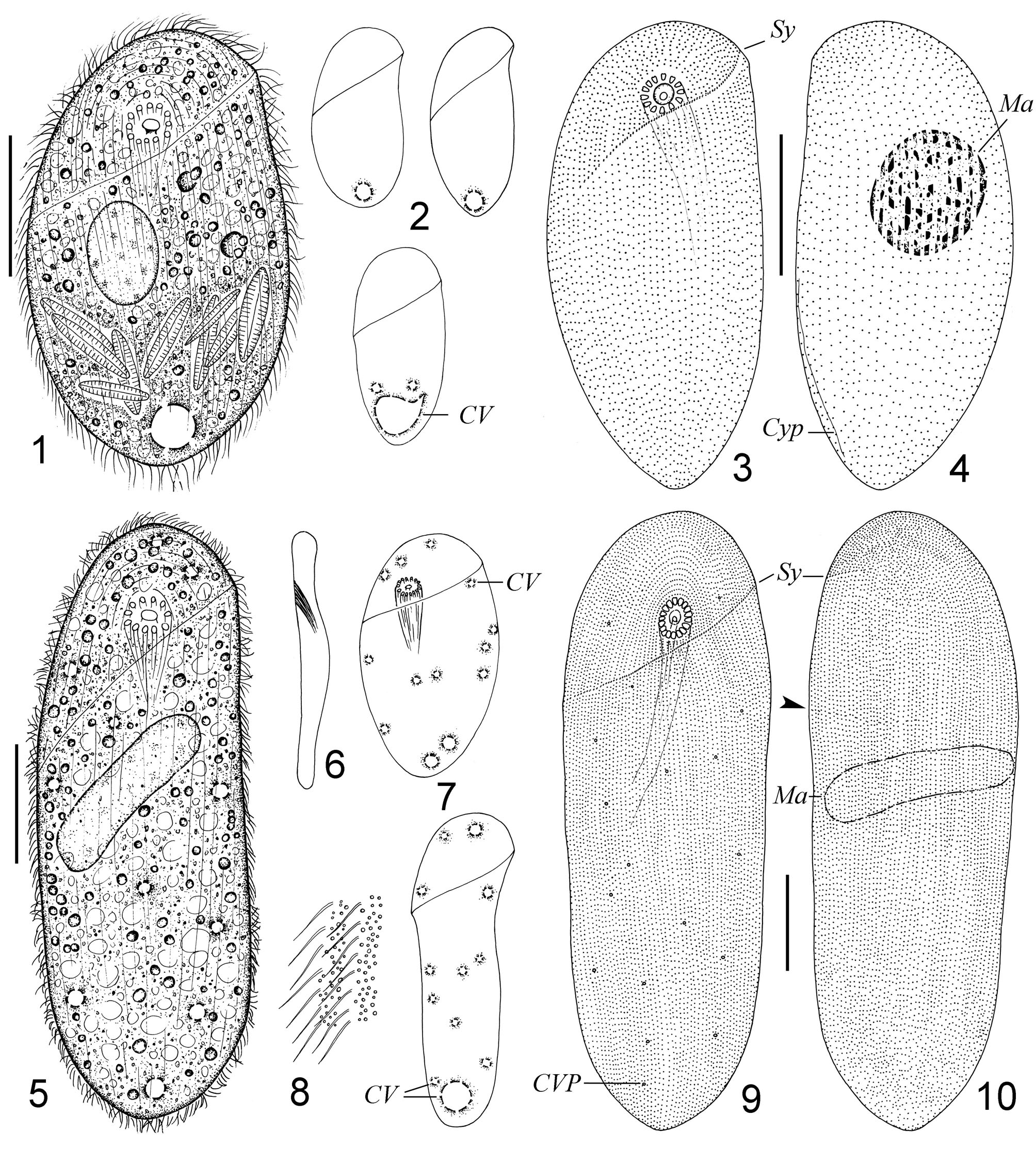
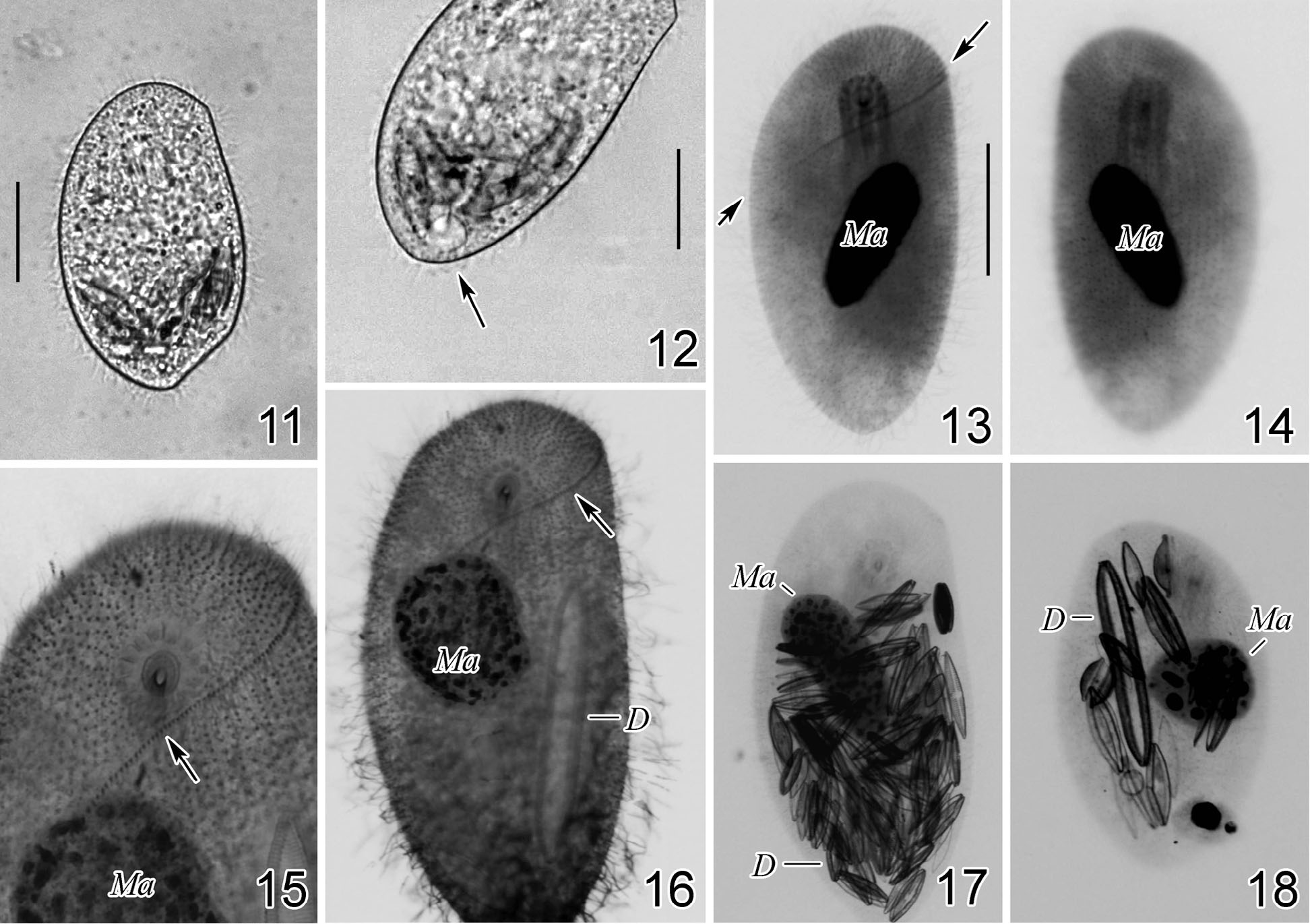
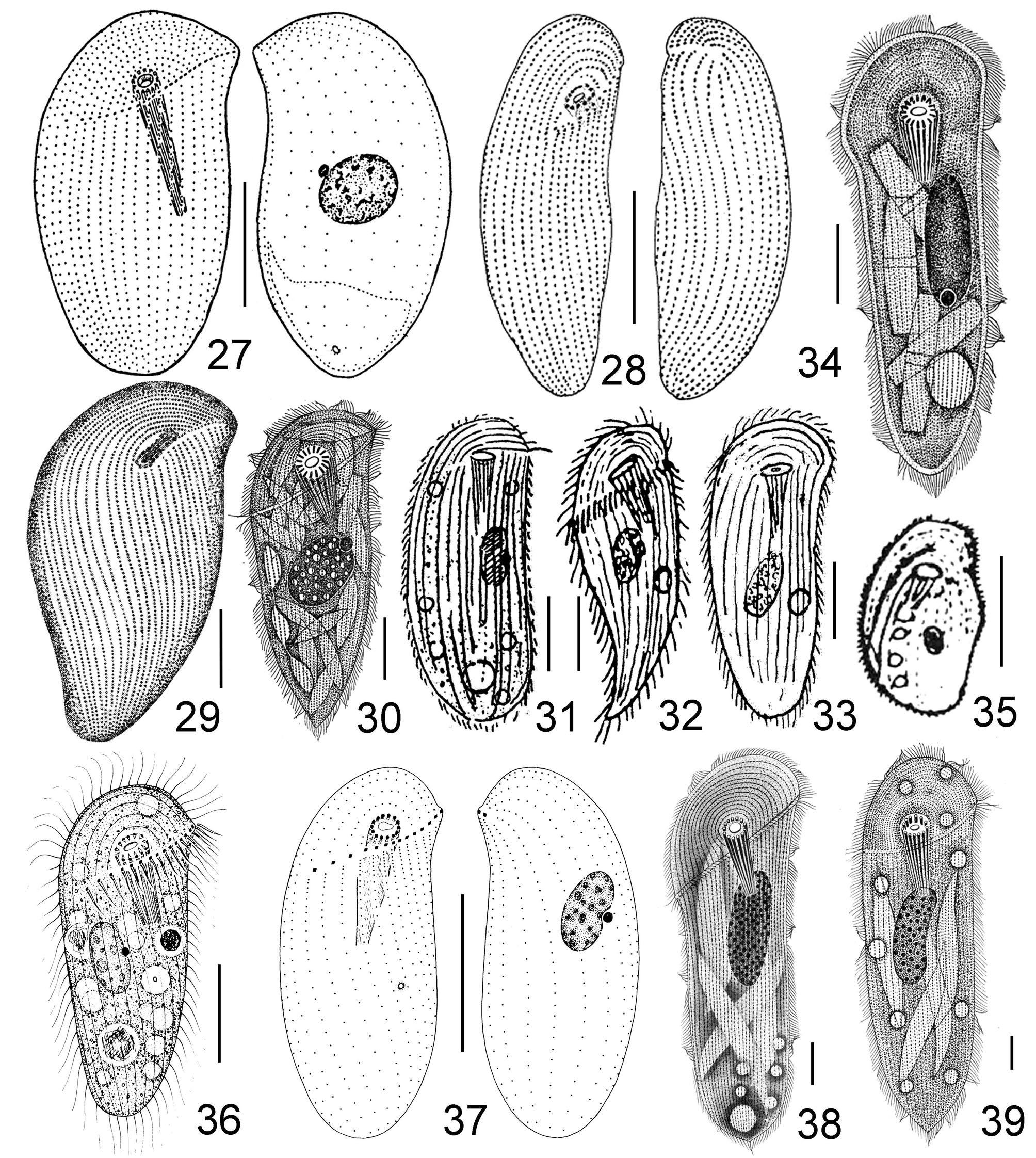
TABLE 1. Morphometric characteristics of Chilodontopsis simplex and Zosterodasys transverses from protargol-impregnated specimens. Measurements in μ m.
| Cell length | 92 160 | 152 380 | 127.3 240.5 | 14.64 61.55 | 2.77 11.5 11.85 25.6 | 28 27 |
|---|---|---|---|---|---|---|
| Cell width | 52 68 | 84 136 | 63.2 92.2 | 9.38 17.01 | 1.77 14.8 3.47 18.4 | 28 24 |
| Length of macronucleus | 26 56 | 52 128 | 41.5 85.0 | 7.53 17.73 | 1.64 18.1 3.48 20.9 | 21 26 |
| Width of macronucleus | 20 12 | 40 40 | 27.4 24.3 | 5.40 7.87 | 1.18 19.7 1.54 32.4 | 21 26 |
| Number of ventral kineties | 30 47 | 45 77 | 36.2 64.0 | 4.24 8.17 | 1.00 11.7 1.83 12.8 | 18 20 |
| Number of dorsal kineties | 23 42 | 38 63 | 30.3 56.9 | 4.75 5.41 | 1.12 15.7 1.21 9.5 | 18 20 |
| Number of somatic kineties | 59 105 | 78 138 | 66.6 121.0 | 5.28 9.39 | 1.24 7.9 2.00 7.8 | 18 22 |
| Number of nematodesmal rods | 11 15 | 13 18 | 11.8 16.3 | 0.70 0.96 | 0.15 5.9 0.20 5.9 | 21 23 |
| Number of dikinetids in synhymenium | 45 65 | 90 130 | 55.3 88.5 | 10.84 17.77 | 2.49 19.6 4.44 20.1 | 19 16 |
| Diameter of cytostome | 3 4 | 5 10 | 4 5.7 | 0.43 1.68 | 0.13 10.8 0.43 29.5 | 12 15 |
| Length of cyrtos | 40 56 | 70 112 | 51.6 88.9 | 14.31 16.81 | 6.40 27.7 4.66 18.9 | 5 13 |
No known copyright restrictions apply. See Agosti, D., Egloff, W., 2009. Taxonomic information exchange and copyright: the Plazi approach. BMC Research Notes 2009, 2:53 for further explanation.