
Zosterodasys transverses ( Kahl, 1928 ) Foissner et al., 1994
|
publication ID |
https://doi.org/ 10.5281/zenodo.279782 |
|
DOI |
https://doi.org/10.5281/zenodo.5691543 |
|
persistent identifier |
https://treatment.plazi.org/id/03EDC957-E25E-AD59-FF23-92F9FB96FE11 |
|
treatment provided by |
Plazi |
|
scientific name |
Zosterodasys transverses ( Kahl, 1928 ) Foissner et al., 1994 |
| status |
|
Zosterodasys transverses ( Kahl, 1928) Foissner et al., 1994
( Figs. 5–10 View FIGURES 1 – 10 , 19–26 View FIGURES 19 – 26 ; Table 1 View TABLE 1 )
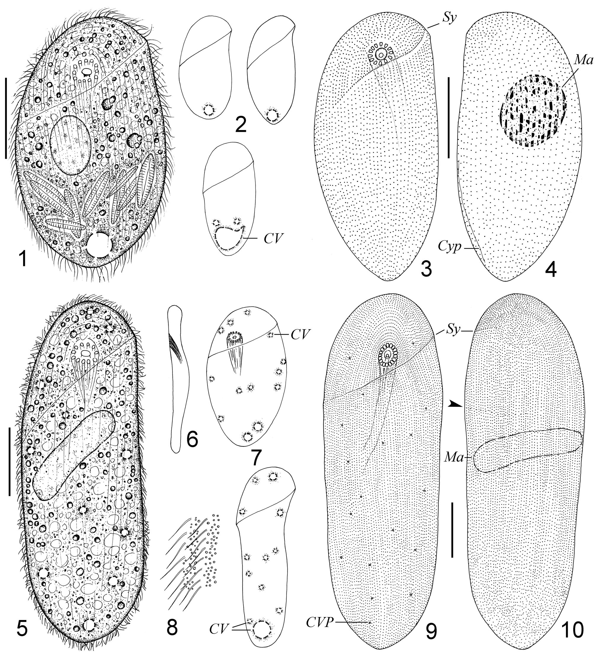
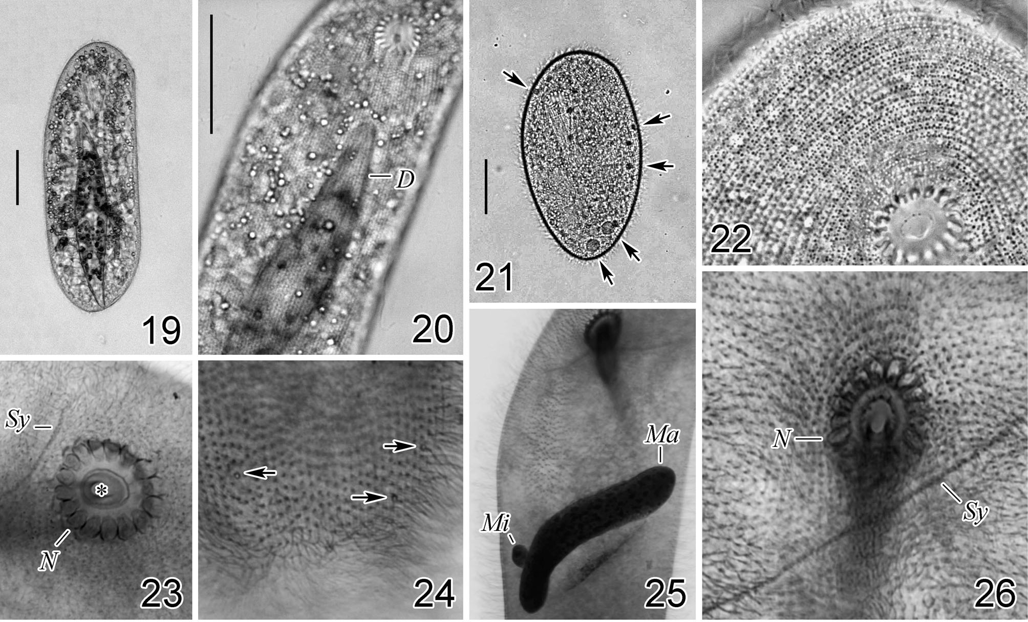
Description of the Korean population. Cell size 160–400 × 80–140 µm in vivo. Cell flexible and non-contractile; shape elliptical to elongate in outline; both ends rounded. Both margins convex with anterior portion slightly projected to left ( Figs. 1, 7 View FIGURES 1 – 10 , 19, 21 View FIGURES 19 – 26 ). Cell slightly dorsoventrally flattened about 3:2 ( Fig. 6 View FIGURES 1 – 10 ). Cyrtos composed of 15– 18 nematodesmal rods ( Fig. 23 View FIGURES 19 – 26 ), each about 80 µm long in vivo. Cytostome at anterior 1/5–1/7 of cell length. Cytoplasm colorless, floc-like, with numerous yellowish oil balls (diameter 5–9 µm), food vacuoles (7–8 µm across) and ingested diatoms ( Fig. 20 View FIGURES 19 – 26 ). Macronucleus usually elongate, sometimes ovoid, positioned at mid-cell, size about 56–128 × 12–40 µm after protargol impregnation ( Figs. 10 View FIGURES 1 – 10 , 25 View FIGURES 19 – 26 ). Micronuclei not observed. Many contractile vacuoles, 4–7 µm in diameter, irregularly distributed on ventral side ( Figs. 5, 7 View FIGURES 1 – 10 , 21, 24 View FIGURES 19 – 26 ). Cilia about 7 µm long in vivo, uniformly distributed on ventral and dorsal surfaces. Numerous fine cortical granules irregularly distributed between ciliary rows ( Fig. 8 View FIGURES 1 – 10 ). Movement moderately rapid, swimming or gliding on substrate.
Synhymenium S-shaped, composed of 65–130 dikinetids, located in a conspicuous suture, running obliquely from anterior 1/7 to 1/3 of cell on ventral and dorsal sides, forming a complete cycle around cell ( Figs. 7, 9, 10 View FIGURES 1 – 10 , 26 View FIGURES 19 – 26 ). In total 105–138 somatic kineties, evenly distributed on both sides. About 20 ventral kineties curving around and anterior to cytostome ( Figs. 9 View FIGURES 1 – 10 , 22 View FIGURES 19 – 26 ).
Remarks. In 1994, Foissner and colleagues transferred Chilodontopsis transversa Kahl, 1928 into the genus Zosterodasys , and gave a detailed description based on a freshwater form, with considering several Chilodontopsis species as junior synonyms ( Foissner et al. 1994; Kahl 1928). Compared with the freshwater form, except that our population is relatively larger in size (160–400 vs. 130–250 µm in length), and has a larger macronucleus (mean 85 × 42 vs. 37 × 23 µm, estimated from drawings), other important characters (e.g. many contractile vacuoles on ventral side, the numbers of somatic kineties and nematodesmal rods, dikinetids in the synhymenium) are almost identical to each other, which, in our opinion, supports the identification of the species. The 18S ribosomal RNA gene (accession number: EU286812 View Materials ) amplified from the marine population described here has been reported in Gong et al. (2009). However, the morphological features of the isolate was lacking. We here also link the morphology to the small subunit ribosomal RNA gene, which may be helpful for future studies, for example, exploring whether the difference in habitats (i.e. marine vs. freshwater) will make them separate species or not.
No known copyright restrictions apply. See Agosti, D., Egloff, W., 2009. Taxonomic information exchange and copyright: the Plazi approach. BMC Research Notes 2009, 2:53 for further explanation.