
Shinkaiya, 2009
|
publication ID |
https://doi.org/ 10.1111/j.1096-3642.2008.00493.x |
|
DOI |
https://doi.org/10.5281/zenodo.5747561 |
|
persistent identifier |
https://treatment.plazi.org/id/03EDCB01-F559-FFCE-FCCD-F8CAFE86FBAA |
|
treatment provided by |
Carolina |
|
scientific name |
Shinkaiya |
| status |
GEN. ET SP. NOV. |
SHINKAIYA LINDSAYI View in CoL GEN. ET SP. NOV.
Diagnosis: As for genus.
Derivation of name: The species is named after the biologist Dhugal Lindsay, who collected the specimen.
Type specimen: The single specimen (and therefore the holotype) was recovered from a push core taken by the Shinkai 6500 submersible from the North Pacific, east of the Japan Trench (38°14.8175′N, 147°00.1885′E; water depth, 5435 m). The major fragment is deposited in the National Museum of Nature and Science , Tokyo (registration number: NSMT-Pr 241) GoogleMaps .
MORPHOLOGICAL DESCRIPTION
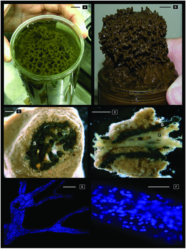
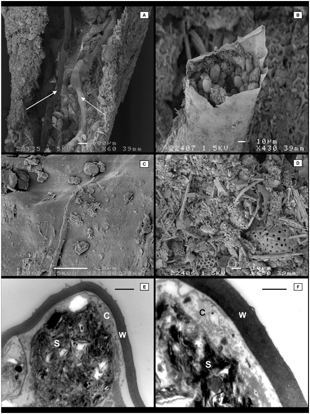
Test form and structure: The test forms a short, approximately cylindrical structure, with a fairly flat upper surface. It measures at least 8 cm in diameter, with ~ 5 cm of test being exposed above the sediment surface. At the base of the test, 1–2 cm long root-like structures extend into the sediment. The test comprises a system of anastomosing branches, typically 0.5 cm in diameter, forming a meshwork with open spaces ranging from 1 to 3 cm in size ( Fig. 2A, B View Figure 2 ). On the upper surface of the test, these branches tend to form an approximately reticulated pattern. The test wall is delicate, soft, and 300-Mm thick. It is light brown in colour, and is mainly composed of fine-grain sediment particles with scattered darker particles, and with occasional larger radiolarian tests (up to 200 Mm in diameter) and quartz grains ( Fig. 3D View Figure 3 ).
Test interior: Usually, the test branches are basically hollow, but there are some internal xenophyae, mainly isolated radiolarian tests, but also quartz grains and sponge spicules ( Fig. 3A View Figure 3 ). Occasionally, the two sides of the test adjoin, leaving almost no lumen.
Cytology: The interior contains prominently developed granellare and stercomare strands, which are intimately intertwined inside the test branches, but without any obvious connections between them ( Fig. 2D View Figure 2 ). The stercomare and granellare are not equally distributed within the specimen. Some test branches contain stercomare but no granellare, although the reverse has not been observed.
The granellare strands are pale yellowish (strawcoloured), and branch in an irregular manner ( Fig. 2D View Figure 2 ). The diameter is highly variable (50– 200 Mm). Sometimes, a thick granellare section gives rise to a cluster of four or five much narrower branches. Where the granellare runs along the length of a test section, the branches may merge; however, these anastomose are not common in the test fragments examined. The organic sheath is very thin, delicate, and has a non-reflective surface ( Fig. 3C View Figure 3 ). No granellae (barite crystals) are visible within the cytoplasm when squashed preparations of granellare fragments are viewed under a high-power microscope. The DAPI staining of the cytoplasm revealed numerous nuclei (roughly 15 ¥ 105 mm-3), of between 2 and 4 Mm in diameter ( Fig. 2E, F View Figure 2 ). Although the cytoplasmic ultrastructure had been largely destroyed by freezing, nuclei and a Golgi apparatus were recognizable in TEM sections, and at least one stercome was present within the cytoplasm. Barite crystals were not observed in any of the sections examined.
The stercomare system occupies a greater volume of the test interior than the granellare ( Fig. 2C View Figure 2 ). The strands are usually attached loosely to the inner surface of the wall, but in places they project into the test lumen ( Fig. 3A View Figure 3 ). They are dark grey, almost black, and the thin organic sheath that encloses the stercomata masses has a distinctly reflective, slightly iridescent surface. The strands range from 30 to 200 Mm in diameter, and their width is often uneven; lobate sections separated by constrictions sometimes develop. Some branches end blindly with rounded terminations. Anastomoses have not been observed, although branches sometimes adjoin without merging. Branching, which is usually dichotomous, may be very frequent. The branches often run in different directions. However, in the more tubular sections of the test, the stercomare strands extend for 100 Mm without branching, and run more or less parallel with the granellare strands.
The stercomata are between 10 and 15 Mm in diameter ( Fig. 3B View Figure 3 ). The TEM observations reveal that the organic envelope enclosing the stercomare is of even thickness: 1-Mm thick ( Fig. 3E View Figure 3 ). The envelope appears rather homogeneous and featureless, except for an outer layer that in places separates from the underlying part to form a loop-like structure ( Fig. 3F View Figure 3 ). An iron peak is evident in the EDAX spectra. The TEM sections of stercomare revealed the presence of cytoplasm associated with stercomata ( Fig. 3E, F View Figure 3 ). The cytoplasm is present around the margins of the stercomare mass, but is inside the organic envelope. Stercomata are composed mainly of flake-like mineral particles. The EDAX microanalysis revealed peaks for silica, aluminium, magnesium, and iron, suggesting that these particles are composed of clay minerals. Barium was also detected within the stercomata. In one case, this element was associated with a crystal. The barite composition of this crystal, however, remains uncertain, because it also yielded a peak for calcium. A sharp peak for titanium was associated with another crystal, presumably rutile.
LIFE POSITION
The specimen was epibenthic. It projected from the seafloor, with the root-like lower part being buried in the sediment. It was found among numerous other unidentified xenophyophores of different sizes.
MOLECULAR CHARACTERIZATION
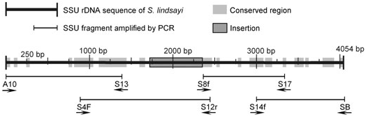
The total length of the SSU rDNA was 4054 bp, and all of the clones sequenced were identical. The sequence alternates between conserved regions and variable regions among foraminiferans, with a long insertion of 624 bp starting in the variable region E23 at position 1744 bp ( Fig. 1 View Figure 1 ). The GC content of Sh. lindsayi sp. nov. (32.2%) is similar to that of Sy. corbicula (34.1%). The sequence divergence between the two xenophyophores is 23.6%, whereas it is 30.2% between Sh. lindsayi sp. nov. and R. algaeformis .
In the phylogenetic tree obtained by the ML method ( Fig. 4 View Figure 4 ), Sh. lindsayi sp. nov. clusters with Sy. corbicula , and the two xenophyophores form a sister group of R. algaeformis . This topology is supported by high bootstrap values (94 and 100%, respectively). The clade consisting of Sh. lindsayi sp. nov., Sy. corbicula , and R. algaeformis branches at the base of polythalamous (multichambered) foraminiferans, including rotaliids, textulariids, and robertinids.
REMARKS
As discussed above, the new species is similar to some species of Syringammina in the construction of the test from reticulated bar-like elements. There is a particular resemblance between Sh. lindsayi sp. nov. and Sy. reticulata in the constant diameter, dimensions, and arrangement of the tubes (compare Fig. 2A View Figure 2 with Gooday, 1996: pl. 7). However, in addition to the differences in wall structure noted above, the overall morphology of the test is distinctly flattened in Sy. reticulata , but is more or less equidimensional in Sh. lindsayi sp. nov.
ELEMENTAL COMPOSITION
Mass spectrometry analyses were performed separately on pieces of the stercomare, granellare, and on intact fragments of the specimen, as well as on environmental samples from the area where the specimen was collected (site 1037) ( Fig. 5 View Figure 5 ). Aluminium, barium, and magnesium were present inside the stercomare, where concentrations were more than 30% higher than in the sediment. These elements were less abundant in the granellare than in the surrounding sediment (site 1037). They occur in roughly the same concentration in the intact fragment (mainly test) and in the environment. Consistent with microscopic observations of barite crystals, barium occurs in the stercomata but not in the cytoplasm. Lead, mercury, and uranium concentrations are also higher (two, four, and six times higher, respectively) inside the stercomare than in the sediment. The concentration of mercury in the granellare is 12 times that in the sediment.


No known copyright restrictions apply. See Agosti, D., Egloff, W., 2009. Taxonomic information exchange and copyright: the Plazi approach. BMC Research Notes 2009, 2:53 for further explanation.
|
Kingdom |
|
|
Phylum |
|
|
Class |
|
|
Order |
|
|
Family |