
Rhynchophorina
|
publication ID |
https://doi.org/ 10.11606/1807-0205/2020.60.special-issue.27 |
|
persistent identifier |
https://treatment.plazi.org/id/03EDCC5D-FF9F-F674-EAFD-FB94668AFEEB |
|
treatment provided by |
Carolina |
|
scientific name |
Rhynchophorina |
| status |
|
Subtribe Rhynchophorina
( Figs. 1-9 View Figure 1 View Figure 2 View Figure 3 View Figure 4 View Figure 5 View Figure 6 View Figure 7 View Figure 8 View Figure 9 )
The following description applies to mature (and submature) larvae of both Dynamis borassi and Rhynchophorus palmarum , which are, respectively, the type species of the genera Dynamis and Rhynchophorus . It is descriptive of the larval stage of the subtribe Rhynchophorina of the current classification of dryophthorines (see Anderson & Marvaldi, 2014).
Mature larva ( Figs. 1-7 View Figure 1 View Figure 2 View Figure 3 View Figure 4 View Figure 5 View Figure 6 View Figure 7 )
Body ( Figs. 1 View Figure 1 A-F) very robust, expanded at middle of abdomen between segments III and VI, narrowing posteriad, with abdominal segments VIII and IX forming a depressed terminal disc ( Figs. 6B View Figure 6 , 7 View Figure 7 A-B). Thoracic and abdominal segments with sternellum present ( Figs. 1B, E View Figure 1 , 6A View Figure 6 ). Cuticle ( Fig. 6 View Figure 6 ) sclerotized around setal groups, elsewhere with coarsely distributed asperities like small spicules; vestiture consisting of sparsely-distributed red-brown setae, mostly short or minute, except longer on terminal disc.
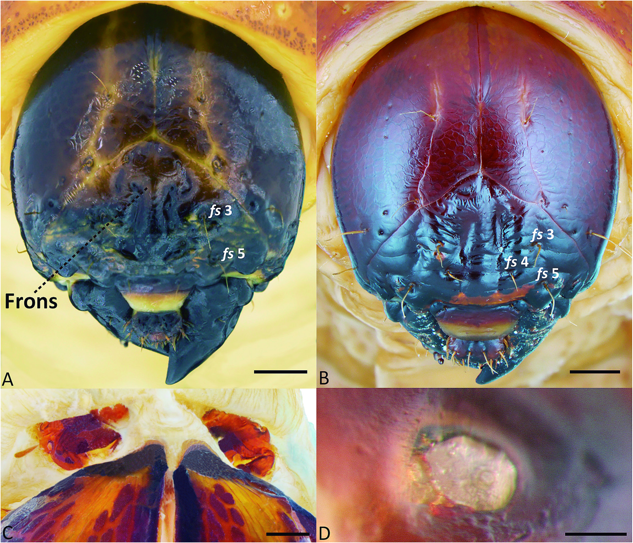
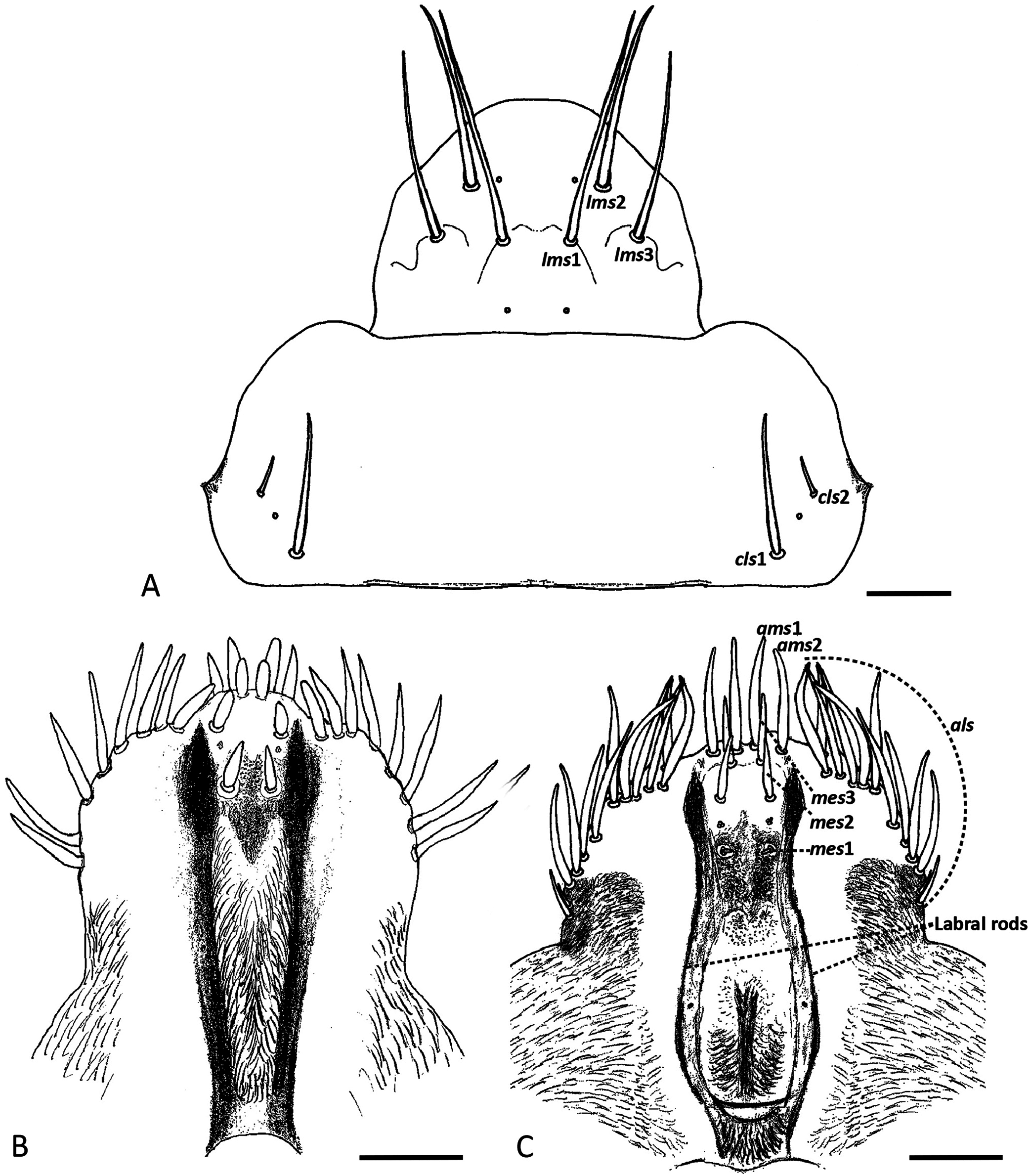
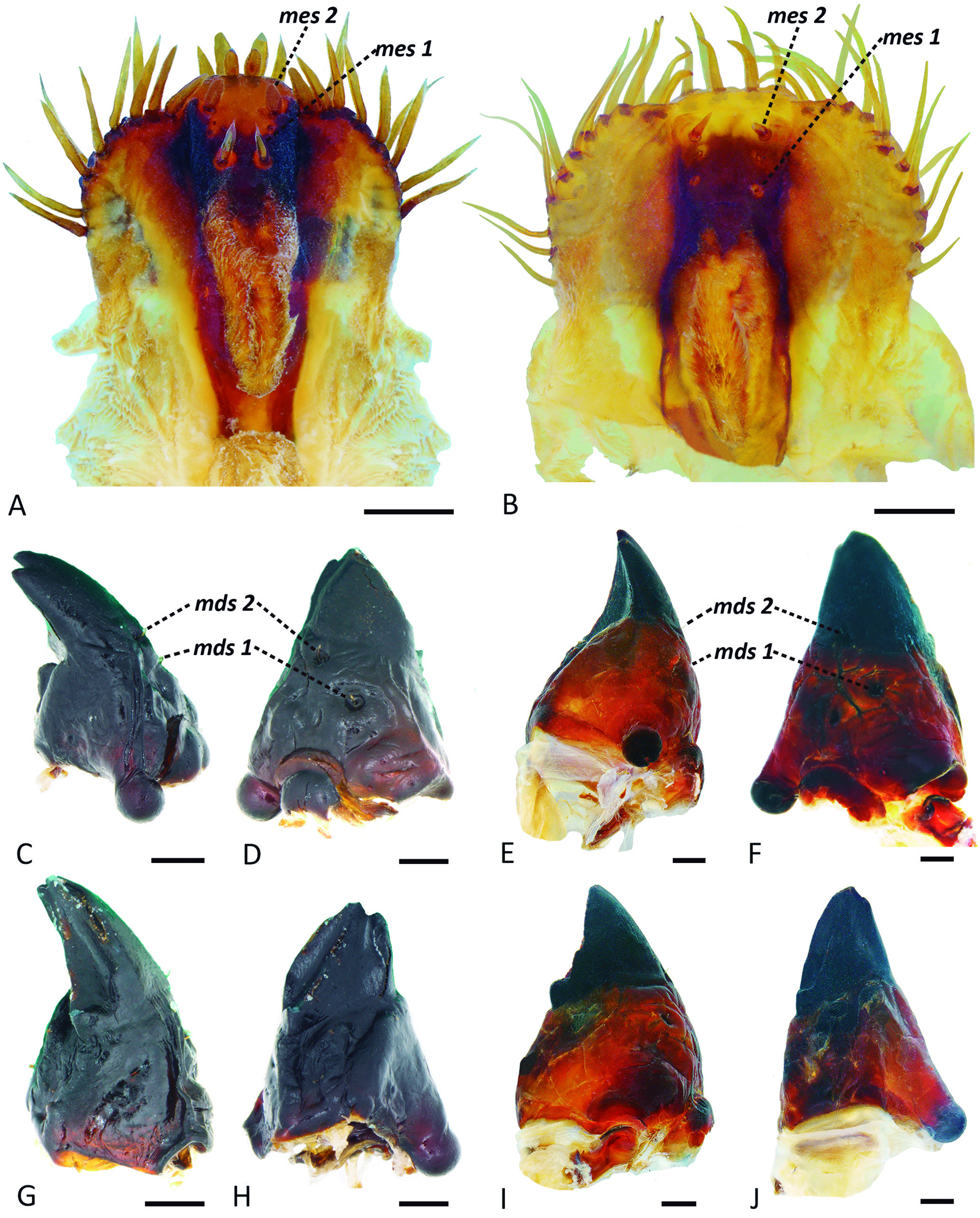
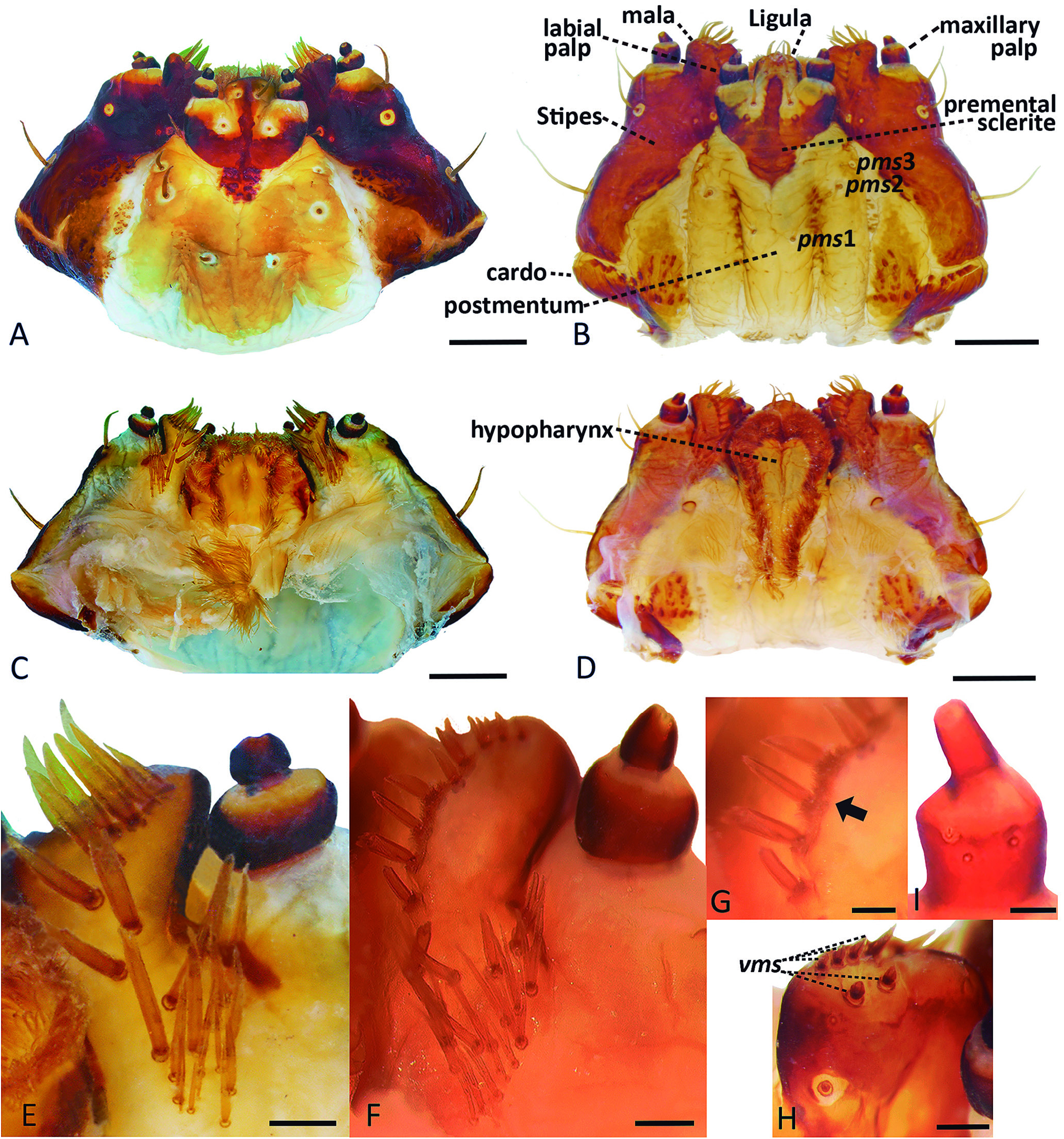
Head ( Figs. 2 View Figure 2 A-B) exposed, longer than wide, strongly sclerotized and deeply pigmented, with paler paramedi- an stripes. Cervix without typical postoccipital condyles, its dorsal side often thickened and with a pair of cervical sclerites on the membrane at joint of head and prothorax ( Fig. 2C View Figure 2 ). Frontal lines distinct, reaching antennae; frons with two medial longitudinal grooves. Endocarina absent. Antennae in oblique position on anterior margin of head; sensorium longer than wide, subconical, circular in apical view ( Fig. 2D View Figure 2 ). Stemmata present, anterior one distinct, near antenna, posterior one faint in older larvae. Five dorsal epicranial setae (des) present; des1, des3 and des5 longest, des2 short and des4 minute; des3 on epicranium,close to frontal line; five frontal setae (fs), fs3 and fs5 longest, fs4 short to well developed, fs1 and fs2 minute; four minute posterior epicranial setae (pes); two lateral epicranial setae (les) unequal in size, les1 much smaller; ventral cranial setae(vcs) unconspicuos.Hypopharyngeal bracon clear. Clypeus ( Fig. 3A View Figure 3 ) with two setae, unequal in size (cls 1 much larger than cls 2), and one sensillum between cls on each side. Labrum ( Fig. 3A View Figure 3 ) pigmented, with anterior margin truncate or slightly produced at middle; labrum with three pairs of setae (lms), with a pair of basal sensilla, and a pair of anterior sensilla, the latter situated near lms2. Epipharynx ( Figs.3 View Figure 3 B-C,4A-B)with simple setae, with five to more anterolateral setae (als); bearing patches of dense pubescence medially and laterally; epipharyngeal sensillum clusters present, a distal pair of three sensillae each, between labral rods, and a proximal pair of two sensillae each;labrum-epipharynx with labral rods elongate, slightly sinuate, joined posteriorly. Mandibles ( Figs. 4 View Figure 4 C-J) robust, dark, often bidentate at apex, with median tooth and a molar area on inner side; with basal sensillum and two short setae on outer surface (mds 1, 2) placed in pits, aligned longitudinally. Maxillae ( Figs. 5 View Figure 5 A-I) with mala truncate and slightly excised apically, bearing several dorsal setae (dms) aligned in a row medially and irregularly placed proximally ( Figs. 5 View Figure 5 E-F); some dms branched, mostly bifid; ventral malar setae (vms) simple ( Fig. 5H View Figure 5 ); maxillary palpi 2-segmented, basal palpomere with two sensilla and one minute seta ( Fig. 5I View Figure 5 ). Labium ( Figs. 5 View Figure 5 A-D) with palpi 2-segmented; premental sclerite trident shaped; postmentum sclerotized and pigmented in mesal and lateral areas, with a lighter stripe between them; three postmental setae (pms), proximal pair (pms1) closer together than pms2 and pms3, median pair (pms2) the longest; hypopharynx ( Figs. 5 View Figure 5 C-D) densely setose in lateral areas, medially smooth with a longitudinal furrow.
Thorax ( Fig. 6A View Figure 6 ). Pronotum ( Figs. 6A View Figure 6 , 1C, F View Figure 1 ) well pigmented, consisting of two sclerotized halves separated dorsally by a lighter band, bearing few pronotal setae (five to seven). Prothoracic sternum with a median depression ( Figs. 1B, E View Figure 1 ). Meso- and metathorax ( Fig.6A View Figure 6 ) with one seta on alar area and several very short spiracular setae (ss); epi- and pleural areas subdivided into two lobes. Pedal areas with six setae, one of them minute, close to the longest one. Thoracic spiracles ( Figs. 6A View Figure 6 , 7C View Figure 7 , F-G) placed on prothorax, narrow-ovate, without airtubes.
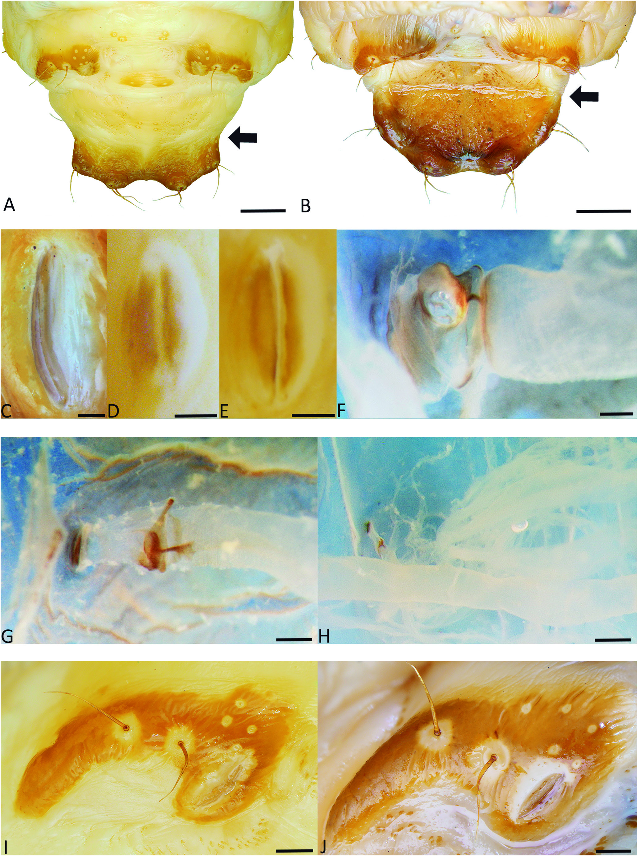
Abdomen ( Fig. 6 View Figure 6 ). Typical abdominal segments ( Figs. 1 View Figure 1 , 6A View Figure 6 ) with four dorsal folds, bearing five postdorsal setae (pds). Segments VIII and IX ( Figs. 1 View Figure 1 , 6B View Figure 6 ) with fewer but longer dorsal setae, forming a sclerotized dorsal disc, with posterior margin extended into two pairs of broadly defined,seta-bearing lobes ( Figs. 6B View Figure 6 , 7 View Figure 7 A-B).Epi- and pleural areas subdivided into three lobes ( Figs. 1 View Figure 1 , 6A View Figure 6 ). Anal region ( Fig. 6B View Figure 6 ) ventral, anus transverse,four anal lobes, the lateral ones bearing one or two small setae.Spiracles narrow-ovate,without airtubes ( Figs. 6 View Figure 6 A-B,7D-E,I-J); abdominal spiracles I to VII ( Figs. 6A View Figure 6 , 7 View Figure 7 D-E) placed laterally near anterior margin of segments, much smaller than the VIII, but functional; spiracles of segment VIII ( Figs.6B View Figure 6 , 7 View Figure 7 I-J) well developed and placed on dorsum,aligned longitudinally.
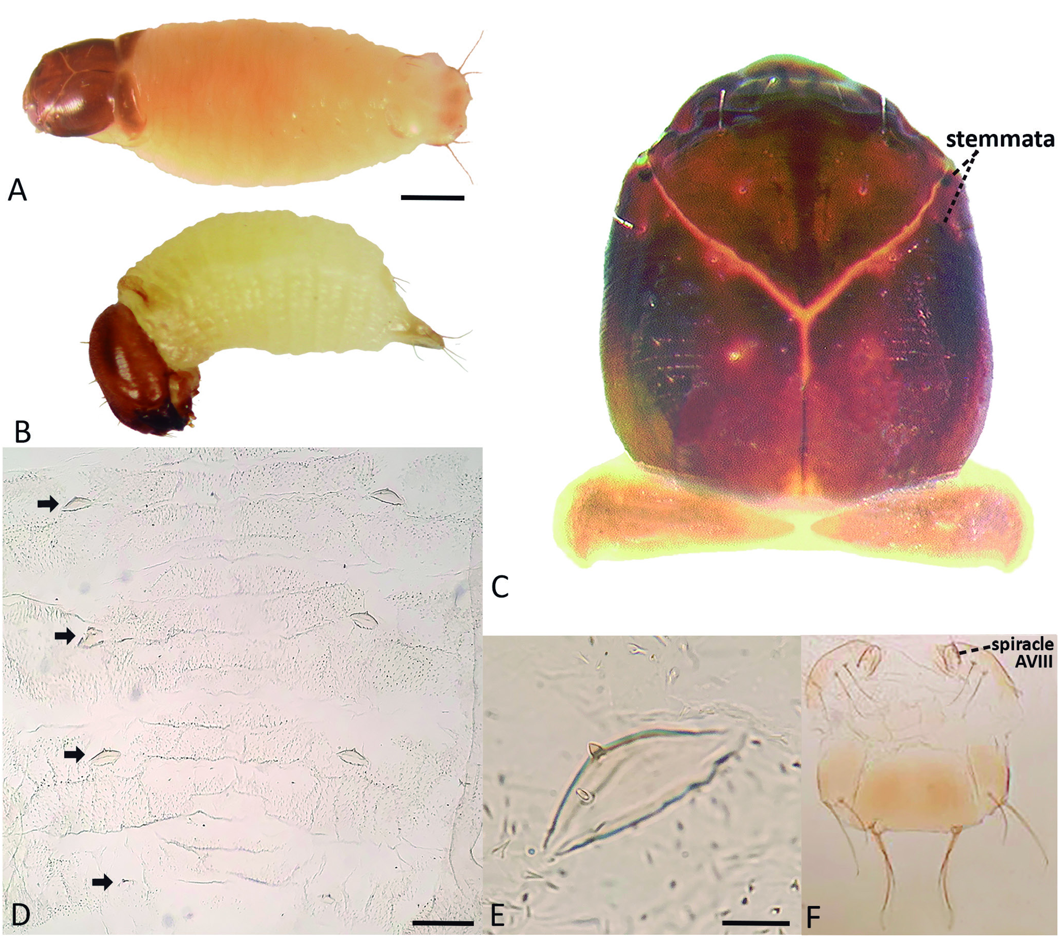
First instar larva ( Figs. 8 View Figure 8 A-F, 9A-C)
Both anterior and posterior stemmata distinct,as dark pigmented spots, anterior larger than posterior ( Fig. 8C View Figure 8 ). Egg-bursters ( Fig. 8D View Figure 8 ) present on abdominal segments IV to VII, on either side in the dorso-lateral region of each segment, the posterior pair smaller than those in AIV-AVI; each egg burster ( Fig. 8E View Figure 8 ) arising on a convex tubercle and consisting of a sub-conical,forwardly directed spine, situated anterior to a blunt seta. Only the spiracles of
thorax and abdominal segment VIII ( Fig. 8F View Figure 8 ) are distinct, abdominal spiracles I to VII absent. Abdominal terminal plate ( Fig. 8F View Figure 8 ) with margins only weakly sinuated.
Comments: The characters mentioned above are exclusive of the first instar. Additional differences between early and older instar larvae involve relative dimension of structures, like the antennae, which are relatively much larger in the first instar; the pigmentation and level of sclerotization of body areas tend to increase in successive instars; the differences in length between se- tae of body areas are higher in early instars; when setae are supernumerary (compared to modal numbers), such as those placed laterally in the epipharynx or dorsally in the maxillary mala, their amount is higher in later instars.


| I |
"Alexandru Ioan Cuza" University |
No known copyright restrictions apply. See Agosti, D., Egloff, W., 2009. Taxonomic information exchange and copyright: the Plazi approach. BMC Research Notes 2009, 2:53 for further explanation.