
Altajosoma kultukensis Mikhaljova, 2019
|
publication ID |
https://doi.org/ 10.25221/fee.374.1 |
|
publication LSID |
lsid:zoobank.org:pub:4467F525-C0C9-46E8-8059-C2B651A0A2FE |
|
persistent identifier |
https://treatment.plazi.org/id/A17B6265-B2BD-40CC-97D6-0B5328307009 |
|
taxon LSID |
lsid:zoobank.org:act:A17B6265-B2BD-40CC-97D6-0B5328307009 |
|
treatment provided by |
Felipe |
|
scientific name |
Altajosoma kultukensis Mikhaljova |
| status |
sp. nov. |
Altajosoma kultukensis Mikhaljova , sp. n.
http/ urn:lsid:zoobank.org:act:A17B6265-B2BD-40CC-97D6-0B5328307009
Figs 1–5 View Figs 1–5
MATERIAL. Holotype – ♂ ( FSCB), Rio Bistraya , 10.IX. 95, Kurtuk, Rusia, F.
Ruano leg. (= Russia: Irkutskaya oblast: Slyudyanskii District, environs of Kultuk village and Bistraya River, 10.IX 1995, leg. F. Ruano).
DESCRIPTION. MALE. The specimen is broken into four separate body portions.
Length about 19, width 2.0 mm with paraterga. Coloration in alcohol including legs and antennae uniformly dark brown (probably the color was distorted by prolonged storage). Eyes black.
Body with 32 segments. Head covered both with relatively long and short setae.
Eye patches subtriangular, with 28–29 ocelli. Collum semicircular. Both collum and somite 2 narrower than head with genae. Somite 2 wider than collum. Body width gradually increasing until somite 6–7, body parallel-sided on somites 21–22,
thereafter gradually tapering. Paraterga beginning on collum, well developed on somites 6–26, reduced on somite 27, onward missing. Metazonital macrochaetae in a transverse row on somites 28–31, like an extended triangle on preceding somites.
Anterolateral and medial macrochaetae subequal in length, caudolateral ones longest, distal part of all macrochaetae filiform, apex pointed. Axial suture well developed.
Legs long and slender. Leg pairs 1–2 typically reduced in size, with tarsal brushes as usual. Legs 3–7 increasingly enlarged toward gonopods. Femora 5–7 swollen and curved.
Leg pairs 3–7 with a small group of funnel–shaped tarsal papillae apically near claw. Claw of leg pairs 3–7 at base with a long setoid outgrowth ventrally but without additional claws dorsally. Postgonopodal legs (including leg pairs 10 and 11) with small group of funnel-shaped tarsal papillae apically near claw, however tarsal papillae gradually reduced toward posterior part of body. Claws of postgonopodal legs (including leg pairs 10 and 11) at base with two small additional claws dorsally and a relatively long setoid outgrowth ventrally, but small additional claws gradually missing toward posterior part of body; at least claws of hindmost legs at base devoid of such additional claw.
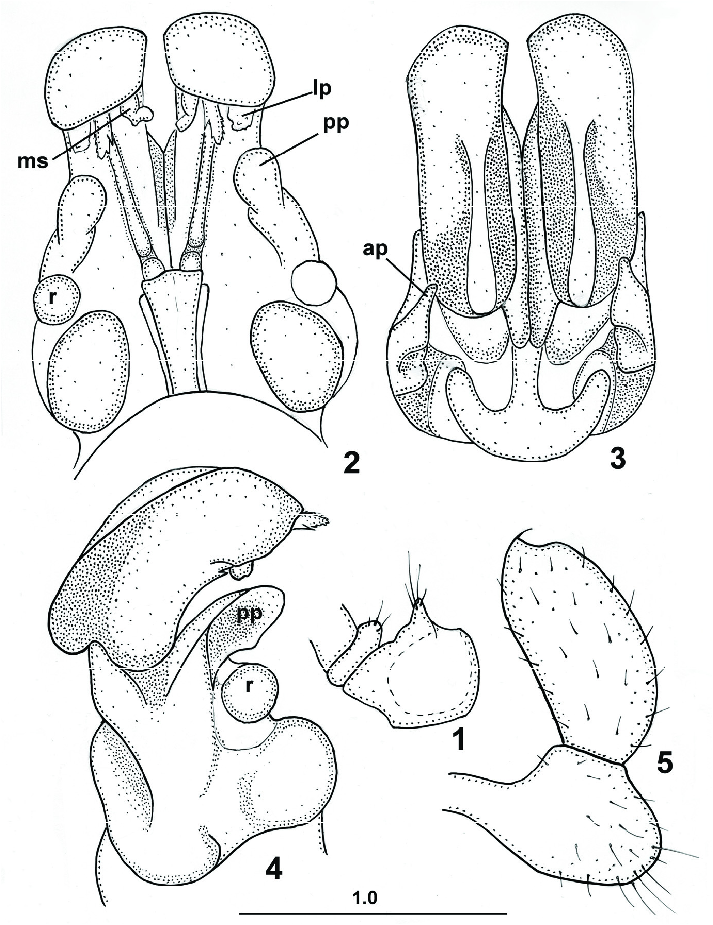
gonopods (caudal view, telopodites of posterior gonopod omitted); 3 – gonopods (front view,
telopodites of posterior gonopod omitted); 4 – gonopods (lateral view, telopodites of posterior gonopod omitted); 5 – telopodite of posterior gonopod; pp – posterior angiocoxal process of posterior gonopod; ap – anterior angiocoxal process of posterior gonopod; ms –
mesal sheath process of colpocoxite; lp – lateral sheath process of colpocoxite; r – remnant of posterior gonopod telopodite. Scale in mm.
Legs 10 and 11 with coxal glands. Coxa 10 with a caudoventral conical process setose apically; trochanter 10 with a ventral low setose outgrowth ( Fig. 1 View Figs 1–5 ). Coxa 11
with a caudoventral low setose outgrowth. Trochanter 11 without processes.
Gonopods large, curved posteriad distally ( Figs 2–4 View Figs 1–5 ; posterior gonopod telopodites broken). Anterior gonopods with subflagelliform 2-segmented telopodites,
latter not hidden inside a narrow sheath groove; pointed tips of second telopodito-
meres beset with cuticular spinules. Posterior gonopod colpocoxites fused to 2/3
extent. Colpocoxites entire. Each angiocoxite with a globule in posterior view.
Posterior angiocoxal processes (pp) large, curved posteriad, rounded at tip, lacking teeth. Mesal (ms) and lateral (lp) sheath processes of colpocoxite small, originating near apex (see Remarks). Angiocoxites with depressions and ridges in anterior view,
each supplied with digitiform process (ap) blunted apically. Each colpocoxite in anterior view with longitudinal crest with broad apical portion. Posterior gonopod telopodite 2-segmented, setose, with a long femur and claw vestige ( Fig. 5 View Figs 1–5 ).
FEMALE unknown.
DIAGNOSIS. New species closely related to Altajosoma shilenkovi (Shear, 1990) ,
but distinguished by the presence of anterior angiocoxal processes, configuration of posterior gonopod colpocoxites, telopodites without subapical triangular lamella,
colpocoxites fused to 2/3 extent, small lateral sheath processes of colpocoxites.
DISTRIBUTION. Russia: Irkutskaya oblast.
ETYMOLOGY. The specific epithet refers to the type locality.
REMARKS. It should be noted that distal parts of colpocoxites of a single examined male-holotype are dark brown and look worn like those of old specimens. As result the shape of mesal and lateral sheath processes of colpocoxite of young individuals may be some differ from that described above. However, to estimate the possible variations of the gonopod processes, additional material needs to be studied.
Altajosoma kultukensis sp. n. can be conspecific with the Craspedosoma armatus
(Gerstfeldt, 1859). Latter was very poorly described from Irkutsk (Gerstfeldt, 1859)
and recorded from Kultuk (Haase, 1880). But in the absence of type material of
Craspedosoma armatum , ultimate solution can be achieved only when strict topo-
types and material studied by Haase become available for examination.
No known copyright restrictions apply. See Agosti, D., Egloff, W., 2009. Taxonomic information exchange and copyright: the Plazi approach. BMC Research Notes 2009, 2:53 for further explanation.
|
Kingdom |
|
|
Phylum |
|
|
Class |
|
|
Order |
|
|
Family |
|
|
Genus |