
Pacifiosoma koiensis Mikhaljova, 2019
|
publication ID |
https://doi.org/10.25221/fee.374.1 |
|
publication LSID |
lsid:zoobank.org:pub:4467F525-C0C9-46E8-8059-C2B651A0A2FE |
|
persistent identifier |
https://treatment.plazi.org/id/03EDCF74-FF84-7931-97EC-FB09FC0693DE |
|
treatment provided by |
Felipe |
|
scientific name |
Pacifiosoma koiensis Mikhaljova |
| status |
sp. nov. |
Pacifiosoma koiensis Mikhaljova , sp. n.
http/ urn:lsid:zoobank.org:act:
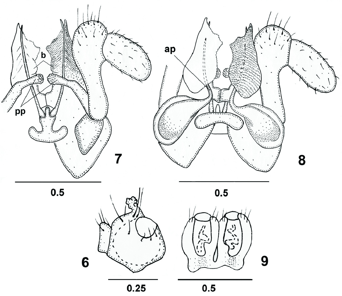
Figs 6–9 View Figs 6–9
MATERIAL. Holotype – ♂ ( FSCB), Russia: Khabarovskii krai (southern):
Sikhote-Alin (Central) range, upper course of River Ko, 47,074° N 136,478° E GoogleMaps ,
700–800 m, fir-birch forest, wet leaf litter, 23–25. V 2015, leg. A. Hansen, M.
Justesen, A. Solodovnikov. Paratypes: 1 male, 1 female ( FSCB), 1 male ( ZMUM) ,
2 males, 1 female, 1 fragment ( ZMUC), the same label as in holotype ; 1 male, 1
juv. ( ZMUC), Khabarovskii krai (southern): Sikhote-Alin (Central) range, upper course of River Ko, 47,074° N 136,478° E, 700–800 m, fir-birch forest, 23–25. V GoogleMaps
2015, leg. A. Hansen, M. Justesen, A. Solodovnikov.
DESCRIPTION. MALE. Length 11–12 mm, width 1.0–1.1 mm with paraterga.
Coloration in alcohol light brown. Venter and lower portion of pleura pale. Legs marbled brownish, darker distad. Antennae brown. Eyes black.
Body with 32 segments. Head covered both with relatively long and short setae.
Eye patches subtriangular, with 27–28 ocelli. Collum semicircular. Both collum and somite 2 narrower than head with genae. Somite 2 wider than collum. Body width gradually increasing until somite 7, body parallel-sided on somites 20–21, thereafter gradually tapering. Paraterga beginning on somite 4(5), well developed on somites
6–27, reduced on somite 28, onward missing. Metazonital macrochaetae in a transverse row on somites 29–31, like an extended triangle on preceding somites. Anterolateral macrochaetae shortest, caudolateral ones longest, all macrochaetae pointed apically, but not very sharply so. Axial suture well developed.
gonopods (caudal view); 8 – gonopods (front view); 9 – vulvae;; pp – posterior angiocoxal process of posterior gonopod; ap – anterior angiocoxal process of posterior gonopod; b –
blade of colpocoxite. Scales in mm.
Legs long and slender. Leg pairs 1–2 typically reduced in size, with tarsal brushes as usual. Other pregonopodal legs slightly enlarged, otherwise unmodified.
Claw of leg pairs 1–2 at base with one small additional claw dorsally and relatively short setiform outgrowth ventrally. Leg pairs 3–7 with a group of funnel–shaped tarsal papillae in the last third of the tarsus; size of papillar tarsal field gradually somewhat increasing toward gonopods. Claw of leg pairs 3–7 at base with a long setoid outgrowth ventrally but without additional claws dorsally. Postgonopodal legs (including leg pairs 10 and 11) without tarsal papillae. Claws of postgonopodal legs (including leg pairs 10 and 11) at base with two small additional claws dorsally and a long setoid filament ventrally, but small additional claws gradually missing toward posterior part of body; at least claws of hindmost legs at base devoid of such additional claws.
Legs 10 and 11 with coxal glands. Coxae 10 with a caudoventral, finger-shaped process covered with tiny knobs ( Fig. 6 View Figs 6–9 ). Coxae 11 without processes. Trochanter
11 with a caudal process rounded apically.
Gonopods as in figs 7–8. Telopodite flagelliform, its distal part with a pointed apex positioned inside sheath groove with elevated edges. Posterior gonopod colpocoxites fused basally, their apices pointed. Each colpocoxite entire. Mesal edge of colpocoxite drawn out into subtriangular blade ( b). Colpocoxites densely covered with low papillae. Colpocoxite sheath groove without evident processes.
Posterior gonopod angiocoxite with a globule in posterior view. Posterior angiocoxal process ( pp) large, with rounded papillate apex. Angiocoxites with depressions and ridges in front view, each supplied with anterior flagelliform process ( ap); distal portion of ap placed inside fold of colpocoxite. Posterior gonopod telopodite 2-
segmented; distal segment large, basal segment with a relatively thick stem.
FEMALE. Length about 12 mm, width 1.0–1.2 mm with paraterga. Body with
32 segments. Ocelli 27–28. Other nonsexual characters as in male. Vulvae poorly sclerotized ( Fig. 9 View Figs 6–9 ). One of the two females is only anterior body portion (13 somites).
DIAGNOSIS. Differs from congeners mainly by the flagelliform anterior angiocoxal processes of the posterior gonopods with distal parts placed inside folds on colpocoxite's front surface as well as by the pointed apices of colpocoxites.
DISTRIBUTION. Russia: Khabarovskii krai (southern).
ETYMOLOGY. The specific epithet refers to the type locality.
No known copyright restrictions apply. See Agosti, D., Egloff, W., 2009. Taxonomic information exchange and copyright: the Plazi approach. BMC Research Notes 2009, 2:53 for further explanation.
|
Kingdom |
|
|
Phylum |
|
|
Class |
|
|
Order |
|
|
Family |
|
|
Genus |