
Astropecten karankawai, Lawrence & Cobb & Herrera & Durán-González & Solís-Marín, 2018
|
publication ID |
https://doi.org/10.11646/zootaxa.4407.1.5 |
|
publication LSID |
lsid:zoobank.org:pub:D3FDB6ED-822C-442B-BAB7-A0641633DC79 |
|
DOI |
https://doi.org/10.5281/zenodo.5962173 |
|
persistent identifier |
https://treatment.plazi.org/id/03EEBD06-FF89-6342-71FE-F9DDA06FA168 |
|
treatment provided by |
Plazi |
|
scientific name |
Astropecten karankawai |
| status |
sp. nov. |
Astropecten karankawai View in CoL n. sp.
Figures 3 View FIGURE 3 , 4c, 4d View FIGURE 4 , 5 View FIGURE 5 , 7, 8 View FIGURE 8 , 11 View FIGURE 11 , 12, 13b View FIGURE 13 , 14; as Astropecten cingulatus: Downey 1973: 115 , pl. 5a,b; Downey, in A.M. Clark & Downey 1992: pl. 6e,f.
Diagnosis. Arms long and narrow, tapering rapidly to a narrow tip. Paxillar area very narrow. Paxillae of disk with thin rod-shaped spinelets surrounding several central granules. Superomarginal plates wide and covered in granules, typically without spines. Inferomarginal plates typically with three or four fringe spines. Inferomarginal plate actinal surface with few or no squamules, resulting in bare patches surrounded by long acute spines. Adambulacral plates in the mid-arm region with three short, pointed furrow spines in the first row adjacent to the adambulacral furrow (furrow spines). Behind this first row of spines in large specimens is an acute central spine that is longer and wider than all other adambulacral spines. Between the furrow spines and this enlarged spine is a distinct bare patch on the actinal surface of the adambulacral plate. The other three sides of the enlarged spine are surrounded by a semi-circle of very short, thin spinelets. In wet specimens, these spinelets appear thicker and conical because of the epithelium. The enlarged spine of the subambulacral spines may be replaced by a pedicellaria in large specimens. Small specimens have fewer subambulacral spines of similar length arranged in two rows of three spines. Pedicellariae common in paxillar area and on adambulacral and actinolateral plates of large specimens.
Material Examined. Holotype: USNM 1422432 (formerly TCRC 3-0938/ FSBC I 134512) 1 dry specimen, R = 79mm, r = 15mm, SM# = 32. SW Gulf of Mexico, 23˚29ˈN, 97˚26ˈW, 208m.
Paratypes: FSBC I 128143 2 wet specimens*, 29˚54.56ˈN, 87˚06.21ˈW, 129m; FSBC I 129419 5 wet specimens, 29˚54.00ˈN, 87˚07.43ˈW, 128m; FSBC I 134511 2 dry specimens, 23˚29ˈN, 97˚26ˈW, 77m; USNM 1422433 (formerly FSBC I 129143) 3 wet specimens*, 29˚54.56ˈN, 87˚06.21ˈW, 129m; TCRC 3-0938 11 dry specimens, 23˚29ˈN, 97˚26ˈW, 208m; TCRC 3-1343 6 dry specimens, 28˚04ˈN, 91˚27.5ˈW, 117m; USNM E22502 View Materials 16 dry specimens, 28˚44.07ˈN, 89˚44.12ˈW, 85m.
*Some specimens include dry fragments.
Other specimens: ICML-UNAM 2.135.0 (n=5) 19˚06ˈN, 92˚39ˈW; ICML-UNAM 2.135.1 (n=5) 19˚54ˈN, 92˚53ˈW; ICML-UNAM 2.135.10 (n=2) 24˚40ˈN, 96˚52ˈW; ICML-UNAM 2.135.11 (n=1) 19˚04ˈN, 92˚43ˈW; ICML-UNAM 2.135.14 (n=2) 18˚52ˈN, 95˚33ˈW; ICML-UNAM 2.135.18 (n=1) 19˚24ˈN, 92˚46ˈW; ICML- UNAM 2.135.19 (n=4) 23˚16ˈN, 87˚39ˈW; ICML-UNAM 2.135.2 (n=5) 18˚35ˈN, 94˚00ˈW; ICML-UNAM 2.135.20 (n=7) 25˚00ˈN, 96˚51ˈW; ICML-UNAM 2.135.21 (n=3) 23˚00ˈN, 97˚29ˈW; ICML-UNAM 2.135.22 (n=18) 24˚37ˈN, 97˚10ˈW; ICML-UNAM 2.135.23 (n=44) 18˚54ˈN, 95˚32ˈW; ICML-UNAM 2.135.24 (n=14) 22˚10ˈN, 97˚43ˈW; ICML-UNAM 2.135.25 (n=24) 18˚48ˈN, 93˚11ˈW; ICML-UNAM 2.135.26 (n=4) 25˚00ˈN, 96˚51ˈW; ICML-UNAM 2.135.27 (n=1) 19˚56ˈN, 91˚51ˈW; ICML-UNAM 2.135.28 (n=1) 23˚24ˈN, 89˚07ˈW; ICML-UNAM 2.135.29 (n=24) 21˚05ˈN, 97˚01ˈW; ICML-UNAM 2.135.3 (n=3) 24˚20ˈN, 97˚00ˈW; ICML- UNAM 2.135.30 (n=1) 21˚05ˈN, 97˚04ˈW; ICML-UNAM 2.135.31 (n=23) 21˚05ˈN, 97˚01ˈW; ICML-UNAM 2.135.32 (n=2) 21˚43ˈN, 96˚50ˈW; ICML-UNAM 2.135.33 (n=3) 18˚47ˈN, 93˚36ˈW; ICML-UNAM 2.135.34 (n=1) 19˚31ˈN, 91˚37ˈW; ICML-UNAM 2.135.35 (n=1) 23˚49ˈN, 97˚37ˈW; ICML-UNAM 2.135.36 (n=1) 18˚23ˈN, 94˚23ˈW; ICML-UNAM 2.135.38 (n=14) 18˚54ˈN, 95˚32ˈW; ICML-UNAM 2.135.39 (n=1) 25˚56ˈN, 97˚02ˈW; ICML-UNAM 2.135.4 (n=133) 18˚58ˈN, 95˚36ˈW; ICML-UNAM 2.135.40 (n=1) 25˚20ˈN, 97˚14ˈW; ICML-UNAM 2.135.41 (n=1) 22˚11ˈN, 91˚34ˈW; ICML-UNAM 2.135.42 (n=1) 22˚10ˈN, 91˚45ˈW; ICML- UNAM 2.135.43 (n=2) 19˚45ˈN, 92˚00ˈW; ICML-UNAM 2.135.44 (n=4) 19˚59ˈN, 92˚01ˈW; ICML-UNAM 2.135.45 (n=3) 18˚37ˈN, 94˚00ˈW; ICML-UNAM 2.135.46 (n=1) 22˚16ˈN, 91˚45ˈW; ICML-UNAM 2.135.48 (n=2) 22˚32ˈN, 90˚47ˈW; ICML-UNAM 2.135.5 (n=605) 25˚31ˈN, 96˚41ˈW; ICML-UNAM 2.135.6 (n=1) 19˚00ˈN, 95˚30ˈW; ICML-UNAM 2.135.7 (n=4) 19˚05ˈN, 92˚43ˈW; ICML-UNAM 2.135.8 (n=2) 19˚26ˈN, 92˚33ˈW; ICML-UNAM 2.135.9 (n=2) 18˚50ˈN, 93˚02ˈW; UF 16346 (n=1) 29˚08ˈN, 88˚42ˈW; USNM E08437 (n=3) 28˚10ˈN, 92˚21ˈW; USNM E12858 (n=3) 28˚47ˈN, 89˚45ˈW; USNM E12859 (n=3) 29˚08ˈN, 88˚51ˈW; USNM E12860 (n=2) 28˚48ˈN, 89˚26ˈW; USNM E12861 (n=1) 27˚56ˈN, 94˚00ˈW; USNM E13820 (n=3) 29˚10ˈN, 88˚48ˈW; USNM E22502 View Materials (n=16) 28˚44ˈN, 89˚44ˈW; USNM E22503 View Materials (n=33) 28˚10ˈN, 91˚30ˈW; USNM E22504 View Materials (n=16) 28˚14ˈN, 91˚41ˈW; USNM E23177 View Materials (n=5) 29˚15ˈN, 88˚37ˈW; USNM E24636 (n=3) 29˚47ˈN, 87˚17ˈW; USNM E24686 View Materials (n=11) 29˚19ˈN, 88˚45ˈW; USNM E34916 View Materials (n=3) 23˚29ˈN, 97˚26ˈW; USNM E38730 (n=1) 19˚08ˈN, 93˚04ˈW; USNM E45660 View Materials (n=3) 29˚12ˈN, 88˚53ˈW; USNM E45675 View Materials (n=2) 29˚10ˈN, 88˚40ˈW; USNM E45675 View Materials (n=2) 29˚10ˈN, 88˚04ˈW; USNM E45676 View Materials (n=1) 28˚09ˈN, 91˚27ˈW; USNM E8437 (n=3) 28˚10ˈN, 92˚21ˈW; USNM E8438 (n=6) 28˚25ˈN, 92˚20ˈW.
Etymology. The species is named for the Karankawa, an Indian tribe that lived on the Gulf of Mexico coast of Texas, off which the specimens in the BRTC collections were collected.
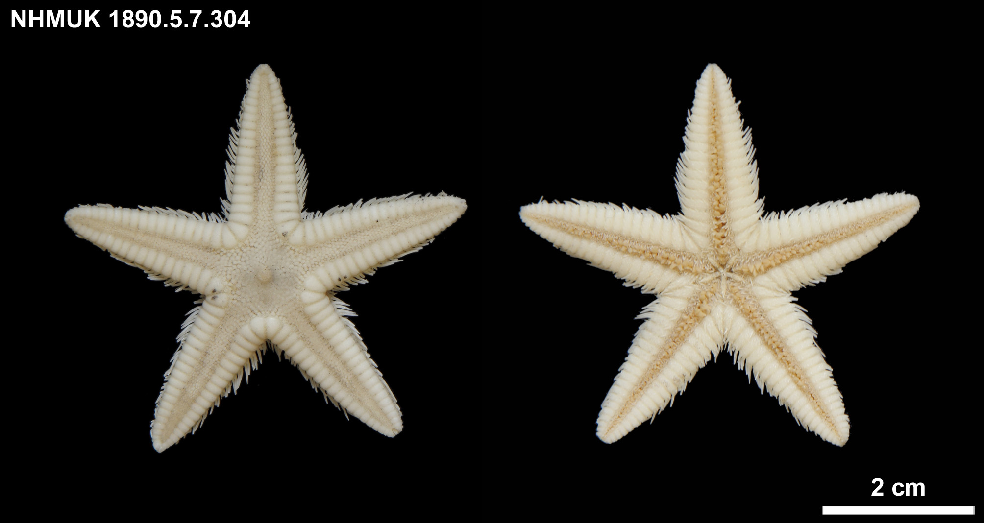
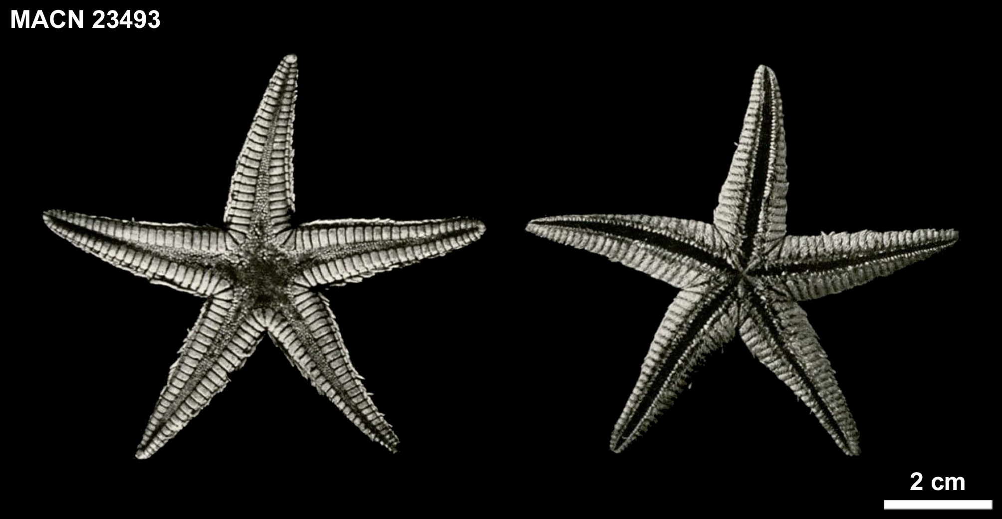
Comparison of Astropecten karankawai and Astropecten cingulatus . Body form. Photographs of Sladen’s small (R = 28 mm) holotype of A. cingulatus are shown in Figure 1 View FIGURE 1 . Figure 2 View FIGURE 2 shows a larger specimen (R = 42 mm) from Argentina in the MACN collections identified by Bernasconi (1941) as A. cingulatus . The disc of the specimen of A. cingulatus from Argentina is large and the arms are long and narrow. A photograph of the holotype (large specimen) and a paratype (small specimen) of A. karankawai are shown in Figure 3 View FIGURE 3 . They have a small disk and long, narrow arms.
Regression equations for r vs R, R:r vs R, AW vs R, and AL:AW vs. R for Astropecten cingulatus and Astropecten karankawai are given in Table 1 All regression equations are similar between species with only minor differences in slope. A difference in slope (F1,83 = 0.107, p = 0.744) and intercept (F1,83 = 2.347, p = 0.129) was not detected for R:r by ANCOVA. The longer, narrower arms of A. karankawai are the result of the narrow paxillar area and an allometric increase in R: r with an increase in R.
Abactinal surface. The paxillar area of A. karankawai ( Fig. 3 View FIGURE 3 ) is narrower than that of A. cingulatus ( Figs. 1 View FIGURE 1 , 2 View FIGURE 2 ). The width of the paxillar area of the specimen Downey (in Clark & Downey 1992) identified as A. cingulatus (her Plate 9 E, F) appears like that of A. karankawai . This difference in PAX can be seen in the regression equations ( Table 1). The narrow paxillar area is responsible for the narrow arm width of A. karankawai . This distinguishes the species.
The paxillae of the type specimen of A. cingulatus has numerous central granules ( Figs. 4A, B View FIGURE 4 ). The paxillae of A. karankawai have thin rod-shaped spinelets surrounding several central granules similar to A. cingulatus ( Figs. 4C, D View FIGURE 4 ). Spinelets are thinner distally and in small individuals. Pedicellariae may be present in the paxillar area of A. karankawai ( Fig. 4D View FIGURE 4 ). We did not observe any pedicellariae in the paxillar area of the A. cingulatus we examined.
Superomarginal plates. The regression equations for SM# and R:SM# vs. R of A. cingulatus and A. karankawai are given in Table 2. SM# is not large in either species and only slightly larger in A. cingulatus than in A. karankawai . As a result, R:SM# is slightly smaller. ANCOVA detected a difference in slopes (F1,83 = 15.437, p <0.001) of R:SM# but no difference in the intercepts (F1,83 = 3.926, p = 0.051).
The SM plates of both A. cingulatus ( Figs. 1 View FIGURE 1 , 2 View FIGURE 2 ) and A. karankawai ( Fig. 3 View FIGURE 3 ) are wide and are casually indistinguishable. When the proximal SMW of both species were compared with an ANCOVA, they did not pass the test of equal slopes (F1,34 = 1.450, p = 0.238) and there was no difference in the intercept of the regression equations (F1,34 = 0.804, p = 0.377).
The SM plates of A. karankawai are covered with uniform granules ( Fig. 4D View FIGURE 4 ) that are less dense and smaller than those of A. cingulatus ( Fig. 4B View FIGURE 4 ). Note the difference in magnification for the photographs of A. cingulatus and A. karankawai . Twenty specimens of A. karankawai were examined for the presence of spines on the SM plates. Three specimens had a single conical spine on the inner margin of 2–4 interradial SM plates ( Fig. 5 View FIGURE 5 ) and one specimen had similar spines on the interradial and non-consecutive proximal SM plates. No spines were observed on the outer margin of the SM plates. No spines were observed on the SM plates of eleven specimens of A. cingulatus we examined. Spination of the SM plates does not distinguish the species.
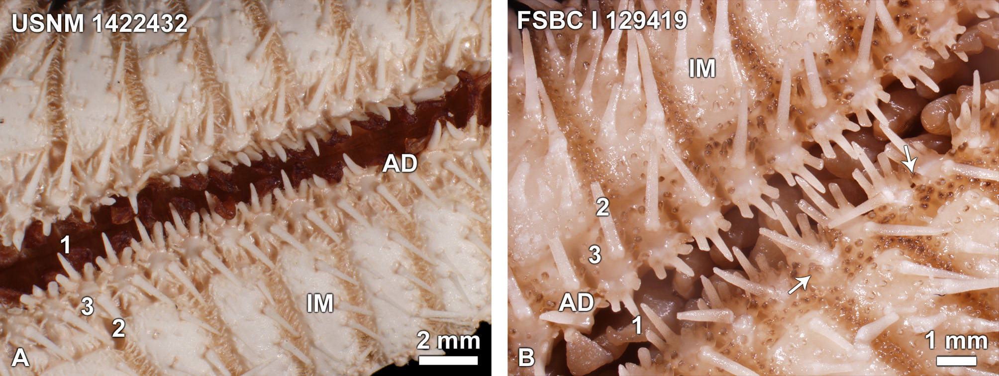
Inferomarginal plates. The holotype of A. cingulatus has two large, moderately long IM fringe spines of equal length on the IM plates ( Fig. 6 View FIGURE 6 ). A tiny third spine often occurs proximally. Fringe spines are round with slightly compressed tips. Some specimens in the NMNH identified as A. cingulatus (e.g., USNM E14309, USNM E18399 View Materials ) have three fringe spines while others (e.g., USNM E18420 View Materials , USNM E18415 View Materials ) have two. Of eleven specimens of A. cingulatus examined from the Caribbean Sea or off the east coast of South America, seven had two fringe spines when R < 30 mm and four had three fringe spines when R> 30mm. The number of fringe spines in A. cingulatus can be two or three.
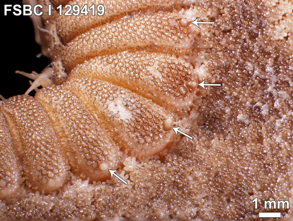
Astropecten karankawai has round to slightly compressed IM fringe spines. Specimens from off the coast of Texas in the TCRC collections have three spines in the mid-arm region. Large specimens from off the coast of Louisiana (FSBC I 128143, FSBC I 129419) have three or four spines ( Fig. 7), although the fourth proximal spine is usually slightly shorter and thinner. Of 51 specimens of A. karankawai examined, two had one fringe spine when R = 8–10 mm, 21 had two fringe spines when R = 15–32 mm, 22 had three fringe spines when R = 22–78 mm, and 6 had 3–4 fringe spines when R = 78–104 mm. Like A. cingulatus , the number of fringe spines of A. karankawai increases with body size. The number of fringe spines in A. karankawai can be two to four spines. The number of fringe spines does not distinguish the species.
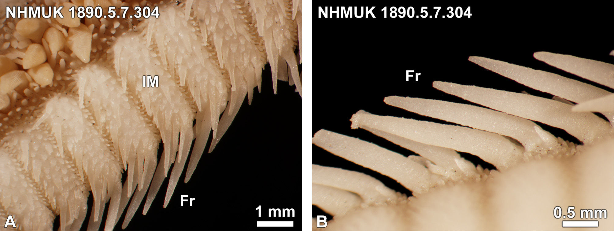
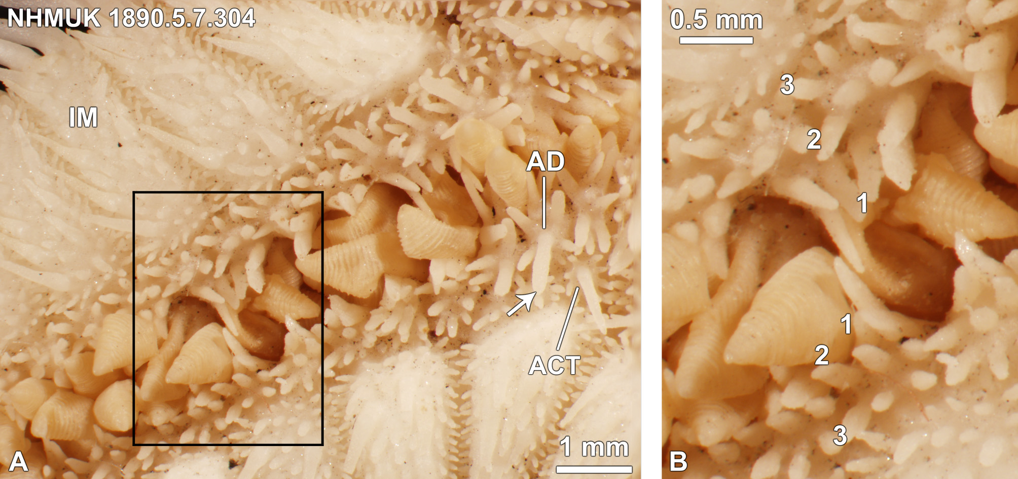
The actinal surface of the IM plates of A. cingulatus is covered with small, flat, widely spaced subcircular squamules with their transverse margins bordered with crowded ciliary spinelets. This is shown in Figure 6A View FIGURE 6 . The actinal surface of the IM plates of A. alligator , A. americanus , A. articulatus , and A. duplicatus are also similarly covered by dense squamules or spines. The actinal surface of the IM plates of A. karankawai has few or no squamules, resulting in bare patches surrounded by long acute spines ( Fig. 8 View FIGURE 8 ). The actinal surface of the IM plates of A. antillensis and A. marginatus also has no squamules, only marginal spines. The lack of squamules on the actinal surface of IM plates of A. karankawai distinguishes the species.
Adambulacral plate spination. A photograph of the AD spination of the holotype of A. cingulatus is shown in Figure 9 View FIGURE 9 and a photograph of a specimen from off the coast of Argentina is shown in Figure 10 View FIGURE 10 . All specimens of A. cingulatus we examined had at least three rows of spines, with a fourth row of slender spines present in some large specimens, such as FSBC I 129674 from Brazil (R = 48 mm). There were three furrow spines as in all North American Astropecten spp., with the middle spine longest and perpendicular to the adjacent spines. The subambulacral spines did not differ greatly in length except for an elongated middle spine in the second row (behind furrow spines), on plates adjacent to the actinal plates. In addition, although three spines were most common in the third or fourth rows, there were occasionally four.
AD spination of A. alligator , A. americanus , and A. nitidus is similar to A. cingulatus . AD plates had 3–4 rows of spines where all spines within a single row were the same length and width as adjacent spines.
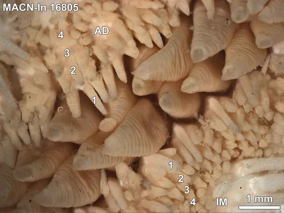
AD spination of A. karankawai is shown in Figure 11 View FIGURE 11 . Both A. karankawai and A. cinglatlus have three furrow spines. However, there are no discrete subambulacral rows. Behind the furrow spines is a very large, long and tapered spine surrounded on the lateral sides and behind by small spines. It is the largest of the AD spines in length and width and is usually rounded to slightly compressed. Between this long spine and the furrow row is a distinct bare patch on the AD plate. Sometimes it or one of the other subambulacral spines is replaced by a pedicellaria ( Figs. 11B View FIGURE 11 , 12). Other subambulacral spines surrounding this spine are short, thin, and spindle-like in dry specimens ( Fig. 11A View FIGURE 11 ). In wet specimens, these spines may appear conical due to the epithelium ( Fig. 11B View FIGURE 11 ). The length of the median spine behind the furrow spines is allometric. Of 47 specimens of A. karankawai examined, three had a median AD spine less than the length of the central furrow spine when R = 8–17 mm, 14 had a median AD spine equal in length or longer to the central furrow spine when R = 15–22 mm, and 31 had a median AD spine longer than the central furrow spine when R = 17–104 mm. This indicates that A. karankawai can be distinguished by the AD spination from A. cingulatus when R is greater than 22 mm.
AD spination of A. marginatus and A. antillensis was similar to A. karankawai except that the spines of the second row in A. antillensis are usually flat, acute and all spines are arranged in discrete rows. In A. marginatus the short spines, behind the furrow spines typically surround the median spine which is larger and wider than all other spines on the plate.
Actinal intermediate plates. Five or six ACT plates per interradial area are present on the holotype of A.
cingulatus ( Fig. 13A View FIGURE 13 ) and up to 10 plates on other specimens from the Caribbean or South America. All specimens of A. cingulatus examined had a large central spinelet on most ACT and on AD plates adjacent to the ACT plates. The ACT plates of A. karankawai are similar to those of A. cingulatus ( Fig. 13B View FIGURE 13 ) in number and spination. Pedicellariae were also frequently observed on ACT plates of A. karankawai .
Color. There is no photo of living A. karankawai . The abactinal surface of specimens preserved in ethanol short period of time before drying is orange with dark shading on the sides of the arms ( Fig. 14). Small dry specimens retain a rust color around the borders of the paxillar area although large specimens lose all color with drying.
Distribution of specimens. Specimens of A. cingulatus examined in this study are illustrated in Figure 15 View FIGURE 15 . Catalog numbers of specimens measured are given in the legend to Table 1. Astropecten cingulatus has been reported from off the coast of North and South America and in the Gulf of Mexico (Downey, in A.M. Clark & Downey 1992). Downey (in A.M. Clark & Downey 1992) gave the depth range of A. cingulatus as 100–1350 m. Specimens of A. karankawai have been collected in the Gulf of Mexico at depths of 20–600 m ( Fig. 15 View FIGURE 15 ).


| USNM |
Smithsonian Institution, National Museum of Natural History |
No known copyright restrictions apply. See Agosti, D., Egloff, W., 2009. Taxonomic information exchange and copyright: the Plazi approach. BMC Research Notes 2009, 2:53 for further explanation.
|
Kingdom |
|
|
Phylum |
|
|
Class |
|
|
Order |
|
|
Family |
|
|
Genus |