
Symphysanodontidae Katayama 1984
|
publication ID |
https://doi.org/ 10.11646/zootaxa.4277.1.4 |
|
publication LSID |
lsid:zoobank.org:pub:EED364DC-47FB-45A5-B0F5-CD71C41ECE2E |
|
DOI |
https://doi.org/10.5281/zenodo.6029139 |
|
persistent identifier |
https://treatment.plazi.org/id/03EF4F05-1A69-3D39-FF17-FB1DE3F5FBD2 |
|
treatment provided by |
Plazi |
|
scientific name |
Symphysanodontidae Katayama 1984 |
| status |
|
Family Symphysanodontidae Katayama 1984 View in CoL View at ENA
( Figure 1 View FIGURE 1 )
Type genus: Symphysanodon Bleeker 1877
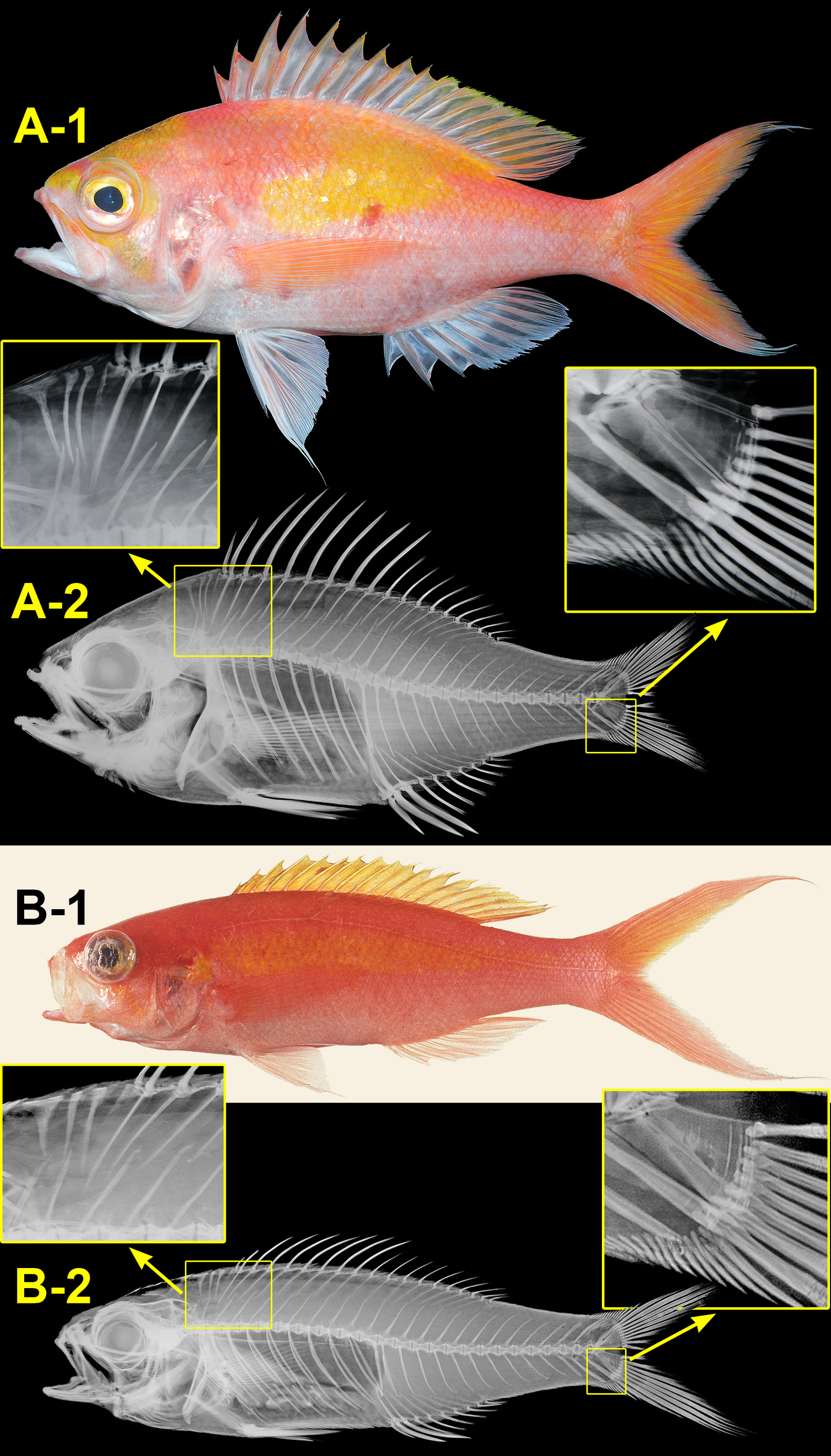
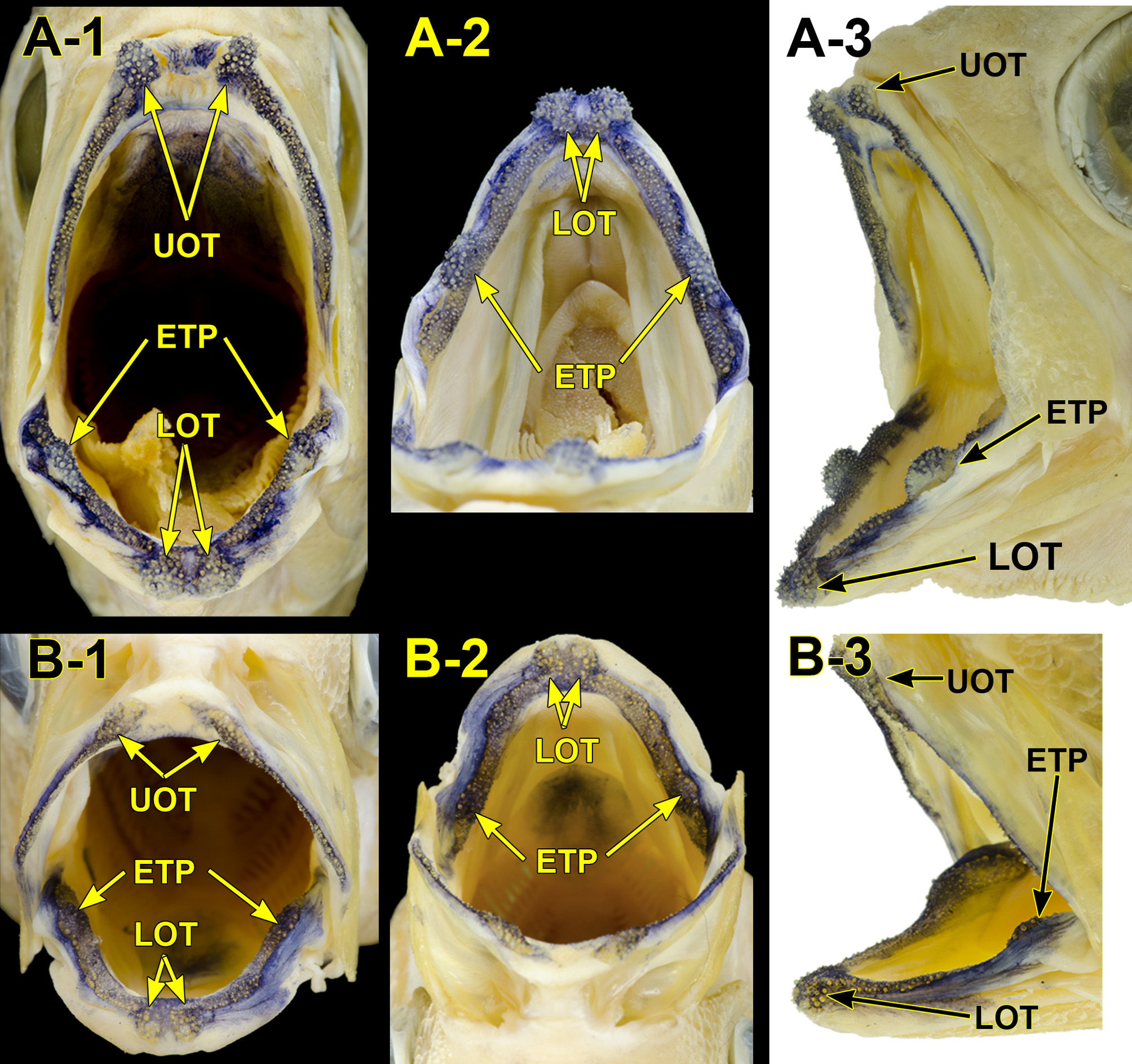
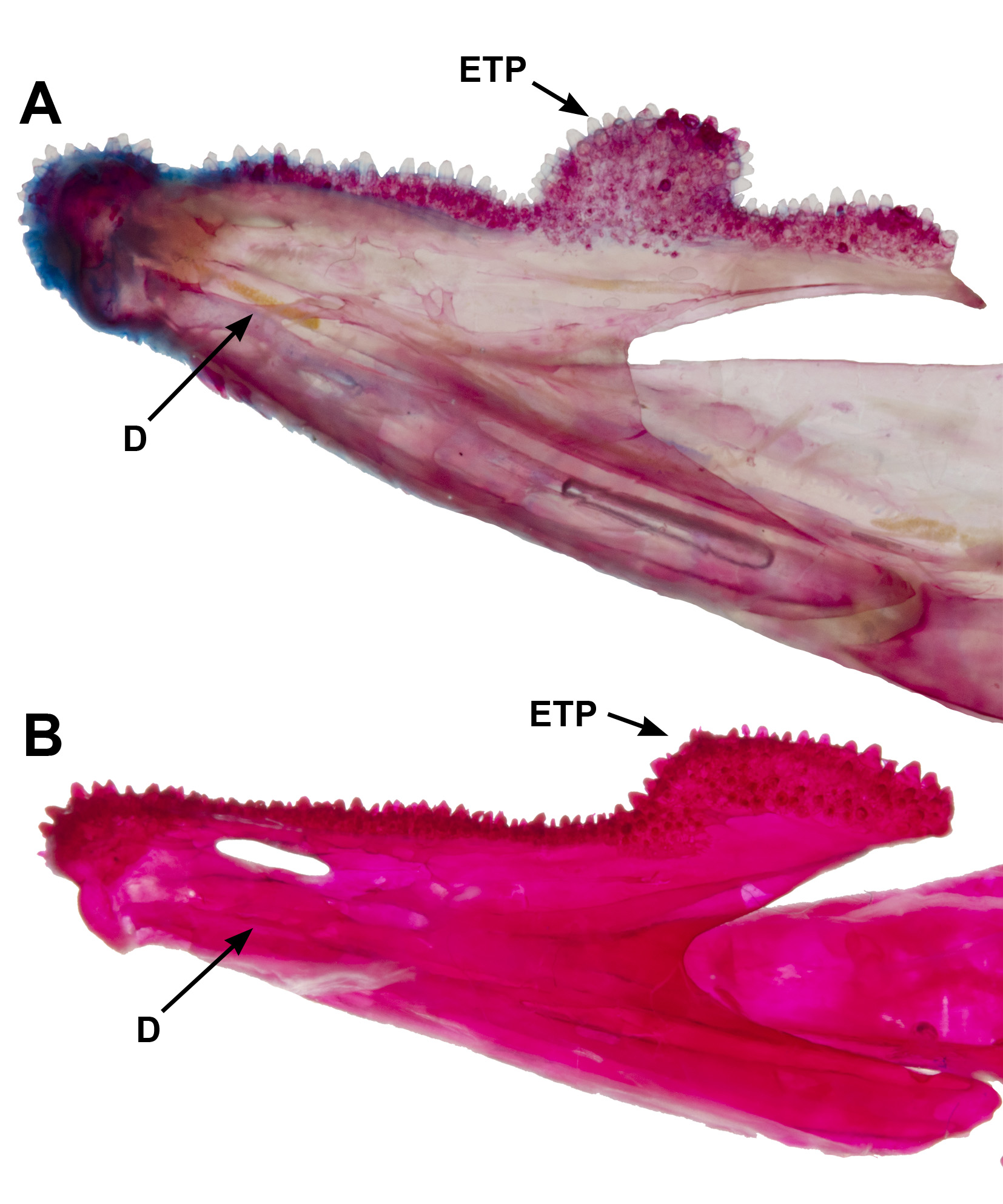
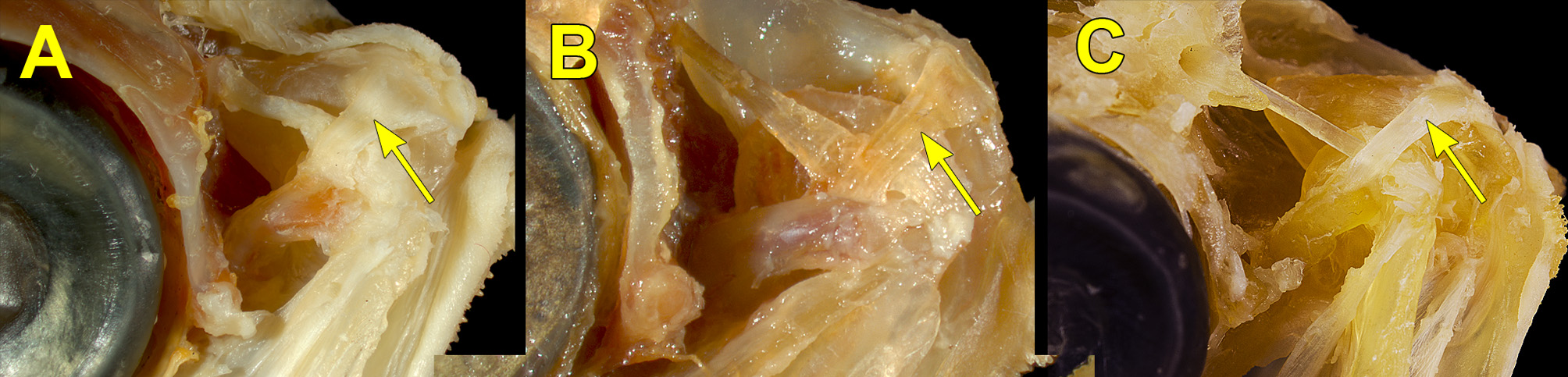
Diagnosis. Jaws subequal or upper jaw slightly projecting; both jaws with large and small conical teeth, no villiform teeth and no canines ( Figure 2 View FIGURE 2 ); anterior tips of both jaws with exposed outer patches of large conical teeth on sides, left and right tooth patches of upper jaw well separated but those of lower jaw close-set in adults ( Figure 2 View FIGURE 2 ); outer tooth patches of the lower jaw fully or partly received into spaces between outer tooth patches of upper jaw; continuous tooth patch present along dorsal and medial surface of most of length of coronoid process of lower jaw (dentary) ( Figures 2 View FIGURE 2 , 3 View FIGURE 3 ); palato-premaxillary ligament passing from anteriormost part of palatine, across head of maxilla, to ascending process of premaxilla (see Johnson, 1981; Figure 4 View FIGURE 4 ); no supramaxilla; suborbital area narrow, first and second infraorbitals slightly covering upper margin of maxilla when mouth closed; fronto-parietal crest developed; preopercle with right-angled corner; posterior margin of preopercle smooth or very weakly serrated; opercle with two flat spines; a single, un-notched dorsal fin with nine or ten spines and nine to eleven soft rays [almost always IX, 10]; anal fin with three spines and seven or eight soft rays; pelvic axillary scale lanceolate; 10 + 15 vertebrae; supraneural and dorsal-fin pterygiophore insertion pattern 0/0/0+2+1/1/ ( S. octoactinus Anderson, 1970 exceptional in having 0/0/0+2/1+1/1/); first supraneural distinctly T-shaped; dorsal procurrent caudal-fin rays 12–14; ventral procurrent caudal-fin rays 10–12; base of penultimate ventral procurrent caudal-fin ray foreshortened; no procurrent spur (sensu Johnson 1975) on posteriormost ventral procurrent caudal-fin ray.
Remarks. Prior to Katayama’s (1984) establishment of the Symphysanodontidae, Fourmanoir (1981) used the family name Symphysanodontidae for Symphysanodon . However, Fourmanoir’s Symphysanodontidae is not available because he did not describe any characters of his Symphysanodontidae (Art. 13.1.1, ICZN, 1999; van der Laan, 2014).
As stated above, members of the Symphysanodontidae are generally similar to the Anthiadinae of the Serranidae , and Fowler (1928) placed Symphysanodon in the Anthiadinae . Munro (1967), Katayama (1968) and Anderson (1970) included Symphysanodon in the Lutjanidae . However, Johnson (1975) found no evidence from osteology and cheek myology for a close relationship between Symphysanodon and the Lutjanidae . Moreover, he noted that the base of the penultimate ventral procurrent caudal-fin ray is foreshortened in Symphysanodon , although there is no spur on the last ventral procurrent caudal-fin ray. He suggested that Symphysanodon is not a lutjanid and may occupy a very primitive position within the Percoidei. Subsequently Johnson (1981) provided a detailed description of Symphysanodon and treated the genus as incertae sedis. Our examinations of the Symphysanodontidae , Lutjanidae and Serranidae have further revealed that the Symphysanodontidae differ from the latter two families in having exposed outer tooth patches on the upper and lower jaws in adults (vs. no outer tooth patches on either jaw in the latter two families), continuous tooth patch present along the dorsal and medial surface of most of the length of the coronoid process of the lower jaw, a lanceolate pelvic axillary scale (vs. triangular or rectangular scale or an undeveloped scale), a unique ( S. octoactinius exceptional) supraneural and dorsal-fin pterygiophore insertion pattern 0/0/0+2+1/1/, and 25 total vertebrae [vs. 24 or 26–28 ( Anderson & Heemstra, 1989; Anderson et al., 1990; present observation)]. The Symphysanodontidae are also distinguishable from the Serranidae by having a well-developed fronto-parietal crest (vs. undeveloped) and from the Lutjanidae by, among other things, having a right-angled preopercular corner (vs. round), as already stated by Katayama (1984), and no attachment of the adductor mandibulae to the subocular shelf ( Johnson, 1981).
The supraneural and dorsal-fin pterygiophore insertion pattern (0/0/0+2+1/1/) that characterizes all but one species of Symphysanodontidae is also found in the African percoid family Dinopercidae (see Johnson, 1984). However, a relationship between those two families is probably rather distant, because the latter differs from the former as follows: lower jaw projecting; no outer patches of teeth at anterior tips of jaws; preopercle with round corner; posterior margin of preopercle serrated; opercle with one flat spine; a single dorsal fin notched with 11 spines and 17–19 soft rays; anal fin with three spines and 11–13 soft rays; no axillary scale; 10 + 16 vertebrae; first supraneural reverse L-shaped; procurrent spur on posteriormost ventral procurrent caudal-fin ray ( Johnson, 1984; present observation).
No known copyright restrictions apply. See Agosti, D., Egloff, W., 2009. Taxonomic information exchange and copyright: the Plazi approach. BMC Research Notes 2009, 2:53 for further explanation.