
Mysorea thaiensis Suzuki, 1985
|
publication ID |
https://doi.org/ 10.11646/zootaxa.3964.3.3 |
|
publication LSID |
lsid:zoobank.org:pub:823F6194-5EAE-4EE5-93A2-FFFDE1EA8A1F |
|
DOI |
https://doi.org/10.5281/zenodo.5615580 |
|
persistent identifier |
https://treatment.plazi.org/id/03EF5248-FFC5-FFE7-8FAB-FE37FE4DF855 |
|
treatment provided by |
Plazi |
|
scientific name |
Mysorea thaiensis Suzuki, 1985 |
| status |
|
Mysorea thaiensis Suzuki, 1985 View in CoL
( Figs. 1–25 View FIGURES 1 – 2 View FIGURES 3 – 11 View FIGURES 12 – 18 View FIGURES 19 – 25 )
Mysorea thaiensis Suzuki, 1985: 102 View in CoL , fig. 19.
Types. Male holotype from Doi Sutep, Chiang Mai Province, Thailand; one female paratype, the same as holotype, deposited in the Zoological Museum, University of Copenhagen (ZMUC), Denmark (not examined).
Material examined. Two males and one female, Laos: Tad Etu [N 15°11´, E 106°06´], November 18, 2012, C. Zhang leg. (MHBU-Opi-12ZC 206–208).
Diagnosis. Scutum piriform in dorsal view. Coxa IV considerably widened, prolaterally with conspicuous hairtipped granules. The middle frontal area in front of ocularium with a spine. Area V with two enlarged median tubercles. Femur of pedipalpus without a setiferous tubercle on the medial distal side. Tarsi III–IV with a pseudonychium and two bare claws. Male femur IV ventrally with three enlarged spines and anal operculum with three enlarged spines; penis with a somewhat rounded distal portion (pars distalis) (dorsal and ventral view).
Redescription. Male habitus as in Figs. 1–2 View FIGURES 1 – 2 . Coloration: entire body yellow, with brown paired patches on the dorsum; median area of carapace with dark brown reticulations; both lateral ridges of the carapace and scutum with blackish brown stripes; free tergites I–III each with a dark brown band; coxae with dark brown reticulations; free sternites with transverse band of dark brown; chelicerae and pedipalps reticulated above; trochanters of all legs pale yellow, femora, patellae, tibiae and metatarsi with black reticulations, tarsi lighter.
Dorsum ( Fig. 2 View FIGURES 1 – 2 ). Dorsal scutum pyriform in shape, widest portion of body at scutal area II. Anterior margin of carapace with two spines at the lateral portion and a single median spine; front margin of the ocularium with a broad median spine, a transverse row of small six tubercles on each side of this median spine. Ocularium small, oval, with a low and conspicuous median tubercle. Opisthosomal region of scutum with five areas. Area I with a median groove and two inconspicuous median hair-tipped granules, areas II–V each with two median tubercles, the larger located posteriorly. Free tergites I–II unarmed, free tergite III with a transverse row of six tubercles, the largest on the lateral. Anal operculum with three enlarged blunt spines.
Venter ( Fig. 14 View FIGURES 12 – 18 ). Surface of coxa I tuberculated, disto-dorsally with a coarse tubercle on anterior, and a row of three tubercles prolaterally, and two rows of tubercles on the ventral surface. Coxa II with a row of marginal tubercles on the prolateral surface, and disto-dorsally with a coarse tubercle on anterior and an enlarged one on both posterior sides respectively. Coxa III with prolateral and retrolateral rows of tubercles. Coxa IV remarkably larger than others, similar length with opisthosomal scutum; prolaterally with many scattered hair-tipped granules, disto-prolaterally with a stout tubercle and disto-retrolaterally with three teeth. Genital operculum with a few hairtipped granules. Free sternites smooth. Spiracles concealed.
Chelicera ( Figs. 3–5 View FIGURES 3 – 11 ). Basichelicerite with distinct bulla, without prominent armaments. Cheliceral hand unarmed, with sparse hairs only. Fingers relatively short, inner edges toothed as illustrated ( Fig. 5 View FIGURES 3 – 11 ): moveable finger with 10 teeth, somewhat square, the proximal one inconspicuous; fixed finger with six teeth, somewhat crested.
Pedipalpus ( Figs. 6–8 View FIGURES 3 – 11 ). Coxa unarmed. Trochanter ventrally with one short and one long distal setiferous tubercle. Femur compressed laterally, widest at the middle of its length, ventrally with a row of six homogeneous setiferous tubercles; dorsally with many low conical tubercles along the entire length; on the medial distal side without a setiferous tubercle. Patella ventromesally and ventroectally only with hair. Tibia ventromesally with two enlarged setiferous tubercles, and ventroectally with a fairly enlarged setiferous tubercle. Tarsus ventromesally with a row of two slightly enlarged and three small setiferous tubercles; and ventroectally expanded, with two slightly enlarged and three small setiferous tubercles. Tarsal claw slightly curved, shorter than tarsus.
Legs ( Figs. 9–11 View FIGURES 3 – 11 ). Short. Trochanters I–IV with small hair-tipped granules on the ventral surface, trochanter IV retrolaterally with a stout tubercle. All femora with hair-tipped granules, but ventrally enlarged; femora III and IV conspicuous curved, femur IV ventrally with three enlarged spines. Tarsi III–IV with a pseudonychium and two bare claws. Tarsal formula (I–IV): 5/9/6/6. Distitarsus I two-jointed and II three-jointed. The remaining legsegments with hair-tipped granules.
Penis ( Figs. 19–25 View FIGURES 19 – 25 ). The shaft slender, sides nearly parallel, then distended in apical portion (pars distalis). The distal end of ventral plate with a median elevation, somewhat bifid crested, two distal setae on each side of the elevation. Ventral plate depressed dorsally. Glans partially sunken into the dorsal depressed portion of the ventral plate, only exposing the margin of the spiny funnel. Spiny funnel with dense small spines, and another transparent membrane ventral bladder (capsula interna), connected to the spiny funnel. Stylus rodlike, blunt. Before eversion of follis (capsula externa), the inverted stylus with capsula interna sunken into the spiny funnel, and all parts mentioned above surrounded totally by the capsula externa (follis). Ventral plate with 16 setae ( Figs. 22, 23, 25 View FIGURES 19 – 25 ): 10 lateral setae, six ventral setae.
Female. ( Figs. 15–18 View FIGURES 12 – 18 ). In general appearance similar to the male but lacking enlarged spines on body and legs. Inner edges of finger of chelicera toothed as illustrated ( Fig. 23 View FIGURES 19 – 25 ): moveable finger with 12 teeth; fixed finger with seven teeth. Tarsal formula (I–IV): 5/8/6/6.
Ovipositor as illustrated ( Figs. 17–18 View FIGURES 12 – 18 ). Ventral surface with four setae and dorsal surface with six setae. Tip of each seta bifurcated.
Measurements. Male (female): body 4.02 (3.36) long, 2.95 (2.60) wide at the widest portion, scutum 3.46 (2.94) long. Ocularium 0.27 (0.24) long, 0.35 (0.34) wide. Pedipalpus claw 0.23 (0.23) long. Penis 1.15 long. Measurements of pedipalpus and legs as in Table 1 View TABLE 1 .
Habitat. The specimens were collected by leaf litter sieving in dark moist places of the forest, under dense canopy.
Variation (the other male). Body ( Figs. 12–13 View FIGURES 12 – 18 ) 3.64 long, 2.76 wide at the widest portion, scutum 3.00 long. Ocularium without median tubercle except for two hair-tipped granules. Areas III with two and area V with one conspicuous median tubercles. Free tergite III with a transverse row of five tubercles.
Distribution. Laos (Champasak Province: Tad Etu), Thailand (Chiang Mai Province: Doi Sutep).
Remarks. Mysorea was originally considered monotypic, established on a single male of the type species M. brevipus Roewer, 1935 from Dekan ( India), and originally placed in Assamiidae : Mysoreinae. There was no further study on this genus until Suzuki (1985), following the Roewer’s classification, described M. thaiensis from Thailand.
Based on the findings presented here, there are some minor differences between our specimens and the original description/illustration of M. thaiensis : the area I without conspicuous median tubercles, the area IV only with a single enlarged tubercle ( Figs. 1–2 View FIGURES 1 – 2 , 12–13, 15 View FIGURES 12 – 18 ; Suzuki 1985: 103, figs. 19A–C), the distal margin of penis with four setae instead of two, the ventral plate of penis with 16 setae rather than six setae ( Fig. 22 View FIGURES 19 – 25 ; Suzuki 1985: 103, fig. 19L).
Except for mentioned above, other external morphology and genital characters of newly collected materials fit well with the original description of M. thaiensis . Apparently, such differences are not adequate to consider both as independent species.
Although Mysoreinae is sometimes considered as a member of Trionyxellidae (e.g., Suzuki 1985), taxonomic validity of Trionyxellidae is uncertain (e.g., Pinto-da-Rocha 2007, Sharma & Giribet 2011). However, based on molecular ( Sharma & Giribet 2011) and morphological data, M. thaiensis should be placed in the Assamiidae . At least, M. thaiensis belongs to the one of five groups proposed by Kury (2007), i.e., the group of “the Sri Lankan/ Indian pseudonychiate genera”.
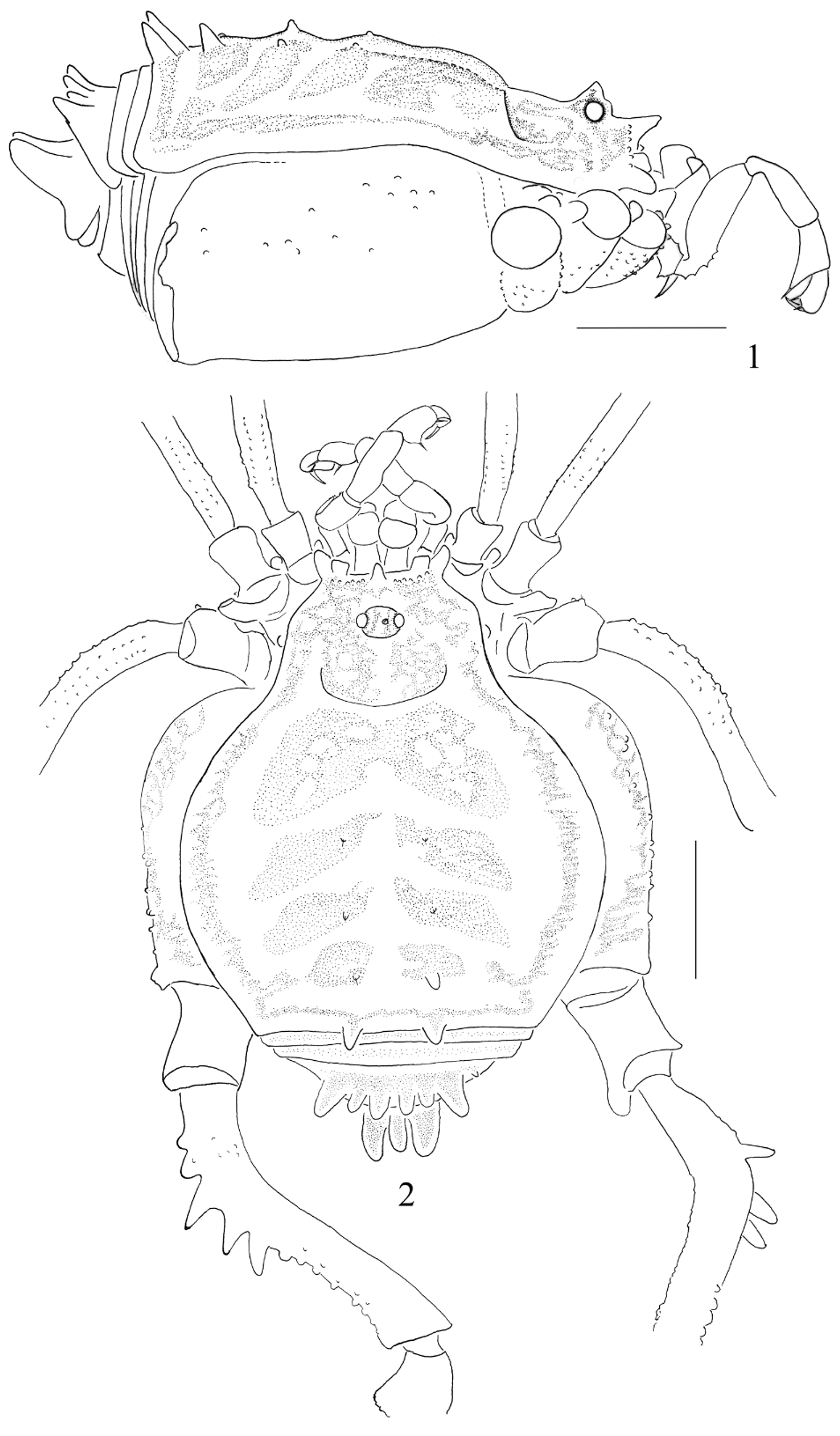
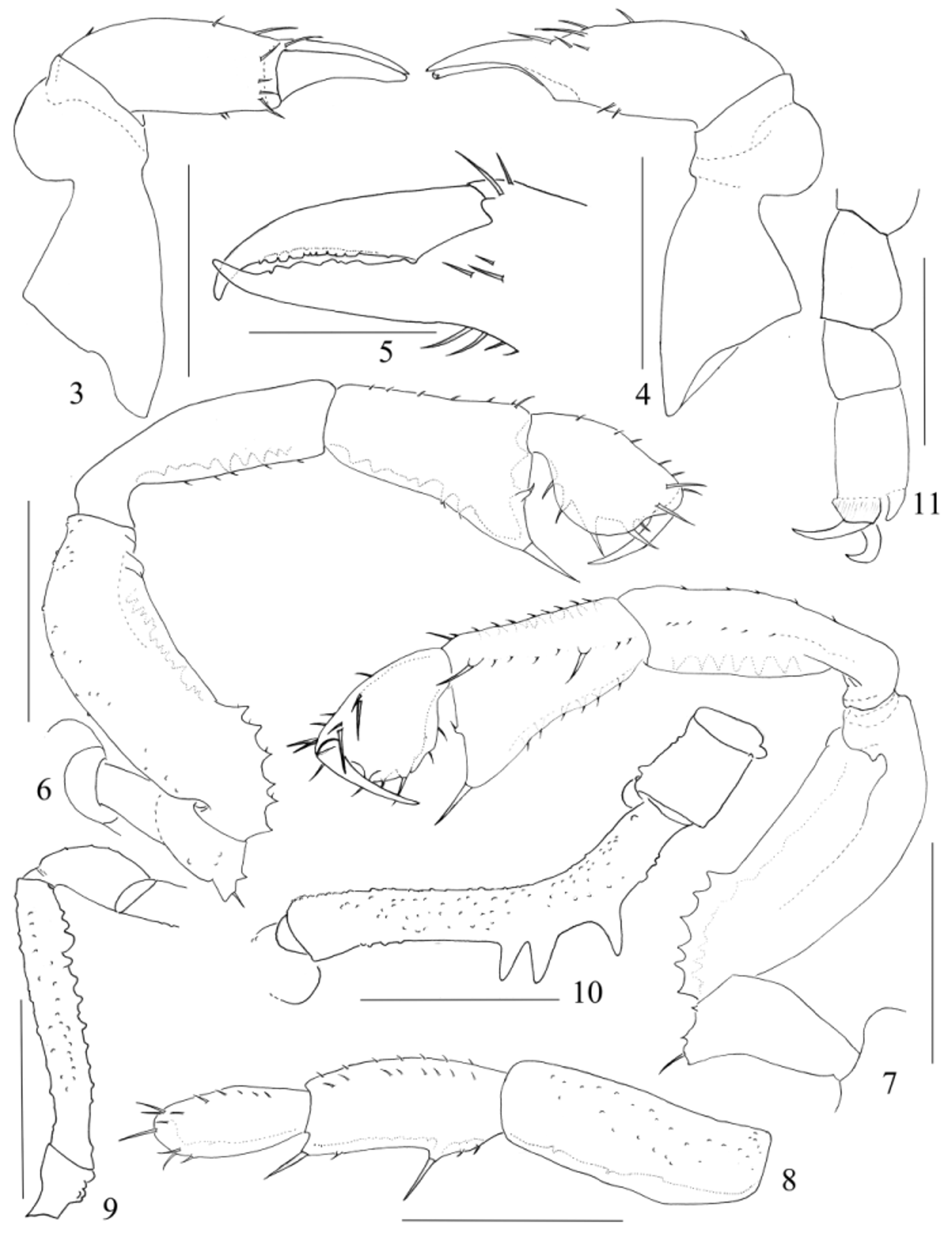
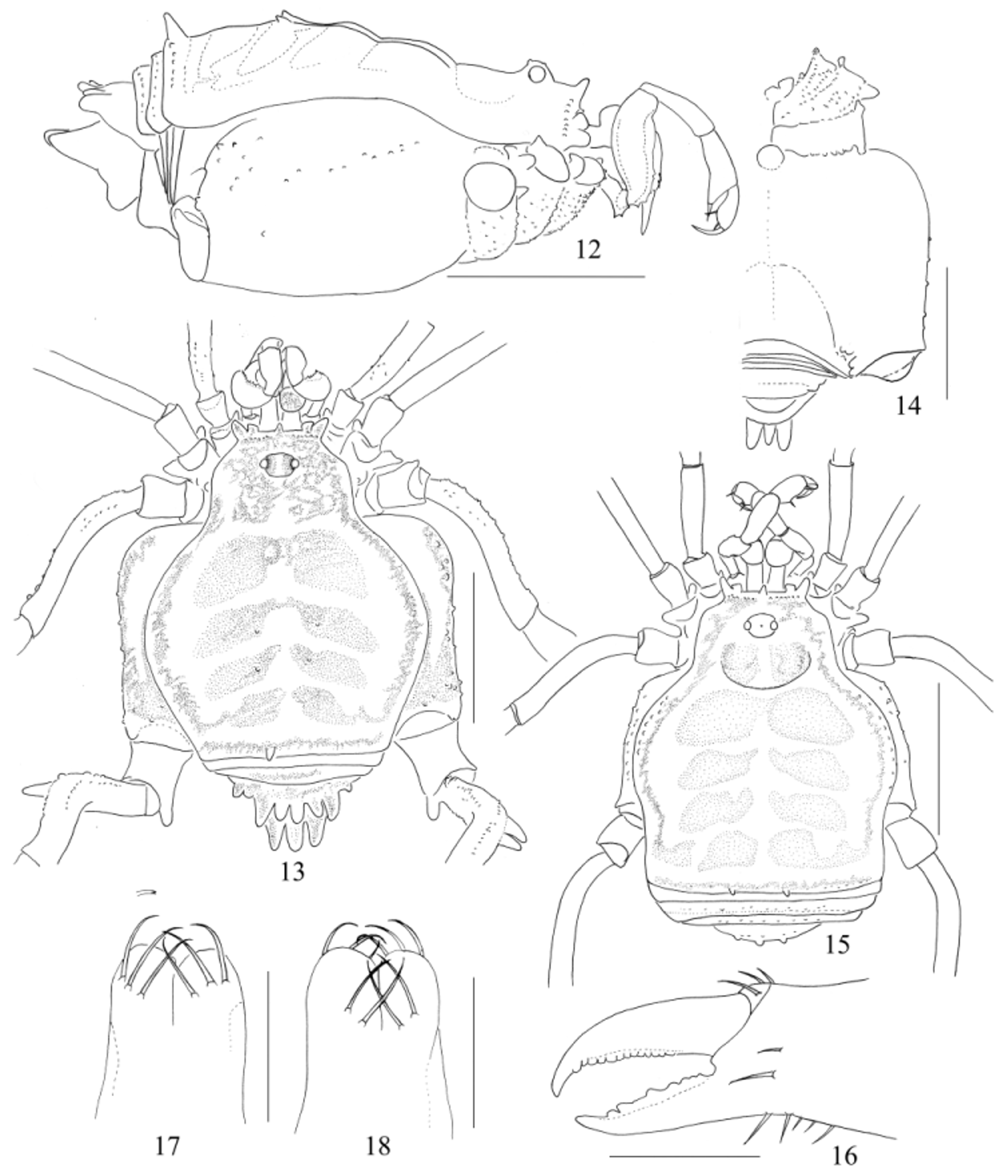
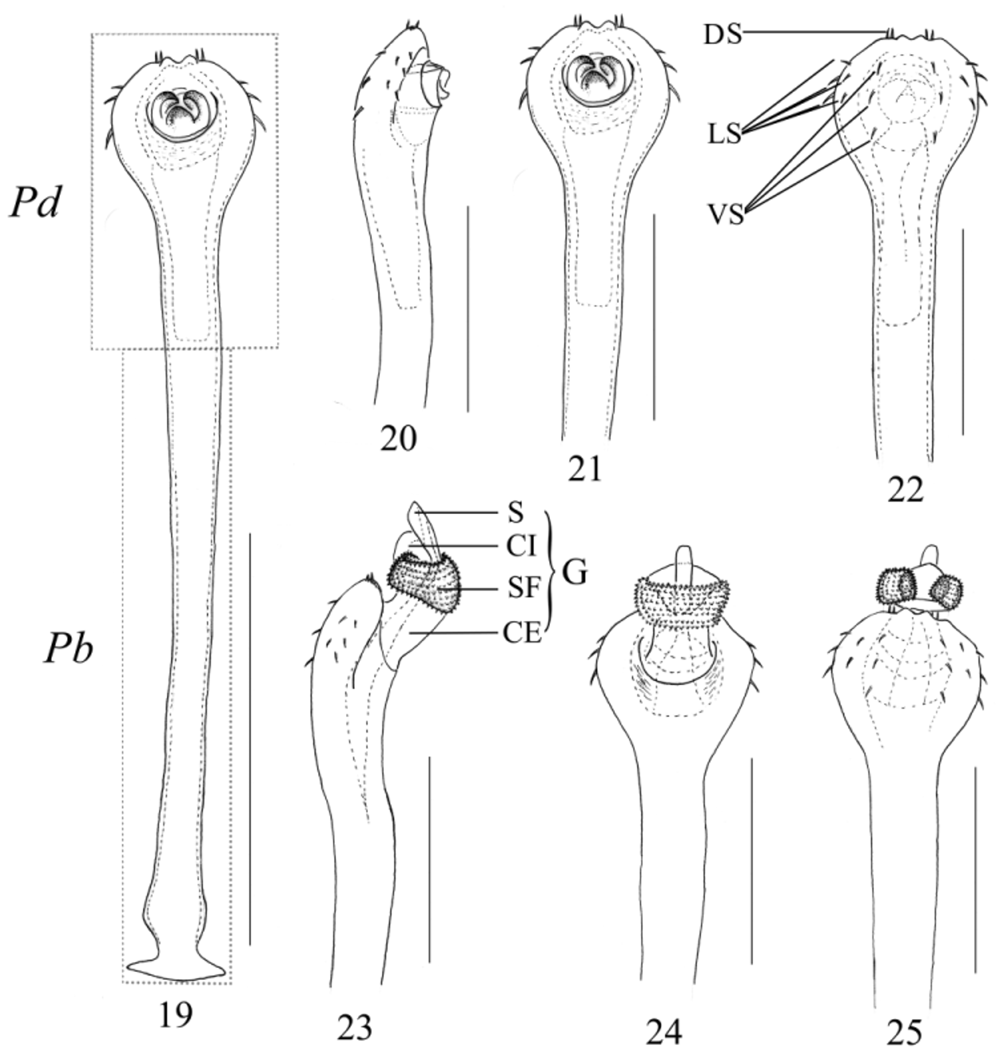
TABLE 1. Pedipalpus and leg measurements of the male (female) of Mysorea thaiensis Suzuki, 1985.
| Trochanter | Femur | Patella | Tibia | Metatarsus | Tarsus | Total | |
|---|---|---|---|---|---|---|---|
| Pedipalpus | 0.41(0.42) | 0.75(0.72) | 0.64(0.61) | 0.43(0.41) | 0.35(0.35) | 2.58(2.51) | |
| Leg I | 0.39(0.32) | 1.31(1.18) | 0.58(0.50) | 0.93(0.83) | 1.76(1.43) | 0.90(0.82) | 5.87(5.08) |
| Leg II | 0.45(0.40) | 2.14(1.90) | 0.70(0.62) | 1.81(1.54) | 2.44(1.86) | 1.90(1.56) | 9.44(7.88) |
| Leg III | 0.45(0.41) | 1.78(1.55) | 0.65(0.58) | 1.28(1.05) | 2.10(1.69) | 0.84(0.77) | 7.10(6.05) |
| Leg IV | 0.63(0.47) | 2.54(2.02) | 0.81(0.70) | 2.30(1.66) | 3.15(2.51) | 1.03(0.94) | 10.46(8.30) |
No known copyright restrictions apply. See Agosti, D., Egloff, W., 2009. Taxonomic information exchange and copyright: the Plazi approach. BMC Research Notes 2009, 2:53 for further explanation.
|
Kingdom |
|
|
Phylum |
|
|
Class |
|
|
Order |
|
|
Family |
|
|
Genus |
Mysorea thaiensis Suzuki, 1985
| Zhang, Chao & Zhang, Feng 2015 |
Mysorea thaiensis
| Suzuki 1985: 102 |