
Squatina occulta Vooren
|
publication ID |
https://doi.org/ 10.11646/zootaxa.3695.1.1 |
|
publication LSID |
lsid:zoobank.org:pub:D68A88C8-654D-4563-BDE2-6D6266D67232 |
|
DOI |
https://doi.org/10.5281/zenodo.6146712 |
|
persistent identifier |
https://treatment.plazi.org/id/03EF8783-FF91-D251-9985-F8BEFED504A3 |
|
treatment provided by |
Plazi |
|
scientific name |
Squatina occulta Vooren |
| status |
|
Squatina occulta Vooren View in CoL and da Silva, 1991
( Figs. 1–10 View FIGURE 1 View FIGURE 2 View FIGURE 3 View FIGURE 4 View FIGURE 5 View FIGURE 6 View FIGURE 7 View FIGURE 8 View FIGURE 9 View FIGURE 10 ; Tabs. 1–2 View TABLE 1 View TABLE 2 )
Squatina occulta Vooren and da Silva, 1991: 589–602, fig. 2 (original description, Southwestern Atlantic ocean, continental shelf of Rio Grande do Sul state, Brazil); Sunye and Vooren, 1997: 86–94, (anatomical description); Gadig et al., 1999: 133–136 (identification); Milessi et al., 2001: 1–7, fig. 2c (identification guide); Gadig and Gomes, 2003: 27 (listed); Vooren and Klipel, 2005, 57–82, figs. 4.1b, 4.3 (ecology); Colonello et al., 2007: 131 (cited); Menni and Lucifora, 2007: 4 (listed); Gomes et al., 2010: 53–55, figs. 56, 58 (identification guide); Stelbrink et al., 2010: 395–404 (molecular phylogeny); Carvalho et al., 2012: 171–183, figs. 2c, 3c, 4c, 6c (description of neurocranium).
Squatina argentina: Bigelow and Schroeder, 1948: 544 –546, figs. 103d, 106 (taxonomic revision); Roux, 1977: 159–168, fig. 8 and pl. 4 (taxonomic revision, in part, only MNHN 1989.133); Roux, 1979: 124–125, fig. 10 (taxonomic account, in part, only MNHN 1989.133) Solé-Cava et al., 1983: 355–358 (isozymic studies, cited as "morphotype II"); Compagno, 1984: 139, 142 (taxonomic account, figured); Solé-Cava and Levy, 1987: 139–144 (isozymic studies, cited as "morphotype II").
Squatina dumeril: Nunan and Senna, 2007: 174 (misidentification, in part, only MNRJ 30191).
Squatina guggenheim: Soto, 2001: 97 (misidentification, listed); Soto and Mincarone, 2004: 83–84 (misidentification, listed); Bernardes et al., 2005: 73 (misidentification, taxonomic account, figured); Compagno et al., 2005: 142–143, pl.18 (misidentification, identification guide).
Squatina squatina :?Schreiner and Miranda Ribeiro, 1903: 80 (taxonomic account);?Miranda Ribeiro, 1907: 169–171, fig. 10 (taxonomic account).
Holotype. MOVI 10195 (formerly MOFURG 06589), adult female (1220 mm TL), continental shelf of Rio Grande do Sul state, Brazil, 33o06’S, 51o40’W (not examined).
Paratypes. MCP 13999, preadult female (903 mm TL), continental shelf of Rio Grande do Sul state, 34º02’S, 52º19’W; MZUSP 41518, preadult male (975 mm TL), continental shelf of Rio Grande do Sul state, Brazil, 33º28'S, 52º01'W.
Additional material examined. 78 specimens (see Appendix 1).
Diagnosis. A species of Squatina diagnosed by its unique dorsal color pattern, composed of a uniform light to dark brown background color, with numerous white to yellowish rounded, small spots on entire dorsal surface, some surrounded by many small black spots, forming irregular ocelli-like markings. Squatina occulta is further distinguished from S. guggenheim by presenting smaller and more numerous white spots on entire dorsal surface (spot size one-half to eye-length vs. less numerous spots about equal to eye to 1.5 eye-length in S. guggenheim ); larger size at maturity (greater than 1000 mm vs. from 730 to 800 mm in S. guggenheim ); dermal denticles over pectoral fins morphologically homogeneous, without any sort of enlarged dermal denticle present (vs. a pair or more of enlarged dermal denticles close to dorsal pectoral origin in Squatina guggenheim ); dorsal midline with denticles morphologically similar to trunk denticles, or denticles over dorsal midline posterior to pectoral insertion slightly enlarged (basal plate 1.5–2 times diameter of other dorsal denticles) and organized in a row in some juveniles of S. occulta ( S. guggenheim with row of enlarged, conical denticles over dorsal midline, morphologically distinct from other dorsal denticles, with basal plate 3–4 times that of other dorsal denticles, and crown with several median ridges and acute non-ridged apex). Squatina occulta is further distinguished from S. argentina by a lighter background color (light to dark brown vs. dark brown to reddish-brown, respectively); presence of a pair of enlarged, conical and morphologically distinct dermal denticles between spiracles (basal plate 3–4 times diameter of other denticles, crown with several median ridges, and smooth and acute crown apex vs. interspiracular surface covered by smaller dorsal denticles, without enlarged denticles, respectively); and lower number of tooth rows, with 18–20 longitudinal tooth rows on upper jaw and 18–22 tooth rows on lower jaw (tooth formula: 9-9 to 10-10 / 9-9 to 11-11 vs. 12-12/12- 12 in S. argentina ); and, in almost all specimens, by anterior margin of pectoral fin straight to slightly convex (vs. anterior margin of pectoral fin convex in S. argentina ). Squatina occulta is distinguished from the similar form recognized here as Squatina sp. by a lower number of vertebral centra (maximum value 138 vs. 150, respectively); more numerous white spots over dorsal surface; and presence of a pair of enlarged dermal denticles with numerous ridges between spiracles (these reduced in size and with fewer ridges in Squatina sp.).
Description. Measurements are presented in Table 1 View TABLE 1 ; meristic data in Table 2 View TABLE 2 . The following description is based on all specimens ( Figs. 1 View FIGURE 1 to 10).
External morphology. Body markedly depressed dorsoventrally from head to origin of caudal fin (head height 5.4 to 9.8% TL). Pectoral-pelvic distance 7.7 to 12.3% TL, width at pectoral origins 12.5 to 17.9% TL, and trunk width 13 to 22.9% TL. Head broad, anterior margin rounded to trapezoidal, and posterior margin straight to concave, projecting obliquely to medial side ( Fig. 5 View FIGURE 5 a). Dermal folds extend along entire lateral margin of head, from distal end of ventral labial furrow to superior margin of first gill opening. Dermal fold margins straight to slightly convex in some specimens, without lobes or projections ( Fig. 53 View FIGURE 53 d). Snout very short. Shape of anterior margin between nostrils slightly convex, straight or concave, with dorsal surface straight to slightly convex. Preorbital length 2.5 to 5.5% TL, without preorbital pit. Eyes small, not greatly protruding above head, elliptical and slightly obliquely positioned (preocular distance 4.6 to 6.4% TL, eye length 1.6 to 3.1% TL and width 0.9 to 1.9% TL). Eyes widely separated, interorbital distance 6.5 to 9.9% TL; interorbital surface slightly bulging. Spiracles well separated from eyes (eye-spiracle distance 2.5 to 3.8% TL), wider than long, about equal or slightly smaller than eyes (spiracle length 1.6 to 2.7% TL), comma-shaped, and with a knob on inner half of posterior margin. Interspiracular distance about equal to interorbital length; interspiracular surface straight. Pseudobranchial folds present on anterior margin of spiracle (generally more than 10, number variable depending on preservation).
Nostrils small, terminal on head, approximately half-length of eye, very spaced apart (5.7 to 7.3% TL), and situated just dorsal to mouth upper lip. Anterior nasal flap very large, overlapping mouth, with three well developed, ventrally directed barbels. Inner and outer barbels elongate, slender and spatulate, larger than nasal aperture; outer barbel slightly wider than inner barbel; medial barbel somewhat square, wider than long, with fringed free margin, about one-length of outer barbel ( Fig. 54 View FIGURE 54 a). Posterior nasal flap fringed, confluent with lateral dermal fold, less prominent than anterior nasal flap, and protruding only sligthly from nasal margin. Mouth large, terminal on head, posteriorly arched, and extending to about level of mid-eye length; mouth width 10.8 to 15.6% TL. Upper labial furrows very large, conjoined, forming a deep preoral groove from symphysis to posterior jaw angle; lower labial furrows smaller than upper, length about one-fourth mouth width. Lower furrow extending from midlength of lower labial cartilage to posterior to jaw angle. Two or three pairs of teeth externally visible in some specimens with mouth closed (e.g. MCP 13999), but entirely covered in others (e.g. NUPEC 1651). Gill openings covered laterally by anterior projection of pectoral fin, however, inner edge of slits visible ventrally. Distance between first and fifth apertures very short (intergill slit length 1.5 to 3.9% TL). Gill slits widely separated, width between first gill openings 7.5 to 10.2% TL.
Pectoral fins very large, two times longer than wide (length 26.7 to 36.6% TL and width 12 to 19.4% TL), with a slender, angular and acute anterior projection. Anterior margin generally straight to slightly convex at its anterior half; three juveniles (MZUSP 110866, NUPEC 2192, MOFURG 493; Figs. 4 View FIGURE 4 a, b) with a prominent convexity (‘shoulder’) on anterior half of pectoral fin. Posterior pectoral margin slightly concave. Inner pectoral margin strongly convex, its length slightly shorter than pectoral fin width (mean inner margin length 16.3% TL vs. mean pectoral fin width 16.9% TL). Pectoral-fin base length large (9.3 to 13.3% TL), positioned anteriorly on body (prepectoral length 18.2 to 23.4% TL). Pelvic fins elongate, triangular to semi-rhomboidal, much longer than wide (length 16.5 to 26.8% TL and width 9.8 to 15.5% TL). Anterior margin of pelvics straight to slightly convex, posterior and inner margins straight, with free rear tip bluntly rounded, not reaching origin of first dorsal. Pelvic fin broad at more or less its midwidth, slender at margins. Distal end of pectoral fin reaches anterior margin of pelvic fin. Pectoral distal end usually extends from close to pelvic origin until half-length of pelvic anterior margin (some specimens with distal end of pectorals reaching close to outer edge of pelvic fin; Figs. 3 View FIGURE 3 a, 4c).
Coloration. Dorsal color pattern (in preservative, with mucous removed; for fresh coloration, see Remarks) with a light to dark brown background, with many small white to yellowish rounded spots on entire dorsal surface, disposed symmetrically on both sides of body. Larger spots about eye-length (on pectoral and pelvic fins) and smaller spots about one-half of eye-length. Some smaller white spots surrounded by many tiny black spots, forming irregular ocelli-like markings on entire dorsal surface. Amount of white spots surrounded by black spots somewhat variable; in paratype MZUSP 41518 ( Fig. 1 View FIGURE 1 a, b), almost all white spots are surrounded by black spots, but NUPEC 1651 ( Fig. 1 View FIGURE 1 c) and MZUSP 42851 ( Fig. 3 View FIGURE 3 a) with very few ocelli-like markings. Pectoral fins with a common pattern of six main larger spots without darker spots (one near pectoral origin, one on pectoral center aligned with its insertion, two near pectoral outer edge, and two close to pectoral inner margin; Fig. 6 View FIGURE 6 ). Pectoral spots vary in shape, from rounded to elliptical or forming a pattern of two opposed horseshoe-shaped spots linked by their edges ( Fig. 6 View FIGURE 6 e). Larger whitish spot without black spots close to pelvic fin origin. On head region, spots proportionally smaller than on trunk, pectoral and pelvic fins. Pigmentation lacking on anterior edge of pectoral fin covered by dermal folds of head. Tail with three pairs of large blackish blotches, width higher than dorsal fin base, disposed laterally on tail (one pair adjacent to pelvic fin tip, another pair adjacent to first dorsal fin, and third adjacent to second dorsal).
Ventral coloration uniform creamy-white on almost entire surface of head and trunk. Pectoral and pelvic fin margins beige to light brown on anterior margin, and beige to greenish on posterior margin; margins with an irregular mottled pattern in adults. In juveniles, anterior beige borders are proportionally smaller, with posterior borders light purple on both paired fins. Margins of cloaca creamy-white. Tail beige in both juveniles and adults from base to origin of lower caudal fin lobe; tail posteriorly with light to dark brown lateral margins similar to dorsal surface. Clasper with dorsal coloration creamy-white on anterior mid-length and posterior outer margin; brownish on distal inner end over cover rhipidion and rhipidion. Ventral coloration of clasper creamy-white on entire length (MZUSP 41518 with beige color on ventral distal end of clasper; Fig. 5 View FIGURE 5 d).
Dentition. Dentition of S. occulta presents a small gradient monognathic heterodonty. Upper jaw teeth proportionally smaller than lower jaw teeth. On upper jaw, teeth of first and second longitudinal medial rows, and that on both distal two longitudinal rows, slightly smaller than teeth on central longitudinal rows. On lower jaw, teeth of first and second longitudinal rows with root width smaller than others; and the teeth on two distal longitudinal rows, not only the root but also entire teeth, proportionally smaller than others. Teeth in 18 to 20 longitudinal rows in upper jaw and 18 to 22 rows in lower jaw (tooth formula 9-9 to 10-10 / 9-9 to 11-11). Three embryos (MZUSP 73171) with fewer longitudinal rows ( Tab. 2 View TABLE 2 ). Tooth row arrangement without overlap (independent dentition of Strasburg, 1963), and with slight differences between upper and lower jaws. Upper arrangement of tooth rows with first three vertical rows densely grouped lateral to symphysis, but other posterior vertical rows slightly less densely arranged (fourth vertical row greatly spaced apart from all other rows, positioned at anterior third of palatoquadrate). Tooth rows equally spaced on Meckel’s cartilages, and also without teeth adjacent to symphysis.
Tooth morphology with main cusp bulky, upright and bent lingually, without serration and accessory cusplets ( Fig. 7 View FIGURE 7 ). Tooth crown base laterally expanded, with prominent heels (Cappetta, 1987), projecting diagonally in most teeth (projecting laterally in first vertical upper tooth row). Cusp taller than width of crown and root. Cutting edges of main cusp and lateral heels sharp, prominent and continuous. Labial face of tooth crown slightly convex. Crown-base with prominent semi-circular apron, labially overhanging root, but not reaching root-base. Crown lingual face convex, contiguous with short, slender and cylindrical uvula covering dorsal surface of lingual protuberance of root. Crown base striated near junction with root. Crown-root junction without evident neck collar. Root laterally elongated, hemiaulacorhize, with root groove partially covered only below lingual protuberance. Root groove with 10 foramina (checked in only one specimen; NUPEC 1452). Distinct mediolingual foramen on distal end of lingual protuberance, visible in lingual view. Root lobes protrude below crown in labial view. Six to seven marginolingual foramina, and one below lateral end of crown heel on each side of tooth, similar to Pseudorhina acanthoderma (Carvalho et al., 2008) . Sexual dimorphism not discernible in teeth.
Dermal denticles. Dermal denticles densely packed covering entire dorsal surface, with exception of posterior borders of unpaired fins and anterior apex of pectoral fin (these usually naked). Mid-width of trunk and tail with dermal denticles usually larger than denticles on head, lateral region of trunk and tail, pectoral and pelvic fins. Denticles of both regions with a posteriorly directed crown, with four ridges over anterior surface extending from crown base to apex; fifth ridge present on some denticles at mid-width, extending from crown base to its midheight. Basal plate morphology slightly variable. Trunk and tail denticles with rounded basal plate, but rhomboidal on pectoral fin; dorsal surface of head with both types of basal plate. In some juveniles ( Figs. 8 View FIGURE 8 , 9 View FIGURE 9 a, b), row of enlarged denticles over dorsal midline sometimes present, usually from origin of tail to origin of first dorsal fin (in NUPEC 1255, this row occurs from mid-length of pectoral base). Morphology of these enlarged denticles similar to others on dorsal surface, presenting only an enlarged rounded basal plate (1.5 to 2 times diameter of basal plate of other dorsal trunk denticles). Specimen NUPEC 1275 ( Fig. 9 View FIGURE 9 a) with a more conspicuous midline row of enlarged denticles with additional central and lateral ridges on crown bases (nine ridges) and four main ridges extending to crown apex.
Enlarged dermal denticles on head symmetrically organized ( Fig. 9 View FIGURE 9 f), with two rows of three to four enlarged denticles between nostrils, from anterior margin of snout to anterior margin of eye (directly over lateral margins of anterior fontanelle of neurocranium); generally two, but sometimes three, enlarged denticles obliquely positioned anterior to each eye, with the anteriormost almost reaching anterior margin of snout (over posterolateral margins of preorbital processes); a pair of enlarged denticles posterior to each eye (over postorbital processes); and a pair of enlarged denticles present between spiracles (over sphenopterotic ridges). Morphology of these enlarged denticles very distinct from other denticles. Basal plate rounded, with diameter three to four times diameter of smaller denticles on dorsal surface. Enlarged denticles with large, conical and posteriorly directed crown, with a smooth base. Crown apex acute and smooth; midheigth of crown with 10 ridges. Enlarged denticles, positioned as described above, less prominent and grouped in clusters of five or more in paratypes (largest specimens studied). Sexual dimorphism absent.
Maturity, female reproductive system and size at birth. No adult males were available for study; largest male analyzed was MZUSP 41518 (paratype), a juvenile 975 mm TL. The clasper in this specimen does not reach the free rear tip of pelvic fin (clasper inner length 3.6% TL and outer length 1.4% TL); however, early stages of formation of clasper groove, rhipidion and cover rhipidion are noticeable ( Fig. 10 View FIGURE 10 ). Our observations are in agreement with Vooren and da Silva (1991) and Vooren and Klippel (2005) in that S. occulta reaches its first maturation at more than 1100 mm TL. Females have prominent ovaries with discernible oocytes since early stages of development (NUPEC 1275, 362 mm TL). Squatina occulta presents only one functional ovary, on left side; its antimere is poorly developed, very reduced and lacking oocytes ( Figs. 36 View FIGURE 36 e, f). According to our material, size at birth in S. occulta varies between 280–300 mm TL, also in agreement with Vooren and Klippel (2005).
Remarks. The holotype could not be studied, but both paratypes were examined. Although fresh specimens were not directly examined, photographs of fresh specimens allowed us to confirm that there are no significant differences in color compared to preserved specimens, only that the background color is generally brighter. Apparently, as in S. guggenheim (described below), the mucous is pigmented exactly over the larger whitish spots ( Fig. 2 View FIGURE 2 c). Accordingly, characters from dorsal coloration are useful to diagnose Southwestern Atlantic Squatina (see diagnosis).
Geographic distribution. According to material examined, Squatina occulta is distributed in the Southwestern Atlantic from Espírito Santo state, Brazil (approximately19o S) to southern Uruguay, near the Rio de la Plata estuary (approximately 34o S) ( Fig. 11 View FIGURE 11 ). We record this species for the first time in latitudes north of 23o S (i.e. from Espírito Santo state, Brazil, based on a single specimen, MNRJ 30191). Vooren and Klippel (2005) proposed that S. occulta might be present along the Argentinean shelf (to 45o S); however, this could not be confirmed.
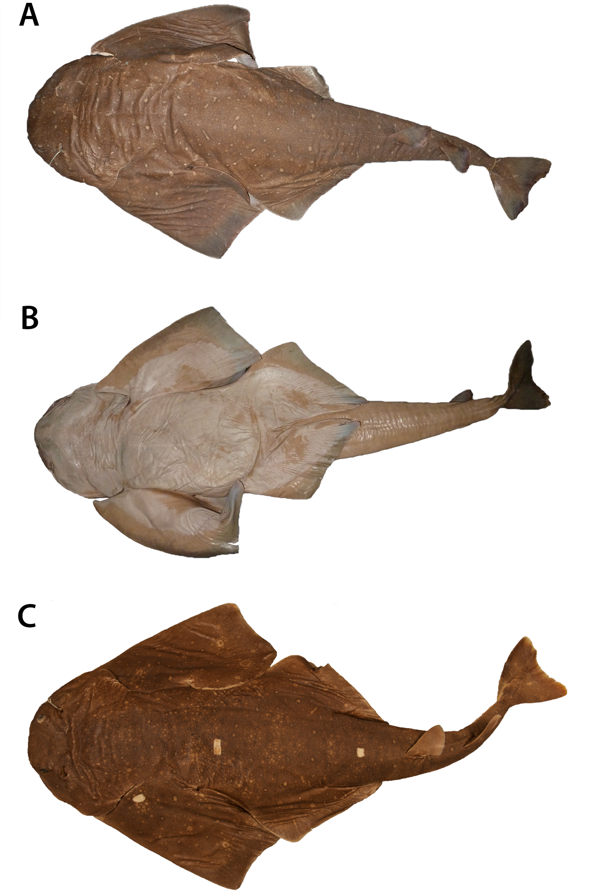


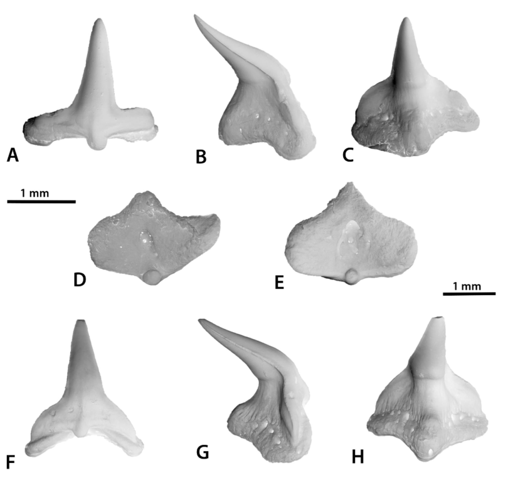


TABLE 1. Measurements for specimens of Squatina occulta. Abbreviations: n, number of specimens; Min, minimum value; Max, maximum value; SD, standard deviation.
| Morphometric character | MZUSP 41518 | MCP 13999 | NUPEC 1651 | MNRJ 30191 | n | Min | Max | Mean | SD |
|---|---|---|---|---|---|---|---|---|---|
| mm % TL | mm % TL | mm % TL | mm % TL | % TL | % TL | % TL | % TL | ||
| Total length (TL) | 975.0 | 903.0 | 743.0 | 808.0 | 47 | ||||
| Pre-caudal length | 845.0 86.7 | 772.0 85.5 | 640.0 86.1 | 683.0 84.5 | 47 | 83.0 | 90.9 | 85.7 | 1.4 |
| Pre-dorsal length | 642.0 65.8 | 585.0 64.8 | 481.0 64.7 | 517.0 64.0 | 47 | 59.6 | 69.4 | 64.4 | 1.6 |
| Pre-pectoral length | 192.0 19.7 | 174.0 19.3 | 144.0 19.4 | 161.0 19.9 | 47 | 18.2 | 23.4 | 20.4 | 1.2 |
| Pre-pelvic length | 386.0 39.6 | 363.0 40.2 | 319.0 42.9 | 327.0 40.5 | 47 | 36.7 | 45.7 | 41.2 | 2.1 |
| Pre-branchial length | 148.0 15.2 | 140.5 15.6 | 111.0 14.9 | 122.3 15.1 | 47 | 13.8 | 18.4 | 15.8 | 0.8 |
| Pre-spiracular length | 86.0 8.8 | 77.7 8.6 | 63.0 8.5 | 67.2 8.3 | 47 | 7.8 | 10.3 | 9.0 | 0.5 |
| Pre-ocular length | 52.0 5.3 | 50.3 5.6 | 39.7 5.3 | 42.4 5.2 | 47 | 4.6 | 6.4 | 5.4 | 0.3 |
| Pre-orbital length | 27.5 2.8 | 29.1 3.2 | 22.0 3.0 | 29.0 3.6 | 47 | 2.5 | 5.5 | 3.5 | 0.6 |
| Head width | 187.0 19.2 | 176.0 19.5 | 148.0 19.9 | 142.1 17.6 | 47 | 15.1 | 22.6 | 19.7 | 1.2 |
| Orbital head width | 129.2 13.3 | 124.5 13.8 | 101.0 13.6 | 99.7 12.3 | 47 | 11.3 | 16.0 | 14.2 | 1.1 |
| Spiracular head width | 207.0 21.2 | 192.0 21.3 | 147.0 19.8 | 149.7 18.5 | 47 | 15.9 | 24.4 | 20.6 | 1.4 |
| Mouth width | 136.0 13.9 | 126.0 14.0 | 103.7 14.0 | 101.0 12.5 | 47 | 10.8 | 15.6 | 14.1 | 0.8 |
| Head height | 78.0 8.0 | 65.0 7.2 | 51.5 6.9 | 60.1 7.4 | 47 | 5.4 | 9.8 | 7.3 | 0.9 |
| Interorbital distance | 81.0 8.3 | 76.6 8.5 | 67.8 9.1 | 67.6 8.4 | 47 | 6.5 | 9.9 | 8.7 | 0.6 |
| Eye length | 15.7 1.6 | 16.8 1.9 | 18.6 2.5 | 15.0 1.9 | 47 | 1.6 | 3.1 | 2.4 | 0.4 |
| Eye width | 12.0 1.2 | 9.4 1.0 | 7.6 1.0 | 11.0 1.4 | 47 | 0.9 | 1.9 | 1.4 | 0.2 |
| Eye-spiracle distance | 34.7 3.6 | 22.4 2.5 | 23.6 4.9 | 24.2 3.0 | 47 | 2.5 | 3.8 | 3.4 | 0.3 |
| Internarial distance | 60.8 6.2 | 57.2 6.3 | 45.0 6.1 | 48.5 6.0 | 47 | 5.7 | 7.3 | 6.4 | 0.4 |
| Interspiracular distance | 74.1 7.6 | 70.9 7.9 | 58.6 7.9 | 59.6 7.4 | 47 | 6.9 | 9.3 | 8.3 | 0.5 |
| Spiracle length | 20.8 2.1 | 22.1 2.4 | 13.6 1.8 | 18.6 2.3 | 47 | 1.6 | 2.7 | 2.2 | 0.2 |
| Intergill width | 98.1 10.1 | 84.0 9.3 | 75.0 10.1 | 75.3 9.3 | 47 | 7.5 | 10.2 | 9.2 | 0.6 |
| Intergill length | 18.6 1.9 | 18.9 2.1 | 15.8 2.1 | 19.9 2.5 | 47 | 1.5 | 3.9 | 2.4 | 0.5 |
| Interdorsal distance | 66.2 6.8 | 54.1 6.0 | 49.0 6.6 | 48.2 6.0 | 47 | 5.2 | 7.5 | 6.3 | 0.5 |
| Dorsal-caudal distance | 67.8 7.0 | 56.2 6.2 | 53.8 7.2 | 55.4 6.9 | 47 | 6.2 | 9.0 | 7.5 | 0.6 |
| Pectoral-pelvic distance | 85.6 8.8 | 80.0 8.9 | 68.0 9.2 | 95.2 11.8 | 47 | 7.7 | 12.3 | 10.1 | 1.2 |
| Pelvic (origin)-caudal distance | 470.0 48.2 | 442.0 48.9 | 363.0 48.9 | 408.0 50.5 | 47 | 40.0 | 51.1 | 46.1 | 2.4 |
| Pelvic-caudal distance | 301.0 30.9 | 293.0 32.4 | 251.0 33.8 | 298.0 36.9 | 47 | 27.8 | 36.9 | 31.7 | 1.8 |
TABLE 2. Meristic data for specimens of Squatina occulta. A: MZUSP 41518. B: MNRJ 30191. C: NUPEC 1651. Abbreviations: n, number of specimens; Min, minimum value; Max: maximum value.
| Character | A | B | C | n | Min | Max | Mode |
|---|---|---|---|---|---|---|---|
| Propterygial radials | 3 | 3 | 3 | 17 | 3 | 3 | 3 |
| Mesopterygial radials | 11 | 11 | 13 | 17 | 10 | 13 | 11 |
| Matapterygial radials | 29 | 26 | 29 | 17 | 21 | 30 | 29 |
| Pelvic radials | 27 | 26 | 29 | 17 | 23 | 29 | 29 |
| Monospondylous vertebrae | 46 | 48 | 46 | 17 | 46 | 49 | 47 |
| Precaudal diplospondylous vertebrae | 56 | 58 | 58 | 17 | 53 | 58 | 56 |
| Caudal diplospondylous vertebrae | 34 | 32 | 31 | 17 | 24 | 34 | 30 |
| Total vertebrae | 136 | 138 | 135 | 17 | 127 | 138 | 134 |
| Upper tooth rows (rigth side) | 9 | 9 | 9 | 35 | 9 | 10 | 9 |
| Upper tooth rows (left side) | 9 | 9 | 9 | 35 | 7 | 10 | 9 |
| Lower tooth rows (rigth side) | 9 | 10 | 10 | 35 | 8 | 11 | 10 |
| Lower tooth rows (left side) | 9 | 10 | 10 | 35 | 9 | 11 | 10 |
No known copyright restrictions apply. See Agosti, D., Egloff, W., 2009. Taxonomic information exchange and copyright: the Plazi approach. BMC Research Notes 2009, 2:53 for further explanation.
|
Kingdom |
|
|
Phylum |
|
|
ParvPhylum |
Chondrichthyes |
|
Class |
|
|
Order |
|
|
Family |
|
|
Genus |
|
Kingdom |
|
|
Phylum |
|
|
ParvPhylum |
Chondrichthyes |
|
Class |
|
|
Order |
|
|
Family |
|
|
Genus |