
Scelidosaurus oehleri, SIMMONS, 1965
|
publication ID |
https://doi.org/10.1111/j.1096-3642.2007.00301.x |
|
DOI |
https://doi.org/10.5281/zenodo.5745352 |
|
persistent identifier |
https://treatment.plazi.org/id/03EF87F4-FFD3-FFBB-754D-FC8EFEE5E31B |
|
treatment provided by |
Carolina |
|
scientific name |
Scelidosaurus oehleri |
| status |
|
SPECIES T. OEHLERI SIMMONS, 1965 ( NOMEN DUBIUM)
Synonymy: T. oehleri Simmons (1965) : 65; S. oehleri Lucas, 1996: 82 .
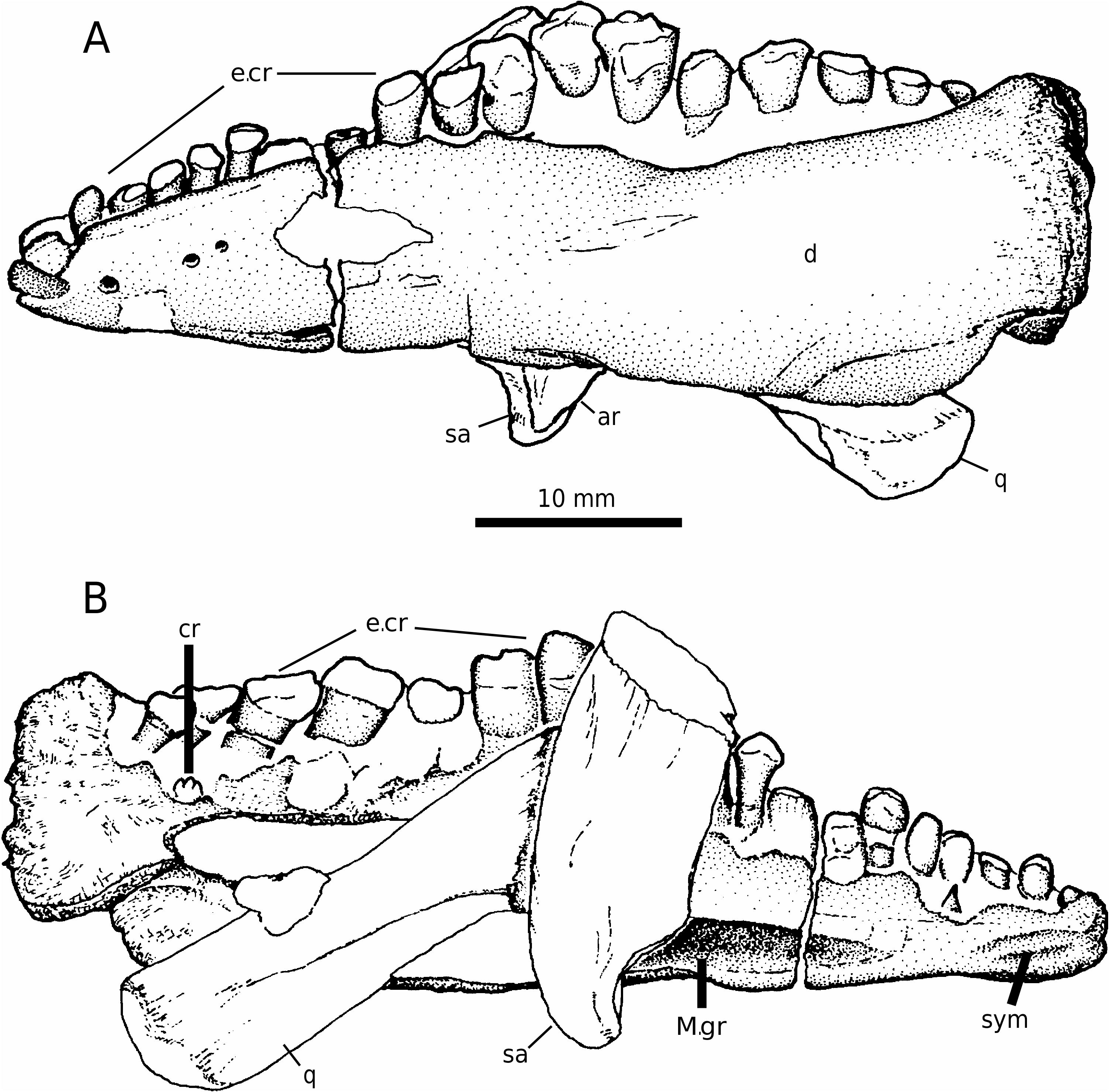
Holotype: FMNH CUP 2088 View Materials . Partial left dentary, with fragments of the quadrate and surangular-articular adhering to its medial surface.
Description: The holotype of T. oehleri Simmons, 1965 ( Fig. 1 View Figure 1 ) comprises a partial (relatively small) left lower jaw measuring just less than 60 mm in length. Fragments of a broken quadrate and of two postdentary bones: the surangular (sa) and what appears to be a portion of the articular (ar) are preserved and firmly attached to the medial surface of the dentary ( Fig. 1A View Figure 1 ). Previous descriptions ( Simmons, 1965: fig. 11; Dong, 1990: fig. 19.2; Lucas, 1996: figs 1–3) have identified many of the salient features of the holotype but have been at variance with respect to the identity of the attached bones. Dong, for example, concluded that the bone was a quadratojugal, whereas Simmons and Lucas interpreted it as a rib fragment.
Dentary: Figure 1 View Figure 1 illustrates, in lateral and medial views, the holotype specimen. The dentary ramus is transversely thick and deep posteriorly at the point where its surface is abruptly truncated. The anterior one third of the dorsal edge of the dentary ramus slopes quite markedly toward the symphysial region; the external (buccal) surface of the ramus has a pronounced, but diminishing, longitudinal bulge that would have formed a ledge, flooring a buccal (cheek) recess, with the posterior cheek teeth positioned along the medial edge of the dentary. At its midpoint along the length of the jaw the dentary ramus appears to be distorted by crushing (many minor fractures and evidence of post-mortem crushing are visible). Toward the symphysis, as the dentary continues to taper, the ramus becomes generally narrower and develops a slight lateral overhang as the lower edge of the dentary twists medially toward its neighbour. The external surface of the ramus is marked by several foramina but, in general, the surface of the dentary is not well preserved and shows few of these more minor anatomical details.
Medially the dentary is partially obscured by the overlying bones ( Fig. 1B View Figure 1 ) and, contrary to the description of Simmons, there is no indication of a splenial bone (which also appears to be the situation as illustrated by Simmons 1965: fig. 11B). A deeply incised Meckelian groove is clearly present (again contradicting Simmons’ original description) and extends almost to the dentary symphysis, but is obscured by a combination of the overlying bones and infilling matrix more posteriorly. The dentary symphysis, which is only separated by a short section of dentary ramus from Meckel’s groove, is marked by horizontal ridges and grooves, and, in the adjacent area near the tip of the jaw and in advance of the first alveolus, there is very little evidence for, or indeed room for, an ornithischian predentary [a broadly similar configuration is also found in the basal thyreophoran Scelidosaurus (D. B. Norman, pers. observ.) and in Emausaurus (SCRM & RJB, pers. observ.]. The ventral edge of the dentary is twisted medially in the proximity of the symphysis, creating a very slightly spout-shaped region to the dentary adjacent to the symphysial region, as observed in ornithischians generally. The lingual wall of the dentary above the Meckelian groove slopes buccally (dorsolaterally) toward the base of the alveolar trough. There is no evidence of an alveolar parapet that supported the lower and medial portions of the dentition ( Fig. 2B View Figure 2 ), but this specimen is poorly preserved and this part of the jaw may easily have been either removed accidentally or eroded away postmortem. The posterior part of the dentary is clearly thick and deep, and hints at the presence of an at least modest coronoid eminence, but there is no clear evidence of sutures for the attachment of the postdentary bones.
Dorsally the relative transverse thickness of the dentary is evident at its posterior end and a pronounced cheek recess is clearly shown. The anterior tapering of the dentary might be expected to be accompanied by a degree of sinuosity of the dentition; however, the teeth appear to be arranged in a more or less linear pattern, but this may also be a post-mortem artefact as the alveolae themselves do seem to indicate some degree of medial curvature as the symphysis is approached.
Dentition: The dentition of Tatisaurus has been described in some detail by both Simmons (1965) and Lucas (1996); however, it must be emphasized that the dentition, as preserved, is heavily eroded and almost completely lacking in detail. The alveolar count of 18 is confirmed, as is the general cadence in tooth size: smaller anteriorly, increasing in size posteriorly before decreasing again in size at the extreme posterior end of the series. The teeth have subcylindrical roots that clearly expand (mesio-distally) into the base of the crown; it is clear from the close packing of the roots of the teeth that the crowns would have been arranged in an overlapping ‘en echelon’ pattern as reported by Simmons. The crowns themselves are heavily eroded and their structure and pattern of wear are not discernible (despite the description provided by both Simmons and Lucas). A single replacement crown tip is visible on the medial surface of alveolus 17 and reveals the presence of coarse denticulations along the mesio-distal margin of the crown; a fragment of replacement crown is present medially in alveolus 1, and a splinter of enamel lies medial to the root of the tooth in alveolus 4 (and might therefore represent the position of the replacement crown). A considerable amount of matrix is still present in the alveoli and around the bases of the much-eroded functional teeth, so the specimen might benefit from either further skilled preparation or non-invasive (computerized tomography, CT) scanning.
Additional bony elements: The bones attached to the medial surface of the dentary are poorly preserved and their identification is necessarily tentative. There appear to be two bones: one oblique and somewhat slender, which is crushed up against a vertically orientated somewhat more ‘blocky’ element. The former element has a slightly convex distal articular surface and a rounded shaft that has a sinuous ridge and is tentatively identified as a partial quadrate ( Fig. 1 View Figure 1 , q). The latter element ( Fig. 1 View Figure 1 , sa) resembles that of an ornithischian surangular in some respects: notably the lipped concave facet, which may well represent the lateral portion of the jaw articulation. The medial side of this element (visible when the jaw is viewed laterally) seems to show the presence of a small posterior portion of the articular or splint of prearticular ( Fig. 1 View Figure 1 , ar). It should be noted that there is no evidence of external foramina (which might be expected to be visible adjacent to the glenoidal part of the surangular), and the cross section in the region of fracture (dorsally) does not appear to show a hollowed interior as might be expected.
| FMNH |
Field Museum of Natural History |
No known copyright restrictions apply. See Agosti, D., Egloff, W., 2009. Taxonomic information exchange and copyright: the Plazi approach. BMC Research Notes 2009, 2:53 for further explanation.
|
Kingdom |
|
|
Phylum |
|
|
Class |
|
|
Order |
|
|
Family |
|
|
Genus |
Scelidosaurus oehleri
| Norman, David B., Butler, Richard J. & Maidment, Susannah C. R. 2007 |
S. oehleri
| Lucas 1996: 82 |
T. OEHLERI
| SIMMONS 1965 |
T. oehleri Simmons (1965)
| SIMMONS 1965 |