
Macrobiotus sottilei, Pilato, Giovanni, Kiosya, Yevgen, Lisi, Oscar & Sabella, Giorgio, 2012
|
publication ID |
https://doi.org/10.5281/zenodo.279945 |
|
DOI |
https://doi.org/10.5281/zenodo.6180889 |
|
persistent identifier |
https://treatment.plazi.org/id/03F03A09-9B50-B467-E5F9-67FEFB9024F3 |
|
treatment provided by |
Plazi |
|
scientific name |
Macrobiotus sottilei |
| status |
sp. nov. |
Macrobiotus sottilei View in CoL sp. nov.
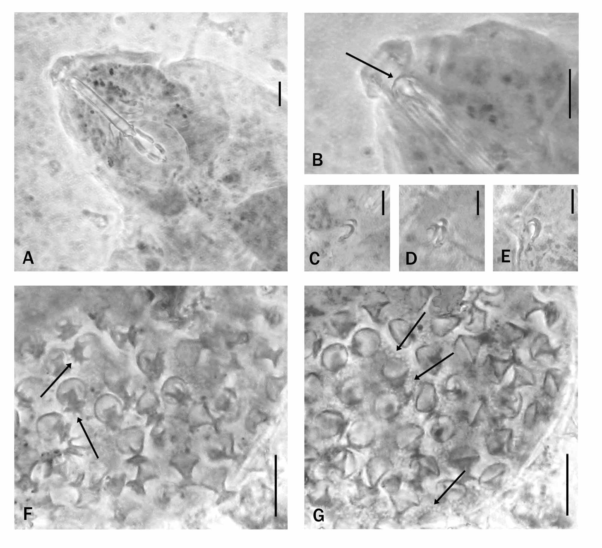
( Fig. 2 View FIGURE 2. A – D )
Type material. Minsk Oblast, vicinity of Zhodino, Pine forest (approximately 54°6ʹ N, 28°21ʹ E), moss on stone collected ( November 2008) by S.A. Dobrovolsky: holotype, 39 paratypes, and 7 eggs; 2 of which with developed embryo.
Type repository: Holotype (slide No. 5428), 16 paratypes and 3 eggs (slides No. 5427, 5428 and 5438) deposited in the collection of Binda & Pilato (Museum of the Department of Animal Biology “Marcello La Greca”, University of Catania, Italy). 23 paratypes and 4 eggs from the same moss sample as well as numerous specimens and eggs from other samples are deposited in the collection of Kiosya (Kharkiv National University, Ukraine).
Specific diagnosis: Cuticle smooth with small pores, eye spots present, dorsal ridges of the buccal armature joined forming a continuous arc, pharyngeal bulb with two macroplacoids and a microplacoid; claws, well developed, of hufelandi - type; accessory points and lunules present; eggs, freely laid, spherical, with trunco-conical processes with indented distal disc. Egg shell with a very dense and regular reticular design with extremely small mesh; that around the base of the processes is not larger than elsewhere on the shell.
Description of the holotype. Body length 337 µm, colourless; eye spots present. Cuticle smooth with small pores and very small dots on the legs.
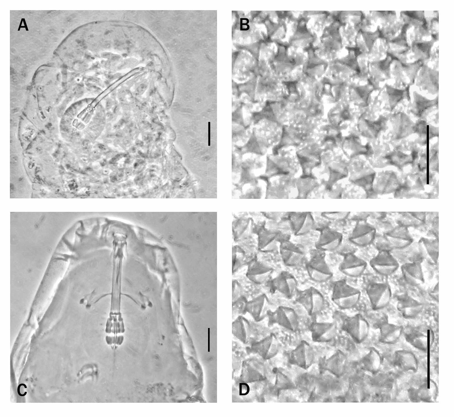
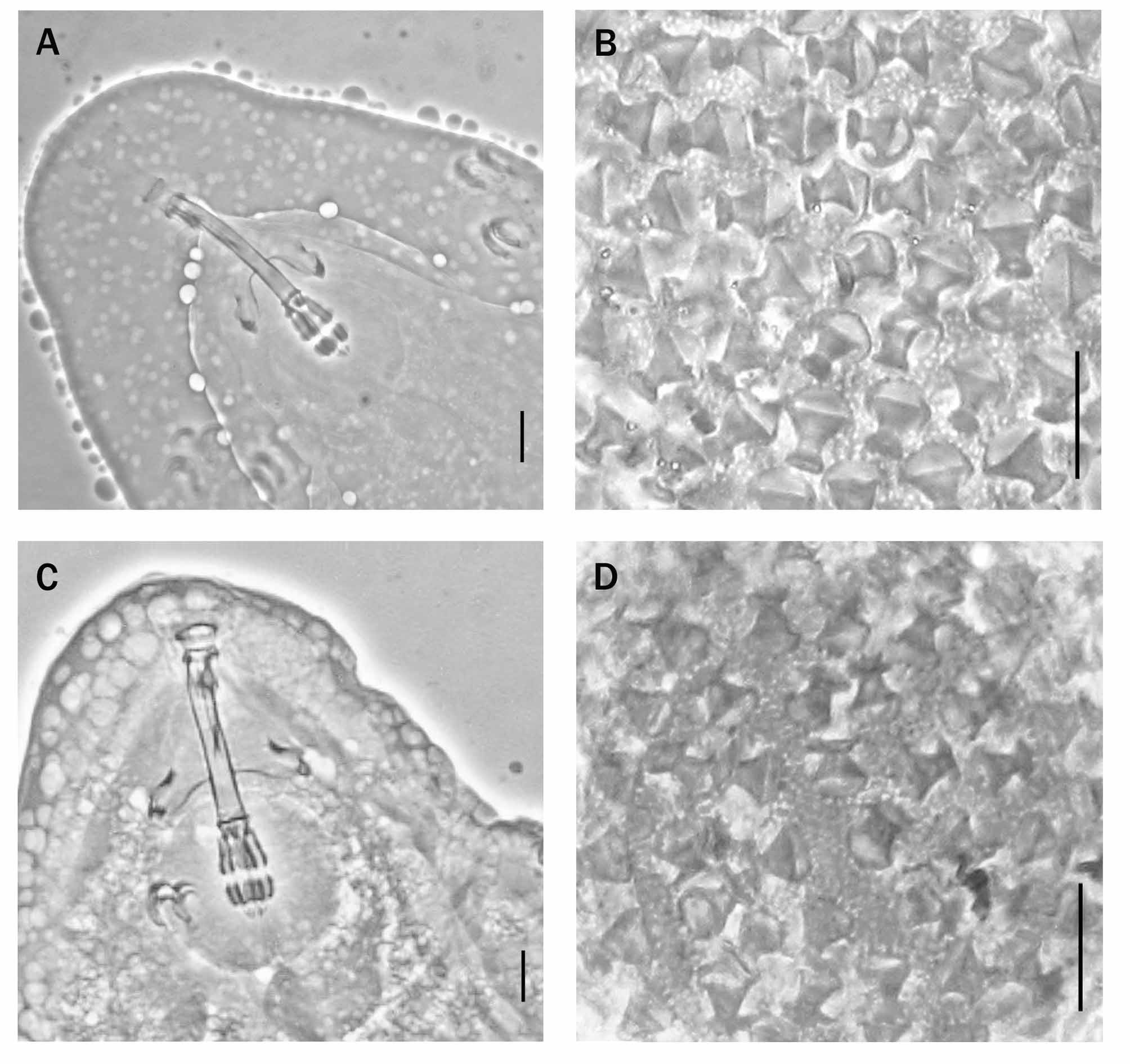
Bucco-pharyngeal apparatus of the Macrobiotus - type ( Fig. 2A View FIGURE 2. A – D ); mouth terminal with ten small peribuccal lamellae, a posterior ring of small teeth and, more caudally, a system of three ventral thin transverse ridges and a continuous dorsal transverse ridge ( Fig. 2B View FIGURE 2. A – D ). Buccal tube 34.1 µm long and 4.4 µm wide externally ( pt = 12.9). Stylet supports inserted on the buccal tube at 76.6 % of its length ( pt = 76.6). Pharyngeal bulb (30.6 µm × 25.4 µm) with apophyses, two macroplacoids, and a microplacoid ( Fig. 2A View FIGURE 2. A – D ). Length of first macroplacoid 9.8 µm ( pt = 28.7), second 6.3 µm ( pt = 18.5), and microplacoid 2.1 µm ( pt = 6.2); entire placoid row 19.3 µm long ( pt = 56.6); macroplacoid row 17.1 µm long ( pt = 50.1).
Claws, well developed, of hufelandi - type ( Fig. 2 View FIGURE 2. A – D C-E) with main and secondary branches diverging after a short common portion; accessory points on the main branches present. External and internal claws of the first pair of legs 11.2 µm long ( pt = 32.8) and 10.0 µm long ( pt = 29.3) respectively; those of the second pair of legs 12.4 µm long ( pt = 36.4) and 10.9 µm long ( pt = 32.0) respectively; length of posterior and anterior claws of the hind legs 12.5 µm ( pt = 36.7) and 11.3 µm ( pt = 33.1) respectively. Lunules present, small and smooth on the first three pairs of legs, slightly larger and with a delicately indented margin on the hind legs. Two cuticular bars present below the claws on each leg of the first two pairs of legs.
Description of the eggs. The eggs, freely laid, were spherical, with trunco-conical processes and indented distal disc ( Fig. 2 View FIGURE 2. A – D F, arrows); we counted 29 processes on the circumference and about 120 on the hemisphere. The diameter was about 72 µm excluding the processes and about 83 µm including. The processes were 5.4–6.2 µm high with a basal diameter of 4.8–5.2 µm; the distal discs have a diameter of 3.6–4.4 µm. The egg shell had a uniform, very dense and regular reticular design with extremely small mesh ( Fig. 2 View FIGURE 2. A – D G, arrows), that showed no distinction between that around the base of the processes and the between process egg shell.
Remarks. The paratypes were similar to the holotype in both qualitative and metric characters (we referred to the pt index values when the specimens had different body length). The measurements of the holotype and two paratypes are presented in Table 2.
Etymology. The specific name is in honour of the histologist Lorenzo Sottile, who has recently died and who had been a former scholar and colleague of G. Pilato.
Differential diagnosis. Within the genus Macrobiotus 11 species were known to have two macroplacoids and a microplacoid and, in the buccal armature, the dorsal transverse ridges joined to form a continuous arc. Three, M.
echinogenitus (according to Binda, 1988), M. semmelweisi and M. drakensbergi , are clearly different from M. sottilei sp. nov. in characters of both the specimens and eggs: M. echinogentus has more visible teeth in the buccal cavity wall and eggs with large conical processes. M. semmelweisi has bands of dorsal gibbosities, stylet supports inserted on the buccal tube in a more cephalic position ( pt = 72–73 in M. semmelweisi , 75.3–76.6 in M. sottilei sp. nov.), claws stout and eggs with slender conical processes. Macrobiotus drakensbergi has strongly indented lunulae and eggs with conical processes.
Comparing adults of, where possible, comparable size the new species can be from the other species (Table 2) as follows:
Characters of the eggs easily distinguish two species, M. diversus and M. seychellensis , from M. sottilei sp. nov.
The eggs of M. diversus had egg processes shaped as a bowling-pin with cup-shaped extremities (the diameter of which was clearly shorter than the basal diameter of the processes); the egg shell was rough but without a true reticular design. In M. sottilei sp. nov. the distal disc of each egg process was larger and had a clearly indented margin; the egg shell had a dense reticular design.
The egg processes of M. seychellensis had a terminal disc with a diameter greater than the basal diameter and the terminal disc margin prolonged into obvious lobes, very different from M. sottilei sp. nov. where the distal disc of each egg process was shorter than the basal diameter and the margin indented ( Figs 2 View FIGURE 2. A – D F and 3B, arrows). The reticular design of the egg shell of M. sottilei sp. nov. was more dense than the eggs of M. seychellensis , having a clearly smaller mesh ( Figs. 2 View FIGURE 2. A – D G and 3B). In addition, with respect to adult animals, M. sottilei sp. nov. had a longer second placoid, and claws with a shorter common portion with respect to the total length of the claws than seen in M. seychellensis .
M. sottilei sp. nov. differed from M. biserovi in having a longer second macroplacoid, more slender claws and clearly different eggs, with smaller processes, distal disc diameter shorter than the basal diameter, margins of distal discs clearly indented, and a more dense reticular design of the egg shell.
M. sottilei sp. nov. differed from M. sapiens in having a narrower buccal tube, stylet supports inserted on the buccal tube in a more cephalic position (Table 2; Figs. 2A View FIGURE 2. A – D and 3 View FIGURE 3. A, B C), and a more dense, smaller mesh, reticular design of the egg shell ( Fig 2 View FIGURE 2. A – D G and 3D).
The new species differed from M. modestus in having a wider buccal tube; stylet supports inserted on the buccal tube in a more caudal position; longer placoids (Table 2; Figs 2A View FIGURE 2. A – D and 4A View FIGURE 4. A, B ), and the reticular design of the egg shell was more delicate and less visible ( Figs. 2 View FIGURE 2. A – D G and 4B).
M. sottilei sp. nov. differed from M. humilis in having the stylet supports inserted on the buccal tube in a more caudal position; longer placoids (Table 2; Figs 2A View FIGURE 2. A – D and 4 View FIGURE 4. A, B C); longer and more slender claws (Table 2) with shorter common tract; and less visible reticular design of egg shell ( Figs. 2 View FIGURE 2. A – D G and 4D).
The new species differed from M. madegassus in having eye spots; slightly wider buccal tube; stylet supports inserted on the buccal tube in a more caudal position, longer placoids (Table 2; Figs 2A View FIGURE 2. A – D and 5A View FIGURE 5. A, B ); slightly longer and more slender claws with shorter common tract; margins of the distal discs of the egg processes more clearly indented; and a smaller mesh reticular design of the egg shell ( Figs. 2 View FIGURE 2. A – D F, G and 5B).
M. sottilei sp. nov. differed from M. iharosi in having a slightly wider buccal tube; longer placoids (Table 2; Figs 2A View FIGURE 2. A – D and 5 View FIGURE 5. A, B C); and a smaller mesh reticular design of the egg shell ( Figs. 2 View FIGURE 2. A – D G and 5D).

No known copyright restrictions apply. See Agosti, D., Egloff, W., 2009. Taxonomic information exchange and copyright: the Plazi approach. BMC Research Notes 2009, 2:53 for further explanation.
|
Kingdom |
|
|
Phylum |
|
|
Class |
|
|
Order |
|
|
Family |
|
|
Genus |