
Globicornis (Hadrotoma) corticalis ( Eichhoff, 1863 )
|
publication ID |
https://doi.org/ 10.11646/zootaxa.3686.5.4 |
|
publication LSID |
lsid:zoobank.org:pub:8CF2B2D6-7AED-4DBA-8DA2-B28E01D70A70 |
|
DOI |
https://doi.org/10.5281/zenodo.6146123 |
|
persistent identifier |
https://treatment.plazi.org/id/03F087FD-5A6B-FFAE-3EAD-FE2BFC02F0F1 |
|
treatment provided by |
Plazi |
|
scientific name |
Globicornis (Hadrotoma) corticalis ( Eichhoff, 1863 ) |
| status |
|
Globicornis (Hadrotoma) corticalis ( Eichhoff, 1863)
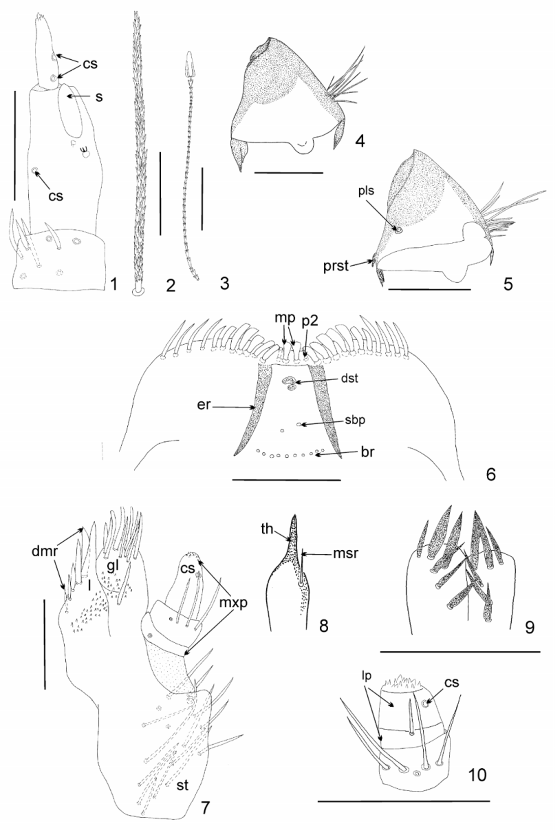
( Figs. 1–16 View FIGURES 1 – 10 View FIGURES 11 – 16 )
Syn.: Hadrotoma corticalis Eichhoff, 1863 Globicornis corticalis: Reitter, 1891
Material examined. 15 exemplars (larvae and exuviae of final instar): Polonia, 8.1.1954, Żednia, distr. Białystok, leg. R. Bielawski. All material deposited in collection of Division of Invertebrate Biology, Evolution and Conservation, Department of Biology, Evolution and Ecology, University of Wrocław, ul. Przybyszewskiego 63/ 77, 51–148 Wrocław, Poland.
Description. Last larval instar. Length 0.9–2.3. Body fusiform, and relatively long, rather flat. Integument of head brown; nota and terga brown with darker pigmented areas ( Figs. 11, 13 View FIGURES 11 – 16 ); all tergal plates sclerotized, all sternal plates hyaline with numerous brown spicisetae. Tufts of prominent brown spicisetae present between legs. Coxae and femora brown; tibiae lighter. Spicisetae and hastisetae on terga and sterna brown. Antennae oriented anterolaterally and composed of 3 antennomeres ( Fig. 1 View FIGURES 1 – 10 ). Terminal antennomere almost 4 times as long as wide. Three appendages and two campaniform sensilla present on last joint ( Fig. 1 View FIGURES 1 – 10 ). Ratio of length of terminal antennomere to length of one and two antennomeres combined 0.3:1.0. Sensorium arising proximad to apex of antennomere 2, in latero-apical position, and slightly extending above apex. One campaniform sensillum at almost 1/2 total length of antennomere 2. Basal segment with two setae and two campaniform sensilla ( Fig. 1 View FIGURES 1 – 10 ). Labro– epipharyngeal margin with 11 setae in outer series. Mesal of labro–epipharyngeal setae spatulate while second pair slender. Epipharynx with 10–12 sensory cups in proximal transverse series, epipharyngeal rods present and slightly diverging proximally. Two sensory cups present in subproximal epipharyngeal sensilla. Distal epipharyngeal sensilla arranged in two groups (completely enclosed/encircled by furrow)—one of two, and second of four. Lateral setae on epipharynx absent ( Fig. 6 View FIGURES 1 – 10 ). Dorsal surface of labro–epipharynx with numerous setae. Mandibles brown with dark brown (almost black) apices; apical teeth and ventral accessory process absent. Apical half of mandible heavily sclerotized and sharply delineated from basal half ( Figs. 4–5 View FIGURES 1 – 10 ). Mandibular mola and pseudomola absent. Hyaline lobe at ventral base of mandible absent. Prostheca small, inconspicuous ( Fig. 5 View FIGURES 1 – 10 ); mesal brush of setae near base of mandible absent. Placoid sensillum in basal 1/5 of structure ( Fig. 5 View FIGURES 1 – 10 ). Maxillary palp with 3 palpomeres; terminal palpomere longest. Ratio of length of terminal palpomere to the length of the two proceeding palpomeres combined 1.4:1.0. Basal palpomere with probably one setae, second palpomere with four setae; third palpomere with one campaniform sensillum. Terminal palpomere with group of 9–11 small sensilla apically situated. Lacinia with one (often unvisiable) slightly sclerotized lacinial tooth at apex ( Figs. 7–8 View FIGURES 1 – 10 ). Sclerotization of lacinia as illustrated on Fig. 8 View FIGURES 1 – 10 . Mesal row of setae on lacinia composed of one thick basal seta ( Fig. 8 View FIGURES 1 – 10 ). Straight, thick to slender five setae in dorsomesal row of setae present ( Fig. 7 View FIGURES 1 – 10 ). Galea arising from stipes, extending above apex of lacinia and terminating near apex of terminal palpomere. Apical area of galea densely covered with setae ( Fig. 7 View FIGURES 1 – 10 ). Stipes with 17 long setae near anterio–lateral margin ( Fig. 7 View FIGURES 1 – 10 ). Hypopharynx hyaline. Bridge sclerite (i.e. central part of distal element of hypopharyngeal sclerome) connected medially. Ligula with about 12 spurlike setae; about six on apex, and about nine on middle section between labial palpi ( Fig. 9 View FIGURES 1 – 10 ). Labial palp 2–segmented. Basal segment only slightly wider than terminal segment; 2.0 times as wide as long; with one seta. Terminal segment 1.5 as long as wide, with group of 14 small sensilla in apical area and one campaniform sensillum ( Fig. 10 View FIGURES 1 – 10 ) at half total length of segment. Antecostal suture on notum I absent, but distinct on nota II and III as well as abdominal terga I– VIII; tergum IX without antecostal suture. Acrotergites of notum I without setae ( Fig. 11 View FIGURES 11 – 16 ); acrotergites of abdominal terga I and acrotergites of other thoracic and abdominal segments with setae ( Figs. 13–15 View FIGURES 11 – 16 ). Nota I with long, stout, large spicisetae along anterior (directed anteriorly under head), lateral and posterior margin (directed latero–posteriorly and vertically). Some spicisetae present on central area of disc of
notum I; numerous hastisetae arranged among spicisetae on almost entire area ( Fig. 11 View FIGURES 11 – 16 ). Nota II and III and abdominal terga I–VIII with median row of large spicisetae, mainly directed latero–posteriorly and vertically; hastisetae more numerous than spicisetae, especially on posterior half of terga, mainly along posterior margin ( Figs. 13–15 View FIGURES 11 – 16 ). Hastisetae of abdominal segments forming dense lateral brushes, particularly on posterolateral region of abdominal terga V, VI, VII, VIII densely inserted on posterior part of tergum ( Figs. 14–15 View FIGURES 11 – 16 ). Setal patterns of abdominal tergum I as illustrated ( Fig. 13 View FIGURES 11 – 16 ); abdominal tergum VII as illustrated ( Fig. 14 View FIGURES 11 – 16 ). Abdominal tergum VIII without pair of abdominal pits (oval apertures); setal patterns as illustrated ( Fig. 15 View FIGURES 11 – 16 ). Abdominal tergum IX reduced in size with numerous long spicisetae creating caudal brush and arranged as illustrated ( Fig. 16 View FIGURES 11 – 16 ). Legs covered with many dark brown setae ( Fig. 12 View FIGURES 11 – 16 ). Tarsal claws dark brown. Ratio of length of tibia to length of femur 0.6:1.0. Pretarsus with two narrow lanceolate setae inserted at base. Anterior pretarsal seta the same length as pretarsus. Posterior pretarsal seta equal with anterior pretarsal seta ( Fig. 12 View FIGURES 11 – 16 ).
Pupa unavailable, but probably develops within last larval exuvium.
Biology. Knowledge of the biology of the species is limited and based only on sparse published information ( Mroczkowski 1975). Larvae appear to feed on dead insects and have been observed on pupae of Pseudoclavellaria amerinae Linnaeus, 1758 ( Hymenoptera : Cimbicidae ). Pupation takes place in late autumn in Poland. After pupation, specimens stay inside the last larval skin until the spring.
Distribution. Europe; Kazakhstan; Russia: W. Siberia ( Háva 2013).

No known copyright restrictions apply. See Agosti, D., Egloff, W., 2009. Taxonomic information exchange and copyright: the Plazi approach. BMC Research Notes 2009, 2:53 for further explanation.
|
Kingdom |
|
|
Phylum |
|
|
Class |
|
|
Order |
|
|
Family |
|
|
Genus |
|
Kingdom |
|
|
Phylum |
|
|
Class |
|
|
Order |
|
|
Family |
|
|
Genus |