
Gilpinia amamiana Okutani, 1965
|
publication ID |
https://doi.org/10.11646/zootaxa.4995.3.4 |
|
publication LSID |
lsid:zoobank.org:pub:AF31D6D4-AFD4-46A2-A400-29B0281810E0 |
|
persistent identifier |
https://treatment.plazi.org/id/03F1576F-FFCA-6942-FBB5-FC26FD1CFD8E |
|
treatment provided by |
Plazi (2021-07-01 20:52:32, last updated 2023-11-03 03:31:24) |
|
scientific name |
Gilpinia amamiana Okutani, 1965 |
| status |
|
Gilpinia amamiana Okutani, 1965
( Figs 2A–F, I–K View FIGURES 2 , 3E–G, M–O, V, W View FIGURES 3 , 4I–L, Q, 5G–J, S, 6F–H, 7D–F)
Gilpinia amamiana Okutani, 1965: 76 ; Okutani, 1984: 26; Abe & Togashi, 1989: 545; Azuma et al., 2002: 301; Yoshida, 2010: 28; Hara, 2019: 42; Hara, 2020: 291.
Female ( Figs 2A–D View FIGURES 2 ). Length 7.5–9.0 mm. Head black, yellow on supraclypeal area, narrow area around torulus and clypeus except for narrow lateral part ( Figs 2I, J View FIGURES 2 ); postocellar area narrowly or mostly yellow to brown; vertex sometimes narrowly or widely yellow or brown ( Figs 3M, N View FIGURES 3 ); gena widely or mostly yellow to brown; antenna black, basally narrowly yellow ( Figs 3V, W View FIGURES 3 ); labrum dark brown ( Figs 2I, J View FIGURES 2 ), sometimes yellow; mandible reddish brown, basally darkened, with yellow spot on outer basal part; palpi yellow, often basally brownish. Thorax yellow ( Figs 2A–D View FIGURES 2 ), dark brown to black on narrow anteromedial part of pronotum, wide anteromedial part of median mesoscutal lobe, most of lateral mesoscutal lobe, very narrow anteromedial margin and narrow posterior margin of mesoscutellum (Fig. 4I), mesoscutellar appendage, anterior or most part of metascutum, metascutellum, and wide ventral part of mesepisternum ( Figs 2B, D View FIGURES 2 ); mesopostnotum medially brown; postspiracular sclerite, anteroventral part of anepimeron and katepimeron except for posterior margin sometimes brown to dark brown; metapleuron partly brown. Legs yellow to brown yellow ( Figs 2A–D View FIGURES 2 ); coxae basally brown to dark brown; femora sometimes basally narrowly darkened; hind tibia sometimes narrowly darkened apically; middle and hind tarsi often slightly darkened apically. Wings colorless transparent ( Figs 2A, C View FIGURES 2 ); veins brown to black; most of vein C and bases of vein 1A and 2A+3A yellow; vein R1 basal to stigma sometimes widely pale; stigma brown yellow or brown, black on narrow margins and basal third. Abdomen yellow ( Figs 2A–D View FIGURES 2 ); first tergum mostly yellow or mostly dark brown; second to eighth terga each dark brown posteriorly; ninth tergum marginally dark brown narrowly or widely; tenth tergum laterally and posteriorly brown to dark brown; posterior sterna each darkened along posterior margin; cercus dark brown; ovipositor sheath with valvifer 2 yellow to brown, anteriorly darkened and valvula 3 mostly black.
Postocellar area slightly or distinctly swollen; lateral furrow distinct, vestigial or indistinct ( Figs 3M, N View FIGURES 3 ). OOL: POL:OOCL 0.9–1.1:1.0:0.8–0.9. OOCL 2.3–3.1 × lateral ocellus length. Inner margins of eyes convergent below ( Figs 2I, J View FIGURES 2 ). Distance between eyes at level of torulus 1.8–2.1 × major axis of eye. Supraclypeal area slightly or moderately swollen. Clypeus with ventral margin truncate or very slightly concave medially; median height 1.0–1.3 × torulus height. Malar space 0.9–1.2 × median ocellus width. Antenna with 21–23 antennomeres ( Figs 3V, W View FIGURES 3 ); first flagellomere without serration, with apical breadth in lateral view 0.8–1.1 × dorsal length; serrations around fourth to eighth flagellomeres largest.
Distance between cenchri 0.6–0.7 × major axis of cenchrus (Fig. 4I). Metascutellum in dorsal view not or very slightly angulated posterolaterally with posterior margin convex; median length 0.3–0.5 × major axis of cenchrus. Hind leg with posterior tibial spur with length 0.8–1.0 × dorsal length of first tarsomere, 0.8–1.1 × apical breadth of tibia in lateral view; dorsal length of second and third tarsomeres combined 1.3–1.6 × dorsal length of first.
Valvula 3 in dorsal view about 3 × as wide as cercus, apically with round lateral projection and small acute medial projection ( Figs 5G, I View FIGURES 5 ); scopa about 1.5–2.0 × as wide as cercus, in posterior view long oval, about 2.0–2.5 × as high as wide ( Figs 5H, J View FIGURES 5 ). Lance with posterior projection on processus articularis apically extending to or near most basal annular suture of lance ( Fig. 5S View FIGURES 5 ). Lancet Figs 6F–H View FIGURES 6 ; lamnium with 9 annuli and length from ventral end of first ctenidium to apex 2.1–2.3 × maximum width; ctenidia nearly straight except second arched and third sometimes slightly arched; sclerite before first annulus narrow; serrulae of second and third annuli each with distinct precalcar (precalcar sometimes absent, probably due to severe wear, Fig. 6H View FIGURES 6 ); serrula of second annulus much larger than other serrulae, with precalcar relatively close to postcalcar.
Head and thorax mostly covered with dense punctures ( Figs 3M, N View FIGURES 3 , 4I, J); punctures very often fused; inter- spaces generally narrower than diameters of punctures. Punctures on mesoscutellum much larger than those of mesoscutum. Abdomen microsculptured.
Male ( Figs 2E, F View FIGURES 2 ). As in female, but differing as follows except for usual sexual differences. Length 7.0–8.0 mm. Black, but antenna sometimes mostly dark brown, labrum dark brown, mandible reddish brown except for narrow base, pronotum posterolaterally widely yellow, tegula yellow, medially dark brown, abdomen ventrally yellow, sometimes with laterotergites each narrowly brown along posterior margin and subgenital plate dark brown. Legs yellow; coxae black except for narrow apices; trochanters basally black; hind tibia apically brown yellow.
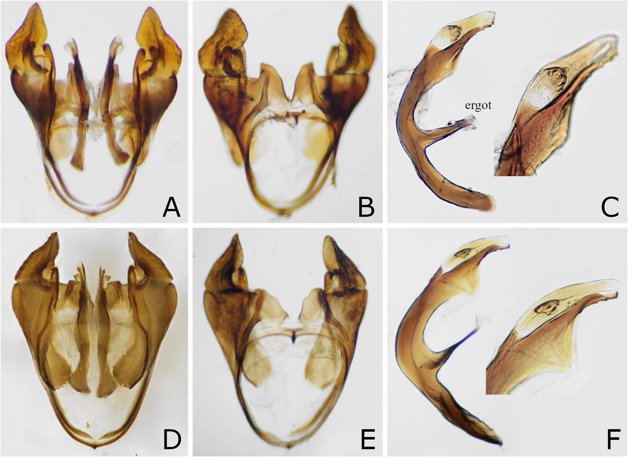
OOL:POL:OOCL 0.8–0.9:1.0:0.6. OOCL 1.6–2.0 × lateral ocellus length. Distance between eyes at level of torulus 2.1 × major axis of eye. Clypeus with median height 0.9 × torulus height. Malar space 1.1–1.2 × median ocellus width. Antenna with 24–25 antennomeres; flagellomeres each with two rami, but most basal and preapical flagellomeres with one ramus and apical most flagellomere without ramus. Distance between cenchri 0.7–1.0 × major axis of cenchrus (Fig. 4K). Metascutellum in dorsal view posterolaterally slightly angulated, with posterior margin slightly convex; median length 0.5–0.6 × major axis of cenchrus. Posterior hind tibial spur.with length 0.8–1.0 × dorsal length of hind first tarsomere, 1.1–1.2 × apical breadth of tibia in lateral view; dorsal length of second and third tarsomeres combined 1.3–1.4× as that of first. Subgenital plate apically nearly truncate in dorsal or ventral view ( Fig. 2E View FIGURES 2 ). Genitalia Figs 7D–F View FIGURES 7 ; in ventral view, parapenis apically truncate, with lateral margin very short, ( Fig. 7E View FIGURES 7 ); penis valve with valviceps exceedingly convex ventrally at apical third ( Fig. 7F View FIGURES 7 ).
Material examined. Holotype ( Figs 2A, B, I View FIGURES 2 , 3E, M, V View FIGURES 3 , 4I, J, 5G, H, 6F): ♀, “Hatsuno Amami 2. April. 1963 Y. Arita”, “ Holotype Gilpinia amamiana Okutani, 1965 ”, “lancet mounted”, “2”, “ Holotype Gilpinia amamiana Okutani ”. The photo of the lancet ( Fig. 6E View FIGURES 6 ) was taken by Smith in 1979. After that, the slide of the lancet probably has been lost.
Other material examined: JAPAN: Amami Islands : 1♂ ( Figs 2E, F, K View FIGURES 2 , 3G, O View FIGURES 3 , 4K, L, 7D–F), Amami Oshima Island , Tatsugo, Nagakumo-toge, 23. VI. 2009, M. Owada ; 1♀ ( Fig. 5S View FIGURES 5 ), Amami Oshima Island , Naze, 28. IX. 1982, S. Yamane ; 1♀ ( Figs 4Q, 6G), Amami Oshima Island, Kinsakubaru, 23. III. 2012, H. Ono ; 2♂, Amami Oshima Is- land, Amami, 28°21'N 129°27'57E, on Pinus luchuensis , 26. III. 2021, H. Hara GoogleMaps ; 1♀ ( Figs 2C, D, J View FIGURES 2 , 3F, N, W View FIGURES 3 , 5I, J View FIGURES 5 , 6H View FIGURES 6 ), Tokunoshima Island , Mikyo, 9. IV. 1998, M. Kimura .
Distribution. Japan: Amami Oshima Island ( Okutani 1965), Tokunoshima Island (new record).
Life history. The adults were collected in March, April, June and September. This sawfly probably has several generations a year. Hara collected two males on a tree of Pinus luchuensis Mayr. The tree species is probably the host plant.
Remarks. Gilpinia amamiana is distinguished from other congeners as stated in the section of the G. hakonensis and similar species and the key above. Okutani (1965) wrote about the female holotype of G. amamiana that “this species is closely allied to G. hakonensis ( Matsumura, 1912) , but easily distinguished by the larger metascutellum, truncate clypeus, hardly defined postocellar area, and yellow legs”. Actually, G. amamiana has the metascutellum not larger than that of the latter (compare Fig. 4I with Figs 4A, C, E), the clypeus not different from that of the latter, and the postocellar area with a distinct anterior furrow and a distinct, vestigial or indistinct lateral furrow ( Figs 3M, N View FIGURES 3 ).
FIGURES 4A–R. A, C, E, G, I, K, M, Posteromedial part of thorax, dorsal view; B, D, F, H, J, L, N, mesepisternum, lateral view; O–R, female seventh abdominal sternum, ventrolateral view. A–H, O, P, Gilpinia hakonensis : A, B, O, lectotype of Lophyrus hakonensis , female; C, D, holotype of G. hakonensis var. laticincta , female; E, F, P, paratype of Diprion fukudai , female; G, H, male, Honshu. I–L, Q, Gilpinia amamiana : I, J, holotype, female; K, L, male, Amami Oshima Isl.; Q, female, Amami Oshima Isl. M, N, R, Gilpinia okinawa , holotype, female.
Abe, M. & Togashi, I. (1989) Symphyta. In: Hirashima, Y. (Ed.), A Check List of Japanese Insects. Entomological Laboratory, Faculty of Agriculture, Kyushu University, Fukuoka, pp. 541 - 560. [in Japanese]
Azuma, S., Yafuso, M., Kinjo, M., Hayashi, M., Kohama, T., Sasaki, T., Kimura, M. & Kawamura, F. (2002) Check List of Insect of the Ryukyu Islands. 2 nd Edition. The Biological Society of Okinawa, Nishihara, xxiv + (2) + 570 pp. [in Japanese]
Hara, H. (2019) Family Diprionidae. In: Editorial Committee of Catalogue of the Insects of Japan (Ed.), Catalogue of the Insects of Japan. Vol. 9. Hymenoptera. Part 1. Symphyta. Entomological Society of Japan, Kyoto, pp. 42 - 46. [in Japanese]
Hara, H. (2020) Family Diprionidae. In: Naito, T., Shinohara, A., Hara, H. & Ito, F., Sawflies and Woodwasps of Japan. Hokkaido University Press, Sapporo, pp. 48 - 53 + 286 - 294. [in Japanese]
Matsumura, S. (1912) Thousand Insects of Japan. Supplement IV. Keiseisha-shoten, Tokyo, 247 pp., 55 pls. [in Japanese and English]
Okutani, T. (1965) Sawflies and horntails from the Ryukyus. Kontyu, 33, 73 - 84.
Okutani, T. (1984) [Symphyta of Japan IX]. Nature and Insects, 19 (1), 23 - 26. [in Japanese]
Yoshida, H. (2010) Checklist of Japanese Symphyta (Hymenoptera) (1). Xyelidae - Diprionidae. Hachi-Kariudo, (2), 13 - 29. [in Japanese]
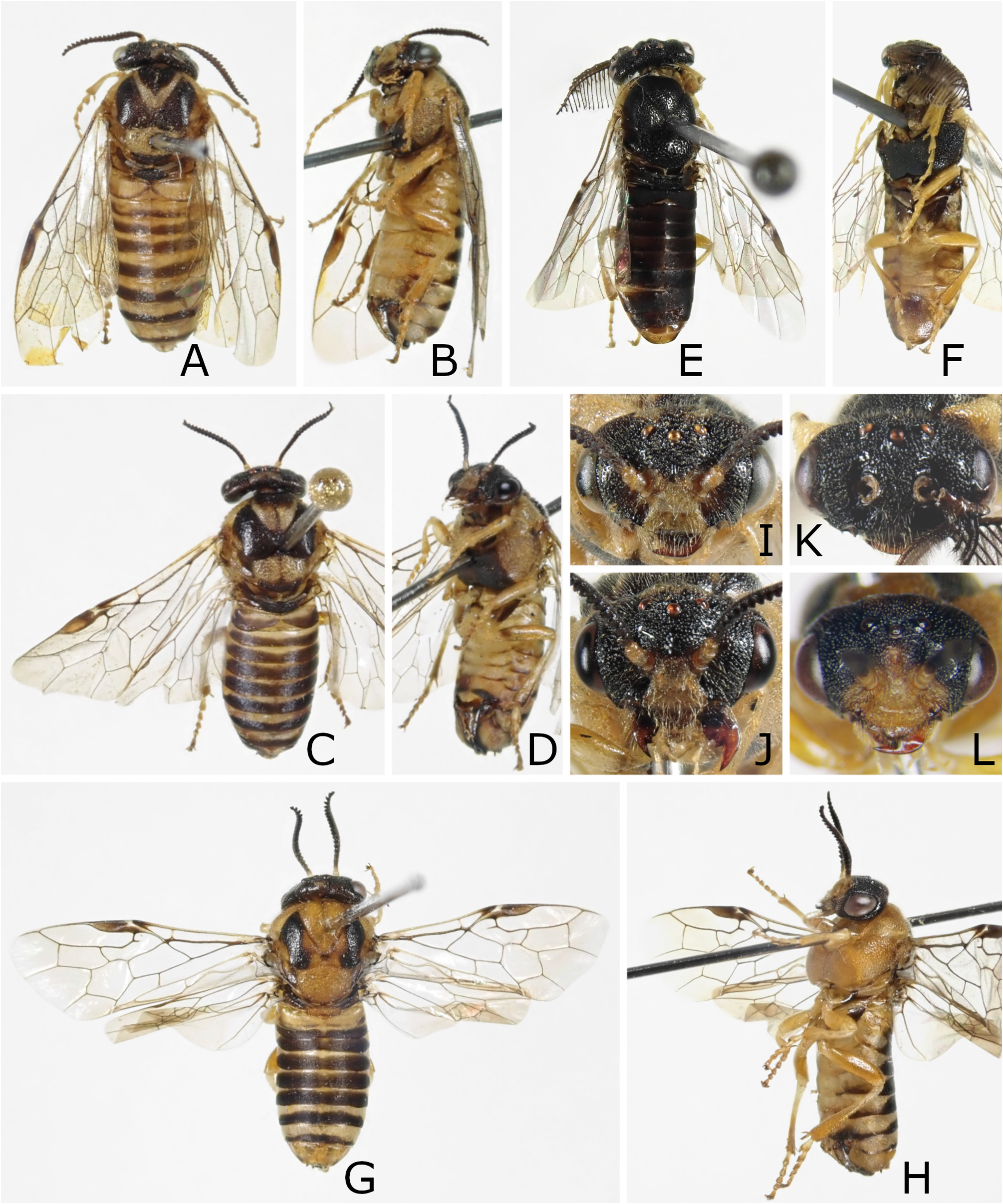
FIGURES 2A–H. A–F, I–K, Gilpinia amamiana: A, B, holotype, female; C, D, female, Tokunoshima Isl.; E, F, male, Amami Oshima Isl. G, H, L, Gilpinia okinawa, holotype, female. A, C, E, G, Dorsal view; B, D, F, H, ventrolateral view; I–L, head, frontal view.
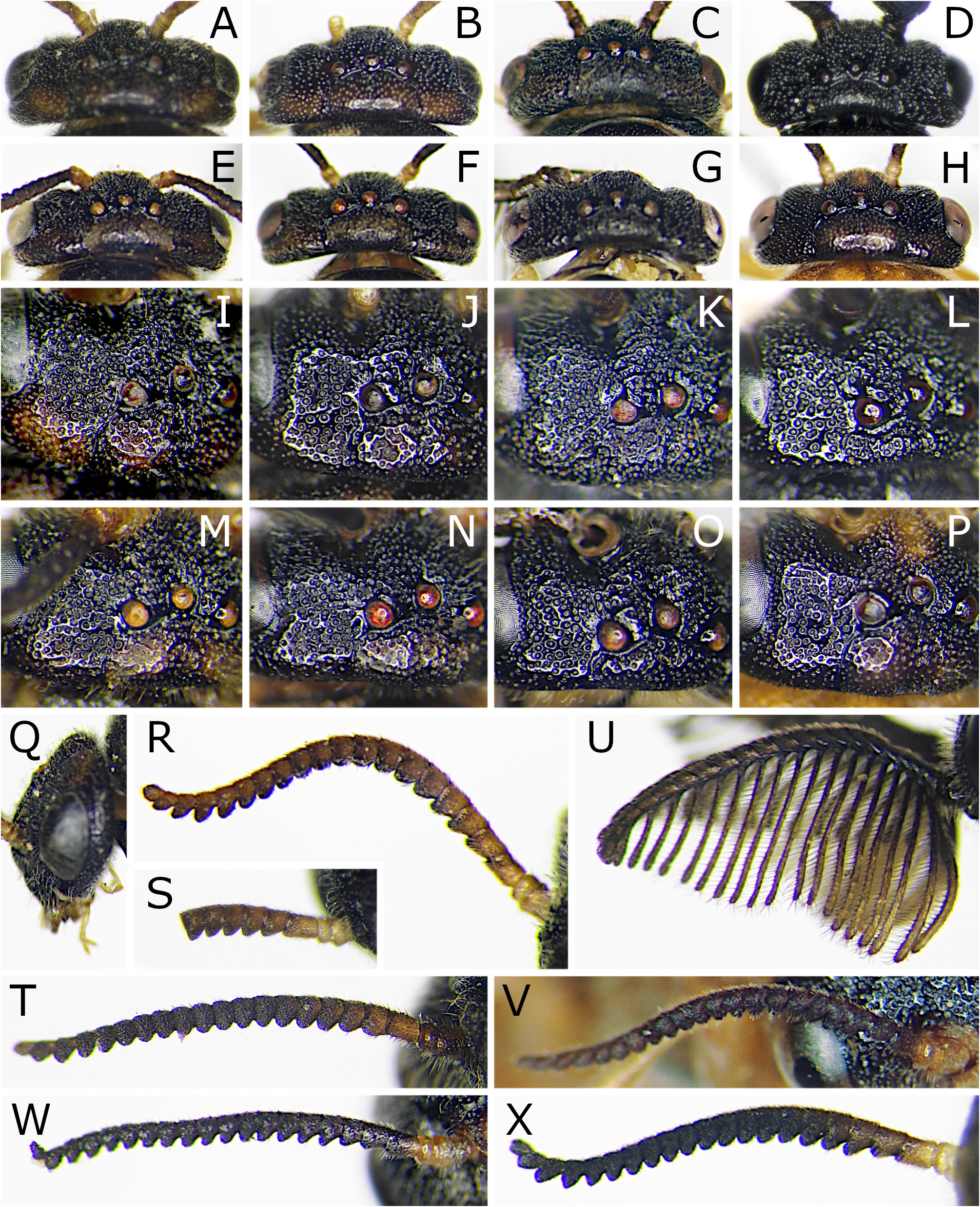
FIGURES 3A–X. A–H, Head, dorsal view; I–P, dorsal part of head, anterolateral view; Q, head, lateral view; R–X, antenna, inner view (R–T, V, W) or outer view (U, X). A–D, I–L, Q–U, Gilpinia hakonensis: A, I, Q, R, lectotype of Lophyrus hakonensis, female; B, J, S, holotype of G. hakonensis var. laticincta, female; C, K, T, paratype of Diprion fukudai, female; D, L, U, male, Honshu. E–G, M–O, V, W, Gilpinia amamiana: E, M, V, holotype, female; F, N, W, female, Tokunoshima Isl.; G, O, male, Amami Oshima Isl. H, P, X, Gilpinia okinawa, holotype, female. T, Reversed.
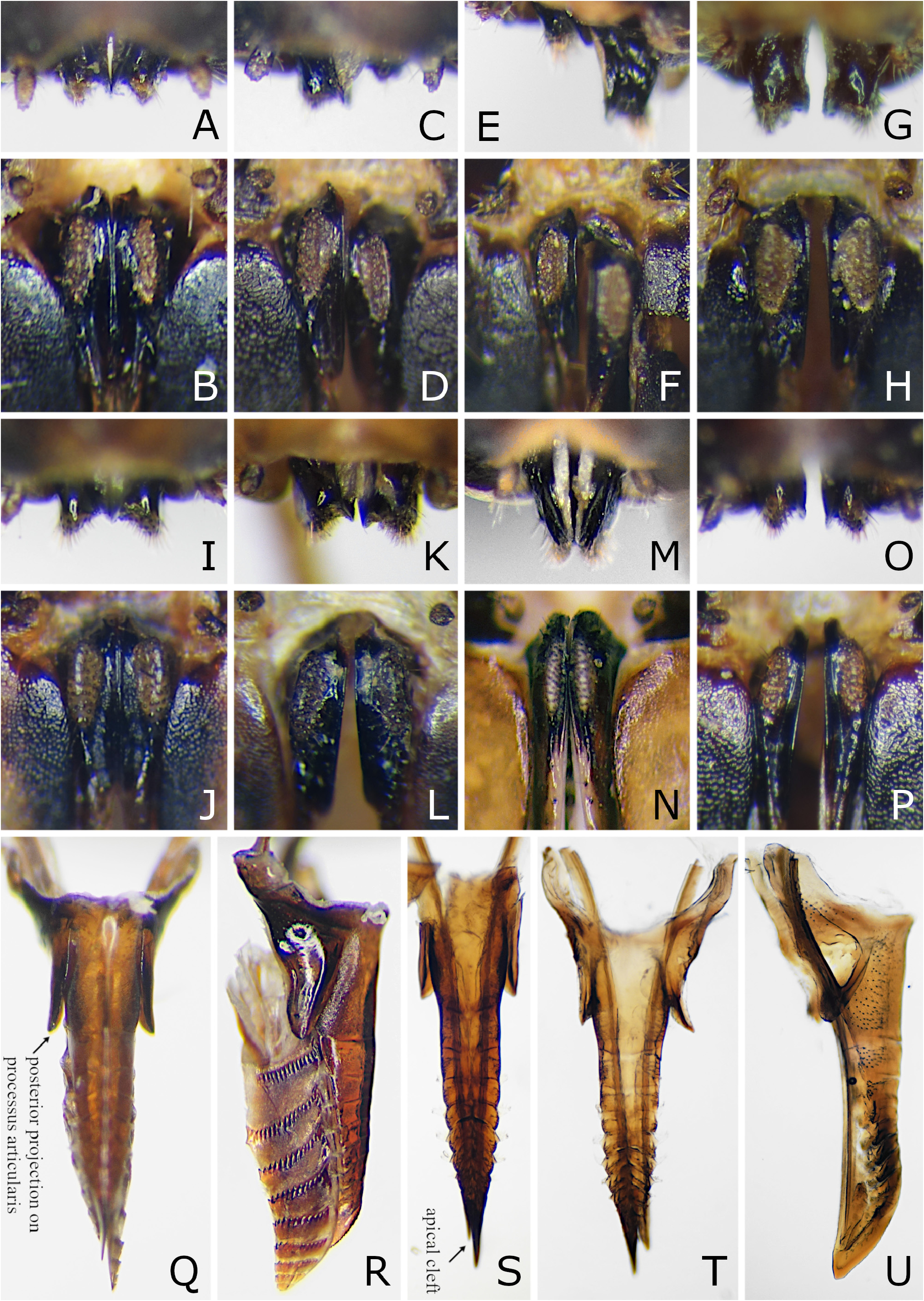
FIGURES 5A–U.A–P, Valvula 3, dorsal and posterior views; Q–U, ovipositor or lance, dorsal view (Q, S, T) or lateral view (R, U). A–F, Q, R, Gilpinia hakonensis: A–B, lectotype of Lophyrus hakonensis; C, D, holotype of G. hakonensis var. laticincta; E, F, P, paratype of Diprion fukudai; Q, R, Honshu. G–J, S, Gilpinia amamiana: G, H, holotype; I, J, Tokunoshima Isl.; S, Amami Oshima Isl. K, L, T, U, Gilpinia okinawa, holotype. M, N, Gilpinia frutetorum, Sweden. O, P, Gilpinia laricis, Germany. R, Reversed.
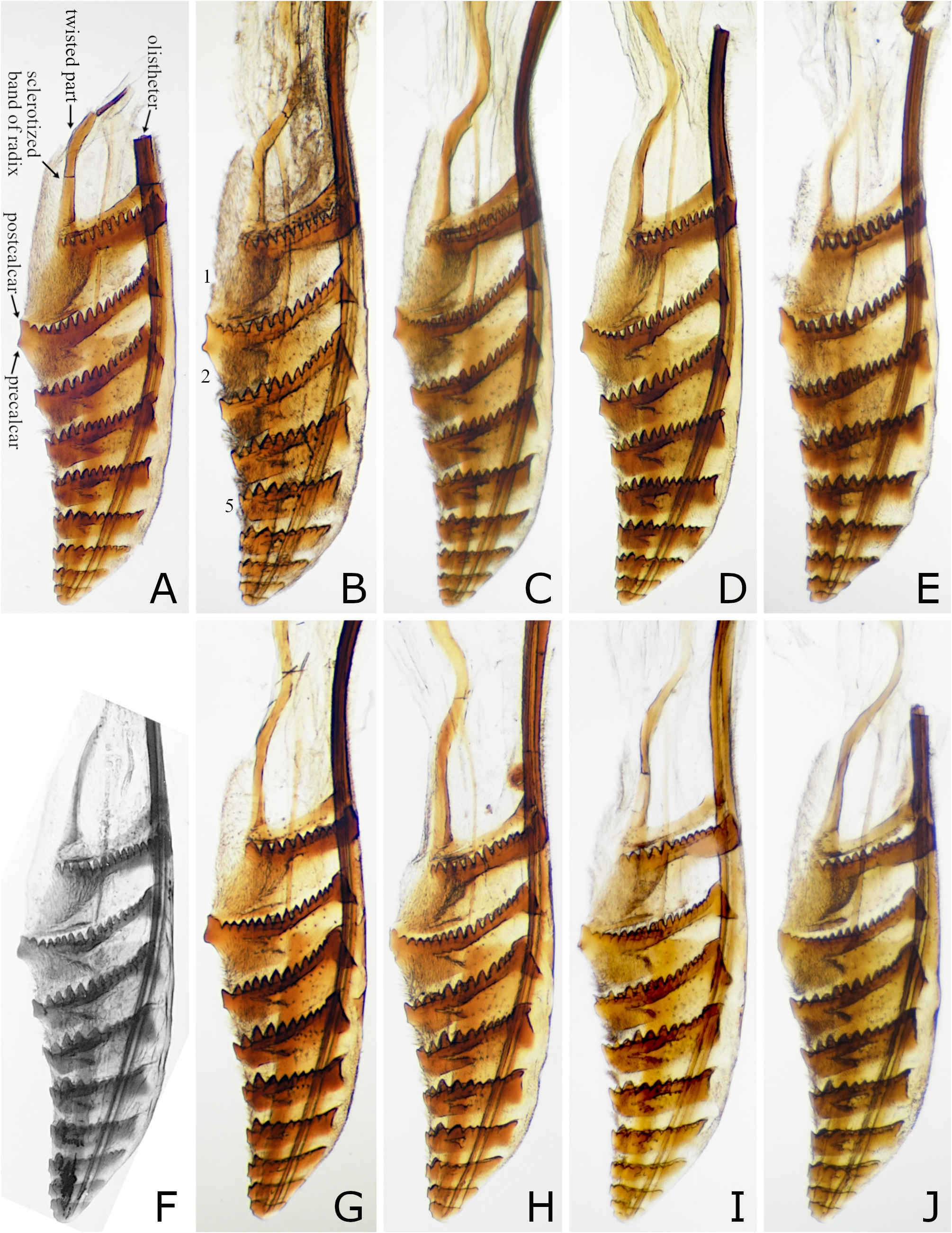
FIGURES 6A–J. Lancet. A–F, Gilpinia hakonensis: A, lectotype of Lophyrus hakonensis; B, holotype of G. hakonensis var. laticincta; C, Kyushu, D, Honshu, E, paratype of Diprion fukudai. F–H, Gilpinia amamiana: F, holotype; G, Amami Oshima Isl.; H, Tokunoshima Isl. I, J, Gilpinia okinawa: I, holotype; J, Okinawa Isl. 1, 2, 5, first (most basal), second and fifth annuli. D–F, H, Reversed.
| VI |
Mykotektet, National Veterinary Institute |
No known copyright restrictions apply. See Agosti, D., Egloff, W., 2009. Taxonomic information exchange and copyright: the Plazi approach. BMC Research Notes 2009, 2:53 for further explanation.
|
Kingdom |
|
|
Phylum |
|
|
Class |
|
|
Order |
|
|
Family |
|
|
Genus |
Gilpinia amamiana Okutani, 1965
| Hara, Hideho, Smith, David R. & Shinohara, Akihiko 2021 |
Gilpinia amamiana
| Hara, H. 2020: 291 |
| Hara, H. 2019: 42 |
| Yoshida, H. 2010: 28 |
| Azuma, S. & Yafuso, M. & Kinjo, M. & Hayashi, M. & Kohama, T. & Sasaki, T. & Kimura, M. & Kawamura, F. 2002: 301 |
| Abe, M. & Togashi, I. 1989: 545 |
| Okutani, T. 1984: 26 |
| Okutani, T. 1965: 76 |