
Prodidactidae
|
publication ID |
https://doi.org/ 10.5281/zenodo.156596 |
|
DOI |
https://doi.org/10.5281/zenodo.5628427 |
|
persistent identifier |
https://treatment.plazi.org/id/03F18792-FFE3-FFD5-EF79-FAC2FC8CF97F |
|
treatment provided by |
Plazi |
|
scientific name |
Prodidactidae |
| status |
|
Prodidactidae View in CoL , New Family
The following description is based on the single species, Prodidactis mystica (Meyrick) and is intended also to function as a redescription of the genus and species. Hence, it includes a combination of familial, generic, and specieslevel characters. The description includes the states for each of the 24 key characters listed in Heppner’s (1998: Table 2) “Family Characters States.” The importance of certain familylevel characters are detailed in the discussion following the description; a comparison of these among the taxa discussed is outlined in Table 1 View TABLE 1 . Choice of characters and definition of character states are based on Epstein (1996), Heppner (1998), and Kristensen (1999). Of the character states described below, the extremely reduced labial palpus and the elongate membranous lobe of the male hindcoxa appear to be of greatest significance, and both are considered autapomorphies for the family.
Adult. Head ( Figs. 1-2 View FIGURES 1 - 4 , 5-7 View FIGURES 5 - 6 View FIGURES 7 8 ): Smooth scaled or only slightly roughened, frons red; vertex pale gray; ocellus absent; chaetosema absent; maxillary palpus well developed, 3-segmented, covered and tufted with large pale scales; labial palpus greatly reduced, 3- segmented, with crimson-red scales; haustellum well developed, unscaled. Area dorsad of haustellum base with a vertical row of pale scales. Antenna filiform, finely ciliated, not differentiated between sexes; antennal pecten absent.
Thorax: Whitish gray ( Fig. 6 View FIGURES 5 - 6 ). Epiphysis well developed in both sexes. Distinct hairpencils on posterior aspect of hind coxae of male associated with free, elongate, membranous, saclike lobes that are asymmetrical ( Fig. 8 View FIGURES 7 8 ); hindtibia with 4 spurs. Pulvillus of pretarsus with long setiform outgrowths on dorsal lobe ( Fig. 21 View FIGURES 15 21 ). Forewing ( Figs. 12 View FIGURES 1 - 4 , 9 View FIGURES 9 11 ) moderately broad; costa weakly arched throughout; apex slightly falcate. Length 1728 mm (females larger). Upper surface buff to yellow buff, faintly rosy tinged in some specimens; anterior 0.66 of costal region suffused whitish, with a few scattered black specks, especially towards dorsal and terminal areas; costa crimson at base; an elongate orange spot near middle of costa; variably developed antemedial, medial, and postmedial zigzag fasciae of pale orange. No costal fold, upraised scales, or other male secondary structures. Length of discal cell approximately 0.55 forewing length, closed; all veins present, free and separate basally; discal cell between M1 and M2 weak, angled inward; Mstem present, weak, ending between M1 and M2; accessory cell present at distal end of discal cell; CuP weak; 1A+2A forked at base; pterostigma absent. Hindwing uniform pale yellow or pale orange yellow. All veins present, separate; discal cell approximately 0.65 length of hindwing, closed; discal cell between M1 and M2 weak; CuP well developed; tuft of long scales arising near base of CuP; pterostigma absent. Wing coupling frenulate, 1 spine in male, 3 in female. Under surface pale yellow.
Abdomen: Apodemes of tortricioidtype ( Fig. 8 View FIGURES 7 8 ); lacking tympanum. Pale orange buff. Male genitalia ( Fig. 10 View FIGURES 9 11 ) with uncus stout, as long as width of tergite, attenuate distally, tapering to a rounded point, densely covered with stout hairs directed anteriorly; attached to tegumen at a partially membranous point of articulation. Gnathos arms moderately broad, united into a single narrow, mesal process, strongly upcurved in distal 0.33, ending in a footshaped sclerite with pointed toe. Socius apparently absent. Valva long, with broad basal portion, slender central portion, and broad apical region; costa strongly sclerotized; inner surface densely covered with long hairs. Aedeagus stout, bent near middle, slightly undulate; attenuate distally with short spine on left side near apex; cornuti absent. Female genitalia ( Fig. 11 View FIGURES 9 11 ) with papillae anales broad, rounded ventrally, moderately sclerotized, densely covered with long hairs; not fused dorsally. Apophyses moderately long, slender. Sterigma a simple plate, ostium bursae simple. Ductus bursae nearly as long as abdomen; corpus bursae small, rounded; signum absent.
Last Instar Larva (n = 3) ( Figs. 34 View FIGURES 1 - 4 , 14 View FIGURES 12 14 , 1719 View FIGURES 15 21 ). General: Length 2226 mm, rather stout. Head red brown with no conspicuous markings. Prothoracic shield dark greenish brown with dorsal separation of halves indicated by a yellow line. Body greenish yellow, with a narrow dark green longitudinal middorsal line and a broad, dark green longitudinal subdorsal line extending ventrad to dorsal edge of spiracles; pinacula conspicuous, yellow; nonsetal bearing pinacula on meso and metathorax red brown; secondary setae absent. Head: Frons extending about 0.7 to occipital foramen; stemmata arranged in an arc except for S5 ( Fig. 19 View FIGURES 15 21 ); spinneret moderate in length, rounded distally ( Figs. 1718 View FIGURES 15 21 ); mandible with three large teeth, one smaller tooth, and a flattened region at inner portion ( Fig. 18 View FIGURES 15 21 ). Thorax ( Fig. 14 View FIGURES 12 14 ): Prothorax with bisetose Lgroup on common prespiracular pinaculum; SD2 seta approximately 0.4 times length of SD1; spiracle on prothorax surrounded by large, clear disk. SVgroup bisetose on prothorax, unisetose on meso and metathorax. Meso and metathorax each with nonsetabearing dorsal pinacula; mesothorax with three naked pinacula, two slightly dorsad of D pinaculum, one on each side, and a third anterad and slightly ventrad of SD pinaculum; metathorax similar, with hint of an additional naked pinaculum anterad and ventrad of first anterior pinaculum. Abdomen ( Fig. 14 View FIGURES 12 14 ): Spiracles surrounded by inconspicuous unsclerotized disk; spiracle on A8 slightly larger than those on A17; D2s on separate pinacula on A9; SD1 well dorsad and slightly anterad of spiracle on A17, conspicuously more anterad on A8; L1 and L2 together, in horizontal row, on large common lateral pinaculum below spiracle on A18; L1+L2 pinaculum on A9 nearly vertical; L3 absent on A9; SV group on 1,2,36,7,8,9 usually 2:3:3:2:2:1; anal fork absent. Crochets 7075 in uniform biordinal circle on A36; crochets 5055 in a semicircle on A10.
Pupa. General ( Figs. 1213 View FIGURES 12 14 , 1516 View FIGURES 15 21 ): Short and broad, compact, zygaeniform. Compound eye unsculptured; haustellum elongate, extending to distal end of front legs; maxillary palpi extending to near end of antennae; labial palpus a small triangle. Abdomen unmodified, without rows of spines on dorsum; spiracle not visible on A1; cremaster absent.
Cocoon. General ( Fig. 20 View FIGURES 15 21 ): Somewhat smooth on one surface and rough on opposite side; oval and flattened, 1.8 cm x 1.0 cm; opening along edge of narrow end, ca. 0.33 length of cocoon.
Life history. Eggs are flat and scalelike (N. Duke, pers. comm.), similar to those of many Tortricidae ( Horak 1999) and Limacodidae ( Epstein 1996) , and are laid on the leaves of the host. Behavior of the early instars was not recorded. Fourth and fifth instars are solitary, apparently feeding from the upper surface on leaves of Nuxia congesta , as supported by photographic evidence ( Figs. 34 View FIGURES 1 - 4 ). The larvae form a shelter in a rolled leaf, but spend more time feeding exposed on the leaf than do tortricids (N. Duke, pers. comm.). Pupation occurs on the host in a dense, tough cocoon spun on or between leaves forming a shelter; overwintering is apparently accomplished in this stage. The pupa does not protrude from the cocoon prior to eclosion. Adults have been collected from October through March ( Janse 1964: 22).
Geographic distribution. According to Janse (1964: 22), P. m y s t i c a is known from Durban Nggeleni (Pondoland), Nkandhla Forest (Zululand), Noordkaap, Barberton, Marieps Mountain, Graskop, Kowyn's Pass, Pretoria, Woodbush, Malta, and Zoutpansberg (all Transvaal). Specimens in the Transvaal Museum (M. Krüger, pers. comm.) provide the following distribution data: SOUTH AFRICA: TRANSVAAL: The Downs, Entabeni Forest, Kownyn's Pass, Pilgrim's Rest District, Uitsoek, Louis Trichardt, Piesangkop NE, Louis Trichardt Mariepskop Forestry, Malta Forest, Woodbush, Barbeton, De Hoek Forestry, Timbadiola Forestry, Pretoria North. NATAL: Ngoya Forest, Mtunzini District, Durban, Nkhandla Forest. EASTERN CAPE: Transkei, the Haven, Nggeleni. SWAZILAND: Mbabane. ZIMBABWE: Vumba Mountains, Lundi.
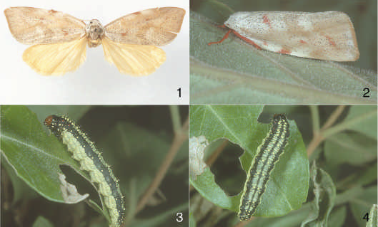

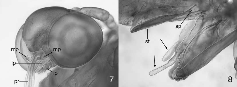
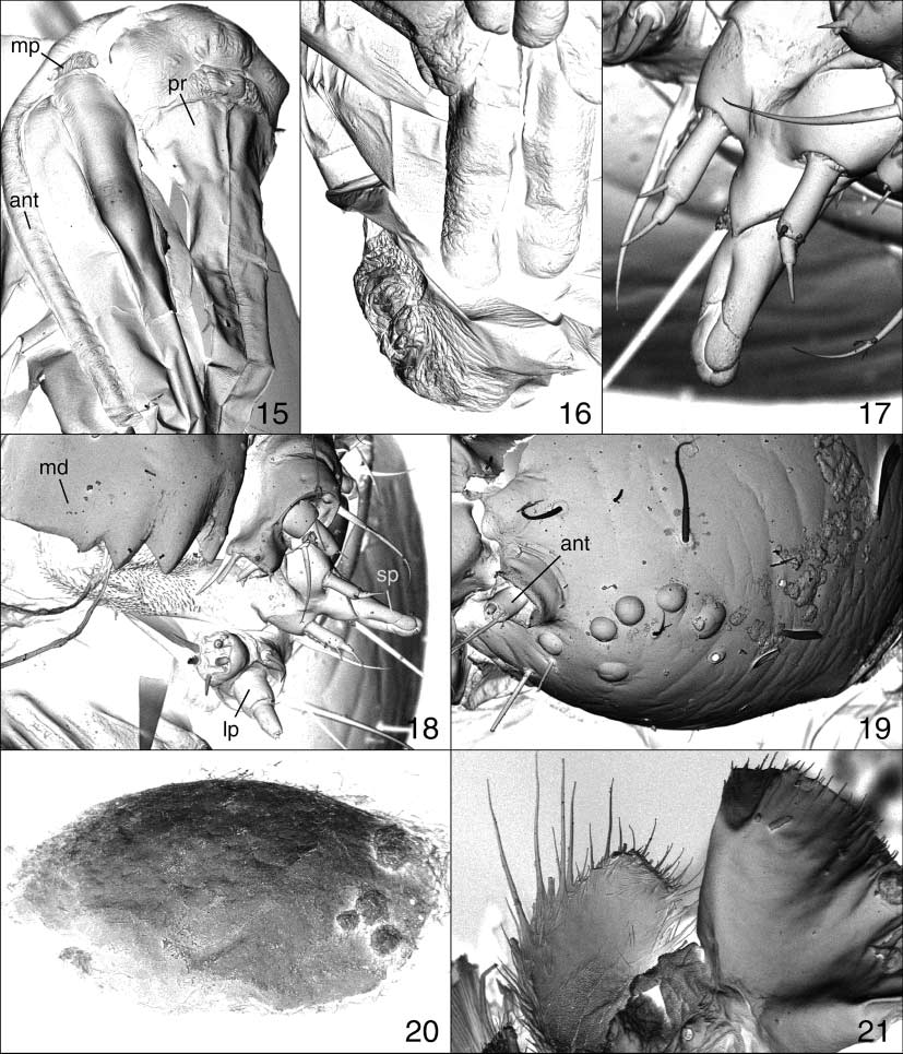
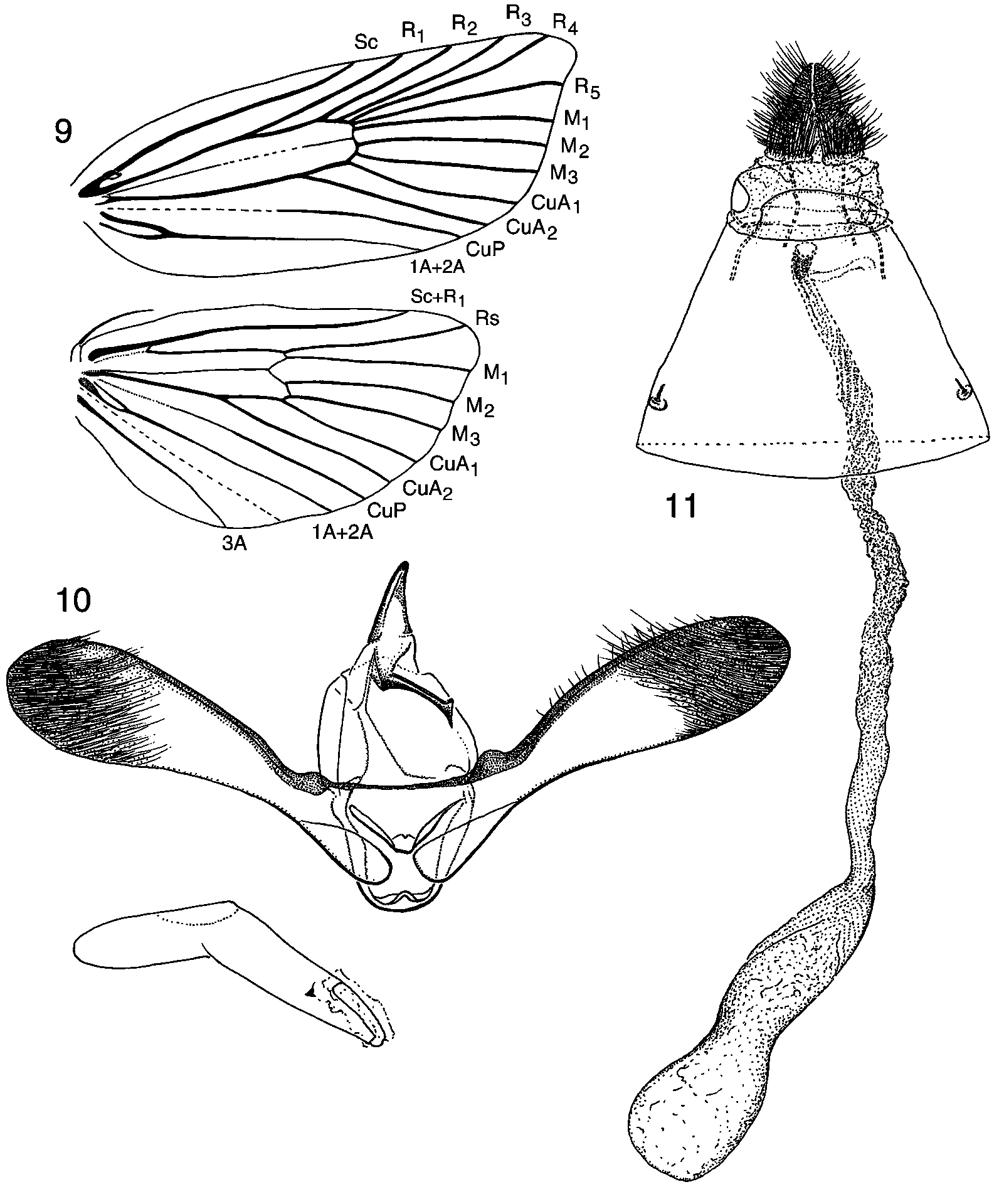
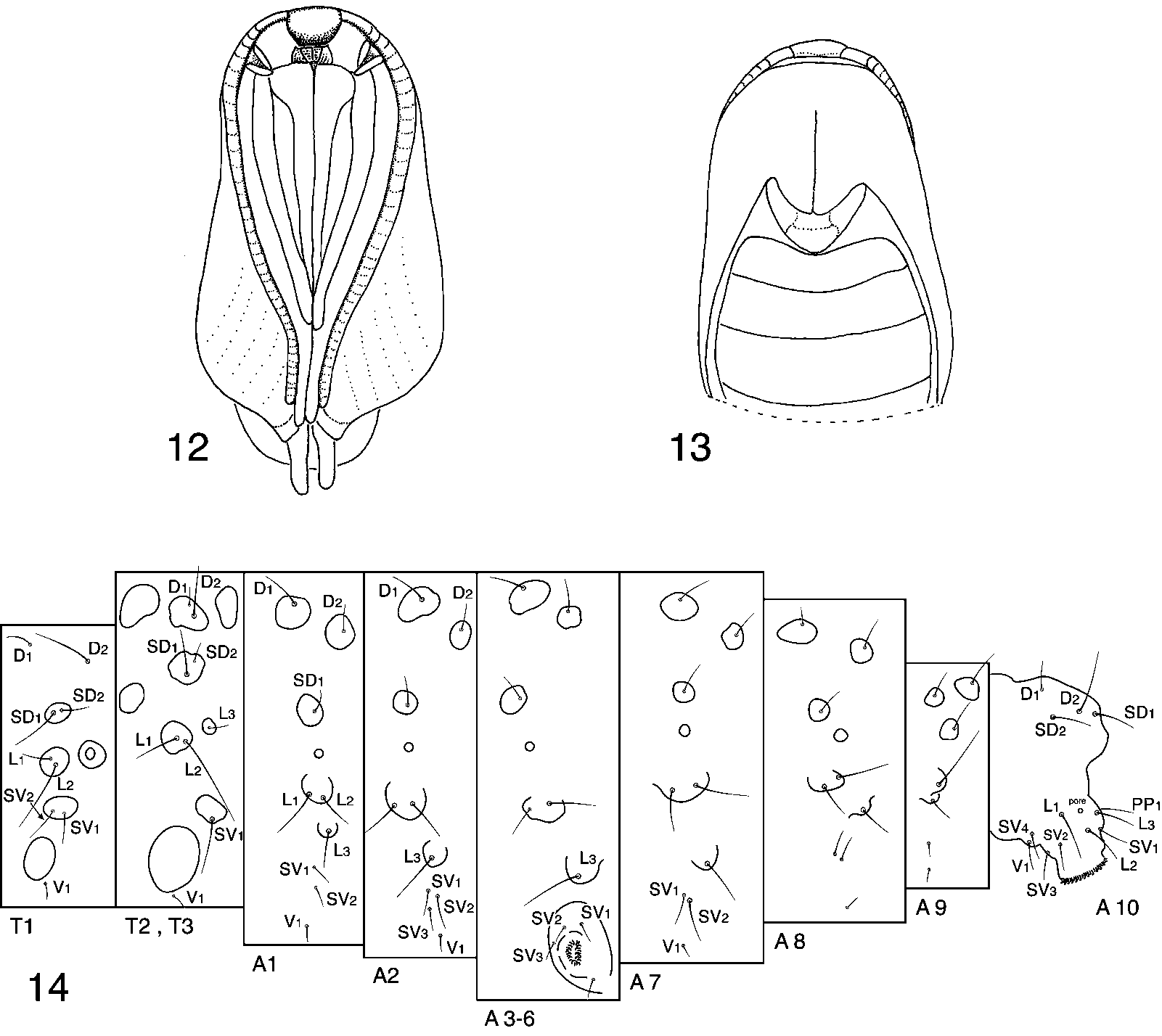
TABLE 1. Comparison of character states of egg, larva, pupa, and adult among families. [Where two states occur, the more common is listed first. * Differs from Heppner (1998)]
| Yponomeutidae | Tortricidae | Zygaenidae | Limacodidae | Immidae | Crambidae | Prodidactidae | |
|---|---|---|---|---|---|---|---|
| EGG | |||||||
| egg type | upright | flat | flat | flat | upright | upright | flat |
No known copyright restrictions apply. See Agosti, D., Egloff, W., 2009. Taxonomic information exchange and copyright: the Plazi approach. BMC Research Notes 2009, 2:53 for further explanation.