
Euconnus hirticollis (Illiger)
|
publication ID |
https://doi.org/ 10.5281/zenodo.282872 |
|
DOI |
https://doi.org/10.5281/zenodo.6166875 |
|
persistent identifier |
https://treatment.plazi.org/id/03F187FC-9246-914E-9BA1-BFF34CD3D4FF |
|
treatment provided by |
Plazi |
|
scientific name |
Euconnus hirticollis (Illiger) |
| status |
|
Morphology of Euconnus hirticollis (Illiger) , the type species of Euconnus Thomson
General body shape ( Figs. 1–2 View FIGURES 1 – 2 ) elongate, moderately slender; body distinctly constricted between head and pronotum but weakly between pronotum and elytra, strongly convex; appendages long and slender, vestiture distinct and not uniform, i.e., various body parts distinctly differ in thickness, length and density of setae.
Head capsule ( Figs. 3–6 View FIGURES 3 – 6 ) divided by occipital constriction ( Figs. 3–4 View FIGURES 3 – 6 ; occ) into large anterior and small posterior part ('neck region'), the posterior part retracted into pronotum. Neck region ( Figs. 3–4 View FIGURES 3 – 6 ; nr) broadening towards foramen occipitale ( Fig. 4 View FIGURES 3 – 6 ; fo); narrowest site of occipital constriction about as wide as half HW. Anterior part of head ( Figs. 3–4 View FIGURES 3 – 6 ) nearly hemispherical. Dorsum of head ( Fig. 3 View FIGURES 3 – 6 ) strongly convex; tempora ( Fig. 3 View FIGURES 3 – 6 ; tm) in dorsal view slightly longer than compound eyes ( Fig. 3 View FIGURES 3 – 6 ; ce); vertex ( Fig. 3 View FIGURES 3 – 6 ; vt) transverse and nearly evenly convex, anteriorly confluent with subrapezoidal and convex frons ( Fig. 3 View FIGURES 3 – 6 ; fr), which is steeply declining anteriorly; supraantennal tubercles feebly developed; genae ( Figs. 3–4, 6 View FIGURES 3 – 6 ; gen) elongate, convex. Clypeus ( Fig. 5 View FIGURES 3 – 6 ; cp) separated from frons and genae by distinct transverse fronto-clypeal groove ( Fig. 5 View FIGURES 3 – 6 ; fcg). Vestiture of head dorsum sparse and composed of long suberect to erect setae, with loose groups on tempora and genae, but without thick bristles. Ventral side of head ( Fig. 6 View FIGURES 3 – 6 ) flattened; gular plate ( Fig. 6 View FIGURES 3 – 6 ; gp) in posterior part with indistinct gular sutures ( Fig. 6 View FIGURES 3 – 6 ; gs), in anterior part with lateral ridges, gular plate indistinctly separated from submentum ( Fig. 6 View FIGURES 3 – 6 ; smn); posterior tentorial pits ( Fig. 6 View FIGURES 3 – 6 ; ptp) elongate and located on anterior part of gular plate.
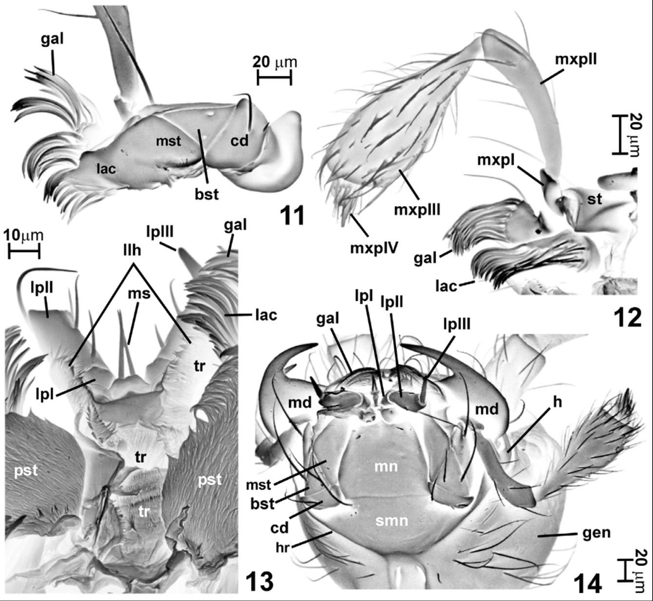
Mouthparts ( Figs. 6–14 View FIGURES 3 – 6 View FIGURES 7 – 10 View FIGURES 11 – 14 ). Labrum ( Fig. 7 View FIGURES 7 – 10 ; lb) transverse, with rounded sides and nearly straight anterior margin, dorsal surface with three rows of long setae. Epipharynx (ventral surface of labrum) ( Fig. 8 View FIGURES 7 – 10 ; eph) with six curved peg-like median marginal sensilla ( Fig. 8 View FIGURES 7 – 10 ; mse) directed anterad and dense lateral groups of trichia ( Fig. 8 View FIGURES 7 – 10 ; lte) directed mesad. Mandibles ( Figs. 7, 9–10 View FIGURES 7 – 10 ; md) symmetrical, moderately large, nearly planar, subtriangular with broad base narrowing distally into slender and strongly curved apical tooth ( Fig. 7 View FIGURES 7 – 10 ; at), with small and sharp subapical mesal tooth ( Fig. 7 View FIGURES 7 – 10 ; sat); prostheca ( Figs. 7, 9–10 View FIGURES 7 – 10 ; pst) composed of numerous dense and short trichia expanded onto dorsal surface of mandible and occupying nearly half of mandibular base. Maxillae ( Figs. 11–12, 14 View FIGURES 11 – 14 ) elongate, with oblique cardo ( Fig. 11 View FIGURES 11 – 14 ; cd) bearing long submedian and short basal seta; basistipes ( Fig. 11 View FIGURES 11 – 14 ; bst) subtriangular, with two long subbasal setae; mediostipes ( Fig. 11 View FIGURES 11 – 14 ; mst) elongate and connected to lacinia; galea ( Figs. 11–12 View FIGURES 11 – 14 ; gal) short, with long and dense trichia along distal margin and long seta on external margin; lacinia ( Figs. 11–12 View FIGURES 11 – 14 ; lac) elongate, with long and dense trichia along distal and mesal margin; maxillary palps ( Fig. 12 View FIGURES 11 – 14 ) long, palpomere I ( Fig. 12 View FIGURES 11 – 14 ; mxpI) small, about 2.5× as long as broad, palpomere II ( Fig. 12 View FIGURES 11 – 14 ; mxpII) strongly elongate and slightly thickening distally, with several long setae, palpomere III ( Fig. 12 View FIGURES 11 – 14 ; mxpIII) longer than I and strongly broadening distally, broadest near distal third, covered with sparse and long setae; palpomere IV ( Fig. 12 View FIGURES 11 – 14 ; mxpIV) minute, subconical, with elongate and pointed apical part, with dense and moderately long setae near base and asetose in distal half. Labium ( Figs. 13–14 View FIGURES 11 – 14 ) with transverse submentum ( Fig. 14 View FIGURES 11 – 14 ; smn) laterally fused to hypostoma ( Fig. 14 View FIGURES 11 – 14 ; h) and bearing a pair of long latero-anterior setae; mentum ( Fig. 14 View FIGURES 11 – 14 ; mn) subtrapezoidal with two pairs of short latero-anterior setae; prementum short, with a pair of moderately long median setae ( Fig. 13 View FIGURES 11 – 14 ; ms); labial palps shorter than mentum, palpomere I ( Fig. 14 View FIGURES 11 – 14 ; lpI) only slightly longer than wide, asetose, palpomere II ( Fig. 14 View FIGURES 11 – 14 ; lpII) strongly elongate and slightly broadening distally, with several submedian and subapical setae, palpomere III ( Fig. 14 View FIGURES 11 – 14 ; lpIII) long and slender, asetose. Hypopharynx (dorsal surface of labium) ( Fig. 13 View FIGURES 11 – 14 ) with elongate lateral lobes ( Fig. 13 View FIGURES 11 – 14 ; llh) densely covered with trichia ( Fig. 13 View FIGURES 11 – 14 ; tr) and exceeding length of prementum, median part covered densely with trichia. Posteriorly and laterally mouthparts demarcated by hypostomal ridges ( Fig. 14 View FIGURES 11 – 14 ; hr) strongly convergent caudad; hypostoma ( Fig. 14 View FIGURES 11 – 14 ; h) anteriorly narrow and elongate, posteriorly fused with submentum.
Antennae ( Figs. 1–2 View FIGURES 1 – 2 ) slender, gradually thickening towards apices, antennomeres compactly assembled in proximal part and loosely in distal part, sparsely covered with suberect to erect setae.
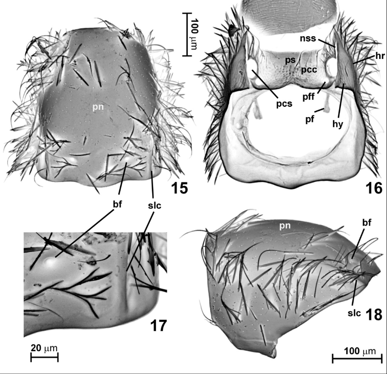
Prothorax ( Figs. 15–18 View FIGURES 15 – 18 ) strongly convex, in dorsal view ( Fig. 15 View FIGURES 15 – 18 ) elongate subtrapezoidal, with rounded anterior and lateral margins and posterior margin distinctly expanded caudad in middle, without lateral carinae or sharp edges but with short and rounded sublateral carina ( Figs. 15, 17–18 View FIGURES 15 – 18 ; slc); anterior and posterior corners of pronotum blunt but well-defined. Pronotum ( Figs. 15, 17 View FIGURES 15 – 18 ; pn) with two small and shallow sublateral basal foveae ( Figs. 15, 17–18 View FIGURES 15 – 18 ; bf), entire surface covered with long and erect bristles, much thicker than those on remaining body parts except sides of mesothorax. Prosternum ( Fig. 16 View FIGURES 15 – 18 ; ps) about 3 times shorter than pronotum, with basisternal part short and not delimited from procoxal cavities ( Fig. 16 View FIGURES 15 – 18 ; pcc); prosternal intercoxal process or carina absent, procoxae contiguous; procoxal sockets ( Fig. 16 View FIGURES 15 – 18 ; pcs) closed by lateral expansions of prosternum; profurcal (postcoxal) foveae ( Fig. 16 View FIGURES 15 – 18 ; pff) relatively large. Hypomera ( Fig. 16 View FIGURES 15 – 18 ; hy) subtriangular, elongate, not expanded mesally, with nearly straight internal (mesal) margins, each delimited from pronotum on entire length by hypomeral ridge ( Fig. 16 View FIGURES 15 – 18 ; hr) and from sternum by straight and entire notosternal suture ( Fig. 16 View FIGURES 15 – 18 ; nss); profurcal arms ( Fig. 16 View FIGURES 15 – 18 ; pf) tubular, with expanded apices.
Mesothorax ( Figs. 19–27 View FIGURES 19 – 24 View FIGURES 25 – 27 ). Mesonotum ( Fig. 19 View FIGURES 19 – 24 ) subtriangular in shape; mesoscutum ( Fig. 19 View FIGURES 19 – 24 ; sc II) strongly transverse, asetose; scuto-scutellar suture ( Fig. 19 View FIGURES 19 – 24 ; sss) barely marked on the surface; mesoscutellum ( Fig. 19 View FIGURES 19 – 24 ; scl II) well visible between bases of elytra in intact specimens, triangular with rounded posterior margin, covered with scale-like sculpture and bearing lateral groups of five thick setae (partly broken off in specimen shown in Fig. 19 View FIGURES 19 – 24 ; ls); posterior and anterior notal processes ( Fig. 19 View FIGURES 19 – 24 ; pnp, anp) prominent.
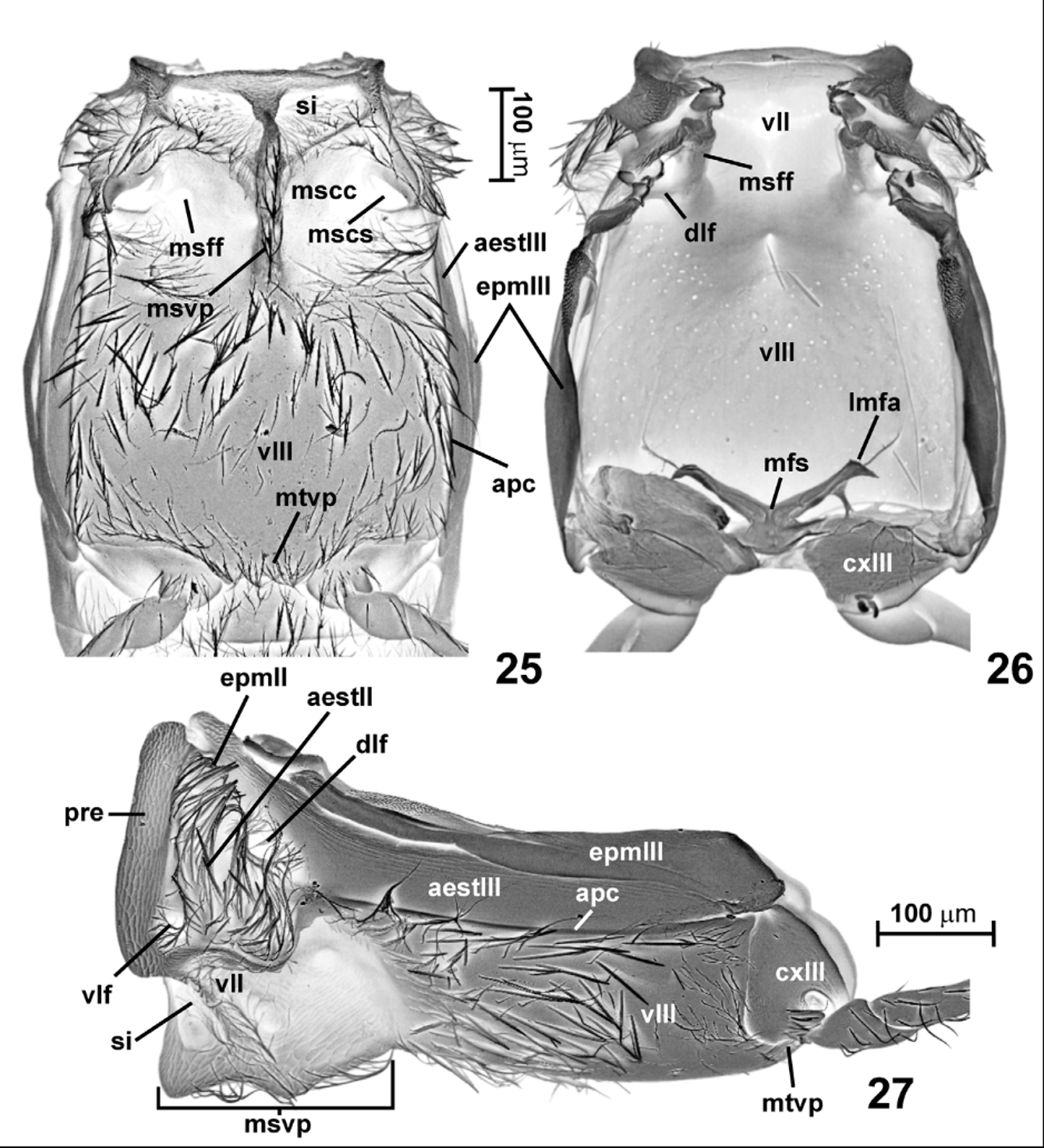
Mesoventrite ( Figs. 20–22 View FIGURES 19 – 24 , 25, 27 View FIGURES 25 – 27 ) relatively short, much broader than long, with broad anterior ridge ( Fig. 21 View FIGURES 19 – 24 ; ar) bearing large and subtriangular median projection ( Fig. 21 View FIGURES 19 – 24 ; par) connected with long, slender and keel-like mesoventral intercoxal process ( Fig. 21 View FIGURES 19 – 24 ; msvp) strongly projecting ventrad ( Figs. 20 View FIGURES 19 – 24 , 27 View FIGURES 25 – 27 ); mesoventrite behind anterior ridge with lateral pair of subtriangular setose impressions ( Fig. 21 View FIGURES 19 – 24 ; se) separated from mesocoxal cavities ( Fig. 21 View FIGURES 19 – 24 ; mscc) by oblique ridge ( Fig. 21 View FIGURES 19 – 24 ; or); mesocoxal sockets ( Fig. 21 View FIGURES 19 – 24 ; mscs) located laterally; mesofurcal foveae ( Figs. 21-22 View FIGURES 19 – 24 ; msff) large and located submedially on lateral parts of mesocoxal cavities, which are largely asetose. Prepectus ( Fig. 21 View FIGURES 19 – 24 ; pre) relatively long, posterior part of mesanepisternum ( Fig.21 View FIGURES 19 – 24 ; aestII) partly visible in ventral view, densely covered with thick and suberect to erect bristles; latero-ventral margins of mesepimera ( Fig. 27 View FIGURES 25 – 27 ; epmII) obscured by dense bristles, possibly mesepimera fused (partly or entirely) with large mesanepisterna ( Fig. 27 View FIGURES 25 – 27 ; aestII) and entirely fused with metanepisterna ( Fig. 27 View FIGURES 25 – 27 ; aestIII). Mesothorax with two pairs of deep lateral foveae ( Figs. 22 View FIGURES 19 – 24 , 26-27 View FIGURES 25 – 27 ): ventro-lateral fovea ( Figs. 22 View FIGURES 19 – 24 , 27 View FIGURES 25 – 27 ; vlf) located latero-ventrally between prepectus and setose part of mesanepisternum and dorso-lateral fovea ( Figs. 22 View FIGURES 19 – 24 , 27 View FIGURES 25 – 27 ; dlf) located latero-dorsally between mesepimeron + mesanepisternum and metanepisternum (aestIII); internally ( Fig. 26 View FIGURES 25 – 27 ) walls of dorso-lateral fovea ( Fig. 26 View FIGURES 25 – 27 ; dlf) forming a tubular structure connected to basal part of thick mesofurcal arm ( Fig. 26 View FIGURES 25 – 27 ; msff) and together constituting latero-ventral internal scaffold of mesothorax.
Metathorax ( Figs. 23–27 View FIGURES 19 – 24 View FIGURES 25 – 27 ). Premetascutum ( Figs. 23–24 View FIGURES 19 – 24 ; prsc III) large, transverse; metascutum ( Figs. 23–24 View FIGURES 19 – 24 ; sc III) very large and transverse, with robust oblique lateral apodemes ( Fig. 24 View FIGURES 19 – 24 ; ap) on ventral side, in middle with broad and impressed median membranous area ( Figs. 23–24 View FIGURES 19 – 24 ; mma); alacristae ( Figs. 23–24 View FIGURES 19 – 24 ; alc) nearly as long as scutum; metascutellum ( Fig. 23 View FIGURES 19 – 24 ; scl III) rudimentary, metascuto-scutellar suture indiscernible; metapostnotum ( Figs. 23–24 View FIGURES 19 – 24 ; psn III) large, strongly transverse.
Metaventrite ( Figs. 25–27 View FIGURES 25 – 27 ; vIII) much longer than mesoventrite, subquadrate in shape, anteriorly fused with mesoventrite, lateral margins slightly rounded, lateral (admetacoxal) parts of posterior margin nearly straight, in middle posterior margin expanded caudad and forming broad and subtrapezoidal metaventral intercoxal process ( Fig. 25 View FIGURES 25 – 27 ; mtvp); median part of ventrite covered with long setae, in anterior part with sparse and thick bristles.
Metanepisterna ( Figs. 25–27 View FIGURES 25 – 27 ; aestIII) partly visible in ventral view, strongly elongate but relatively broad, slightly narrowing caudad; metepimera ( Figs. 25–27 View FIGURES 25 – 27 ; epmIII) elongate, distinctly broadening caudad; anapleural cleft ( Figs. 25, 27 View FIGURES 25 – 27 ; apc) distinct and nearly entire, with only short anterior part obliterated.
Metafurca (metendosternite) ( Fig. 26 View FIGURES 25 – 27 ) with short stem ( Fig. 26 View FIGURES 25 – 27 ; mfs) and strongly divergent lateral furcal arms ( Fig. 26 View FIGURES 25 – 27 ; lmfa).
Elytra ( Figs. 1 View FIGURES 1 – 2 , 23 View FIGURES 19 – 24 , 28 View FIGURES 28 – 29 ) oval, with rounded apices; humeral denticle absent; humeral callus moderately distinct, indistinctly delimited from adsutural region by shallow basal impression; subhumeral line absent; elytral base with two distinct, circular and asetose basal foveae ( Fig. 23 View FIGURES 19 – 24 ; bef); elytral disc sparsely covered with long, curved and suberect setae. Ventral surface of elytron ( Fig. 28 View FIGURES 28 – 29 ) with elongate sutural trichial pad ( Fig. 28 View FIGURES 28 – 29 ; stp) on basal part of sutural rim; oval sublateral trichial pad (sltp) in subbasal region and irregular setal row along lateral subapical ( Fig. 28 View FIGURES 28 – 29 ; lsr) and apical ( Fig. 28 View FIGURES 28 – 29 ; sasr) elytral margin.
Metathoracic wings ( Fig. 29 View FIGURES 28 – 29 ) about twice as long as elytra, with posterior margin bearing dense fringe of long setae; venation highly reduced and homology of few indistinct veins visible only in basal part of wing difficult to interpret.
Abdomen ( Figs. 30–33 View FIGURES 30 – 36 ) elongate, subtriangular; abdominal sternites III–VIII ( Fig. 30 View FIGURES 30 – 36 ; stIII–VIII) gradually narrowing towards abdominal apex, sternite III longest, with shallow metacoxal cavities ( Fig. 30 View FIGURES 30 – 36 ; mtcc) each demarcated posteriorly by curved coxal line ( Fig. 30 View FIGURES 30 – 36 ; cxl); suture between sternite VII and VIII ( Fig. 31 View FIGURES 30 – 36 ) less distinct than between remaining sternites. Propygidium ( Fig. 32 View FIGURES 30 – 36 ; prpg) hidden under elytra, strongly sclerotized and densely covered with transverse rows of trichia ( Fig. 33 View FIGURES 30 – 36 ); pygidium ( Fig. 32 View FIGURES 30 – 36 ; pg) exposed in intact specimens, strongly sclerotized, subtriangular with rounded apex, covered with setae.
Female terminalia ( Figs. 30, 34 View FIGURES 30 – 36 ). Ovipositor lightly sclerotized and fragile; proctiger ( Fig. 34 View FIGURES 30 – 36 ; prc) subtriangular with rounded posterior margin, separated from fused and large paraprocts ( Fig. 34 View FIGURES 30 – 36 ; ppr); valvifers ( Fig. 34 View FIGURES 30 – 36 ; vf) large, distal margin of each with row of setae; coxites fused indistinguishably to valvifers; styli absent.
Spermatheca ( Figs. 35–36 View FIGURES 30 – 36 ) very small, ovoid, thick-walled, in axial view asymmetrically oval, in lateral view flattened and slightly irregular, basal part of ductus spermathecae ( Fig. 36 View FIGURES 30 – 36 ; ds) thick; accessory gland not found.
Male terminalia ( Figs. 37–39 View FIGURES 37 – 39 ). Sternite IX ( Figs. 37–38 View FIGURES 37 – 39 ) large, with arcuate anterior (proximal) margin, lateral margins nearly straight in proximal half and only slightly convergent caudad in posterior half, posterior (distal) margin slightly emarginate; tergite IX ( Figs. 37–38 View FIGURES 37 – 39 ) reduced to lateral apodemes ( Figs. 37–38 View FIGURES 37 – 39 ; ap tIX) fused with latero-distal margins of sternite IX; tergite X ( Fig. 39 View FIGURES 37 – 39 ) subtriangular with rounded apical margin.
Aedeagus ( Figs. 40–45 View FIGURES 40 – 45 ) bulbous, elongate but relatively stout, with rounded base of median lobe and narrowing apically, with lightly sclerotized membranous area ( Fig. 41 View FIGURES 40 – 45 ; ma) on ventral wall ( Fig. 40 View FIGURES 40 – 45 ; vw), strongly sclerotized subtriangular ventral apical projection ( Figs. 40–41 View FIGURES 40 – 45 ; vap) distal to membranous area and distinctly separated from strongly sclerotized parts of ventral wall, and subtrapezoidal dorsal apical projection ( Figs. 41, 43 View FIGURES 40 – 45 ; dap) continuous with dorsal wall ( Fig. 43 View FIGURES 40 – 45 ; dw). Dorsal wall of median lobe bearing lateral groups of minute setae or trichia with impressed bases ( Fig. 44 View FIGURES 40 – 45 ). Parameres ( Figs. 40–44 View FIGURES 40 – 45 ; pm) free (i.e., not fused with median lobe), slender, each with two approximate apical setae ( Fig. 42 View FIGURES 40 – 45 ) and group of 3–4 short and thin subapical setae ( Fig. 42 View FIGURES 40 – 45 ). Basal orifice ( Fig. 43 View FIGURES 40 – 45 ; bo) large, located subbasally on dorsal wall. Internal armature of aedeagus in transparent mounts ( Fig. 40 View FIGURES 40 – 45 ; ia) visible as elongate, paired and darkly sclerotized structure with several unclearly demarcated subcomponents; isolated internal armature in dorsal view ( Fig. 45 View FIGURES 40 – 45 ) is a complex structure composed of central sclerite accompanied at each side by elongate columns connected at base and at apices, with U-shaped median apical subcomponent; base (i.e., proximal part) of internal armature connected with internal surface of dorsal wall, this connection does not seem permanent and in some studied aedeagi the sclerotized internal armature was displaced, usually shifted towards the base of median lobe or slightly rotated.


No known copyright restrictions apply. See Agosti, D., Egloff, W., 2009. Taxonomic information exchange and copyright: the Plazi approach. BMC Research Notes 2009, 2:53 for further explanation.