
Acanthosentis seenghalae Chowhan, Gupta, and Khera, 1988
|
publication ID |
https://doi.org/ 10.11646/zootaxa.4766.1.7 |
|
publication LSID |
urn:lsid:zoobank.org:pub:A8B9071B-1DA9-436C-BD3D-131E526298E3 |
|
persistent identifier |
https://treatment.plazi.org/id/03F2AF17-5220-FF83-FF0C-B92FFC28A7DD |
|
treatment provided by |
Carolina |
|
scientific name |
Acanthosentis seenghalae Chowhan, Gupta, and Khera, 1988 |
| status |
|
Acanthosentis seenghalae Chowhan, Gupta, and Khera, 1988 View in CoL
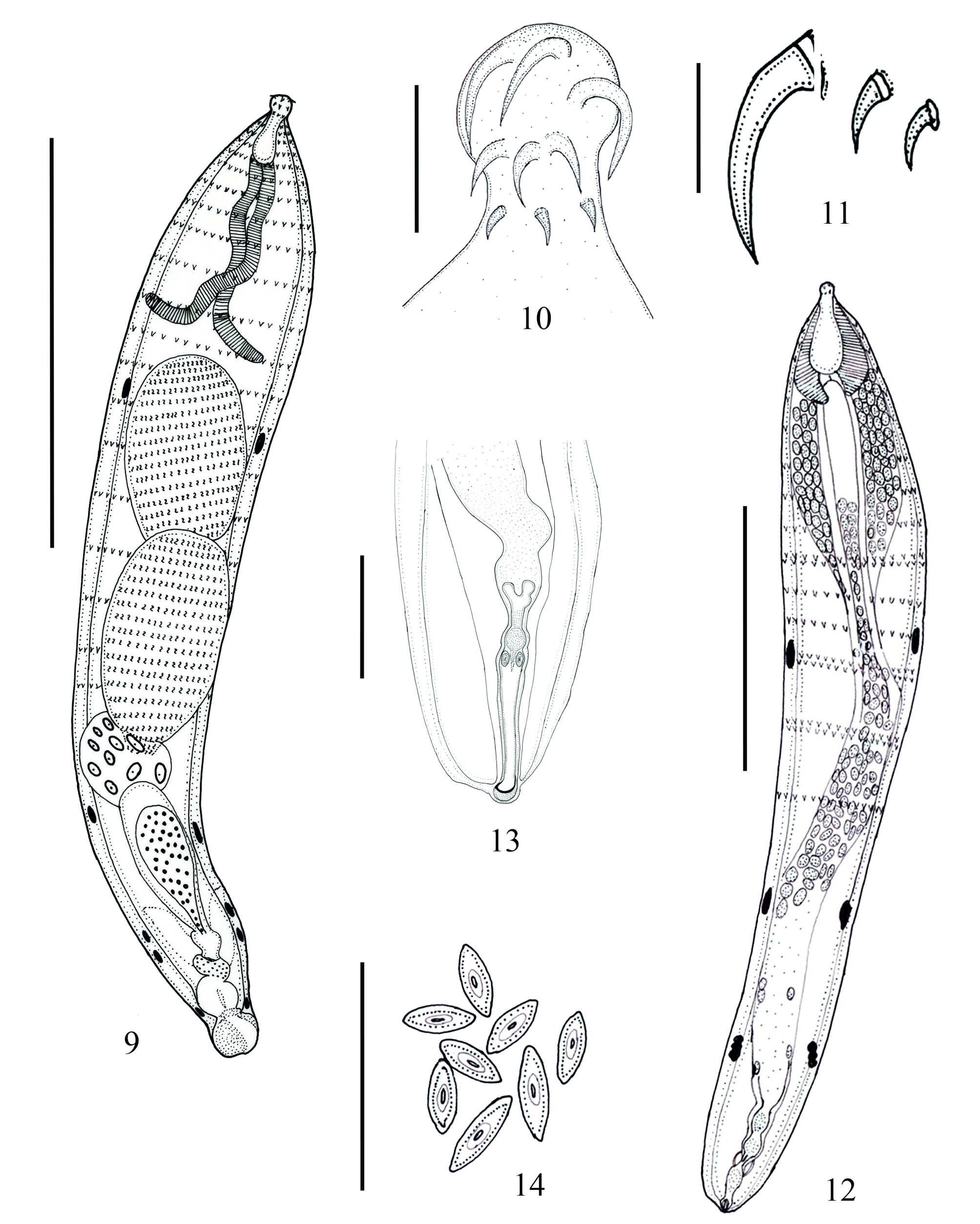
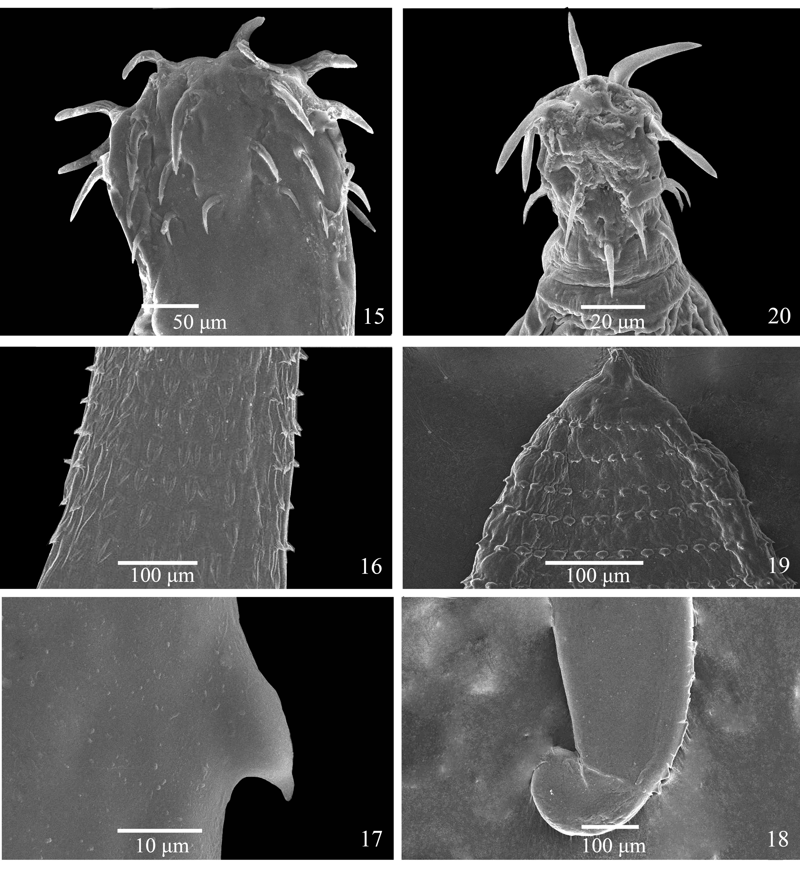
( Figs. 9–14 View FIGURES 9–14 and 19–20 View FIGURES 15–20 )
Type host: Gangetic mystus, Mystus seenghala (Hamilton) ( Siluriformes , Bagridae ).
Type locality: Nabi Panah Pond, Malihabad, Lucknow, Uttar Pradesh, India (26.5830° N, 80.4322° E). Additional host: Pool barb, Puntius sophore (Hamilton) ( Cypriniformes , Cyprinidae ).
Site of infection: Intestine.
Specimens deposited: Voucher male ZSI / GPRC, IV– 4362a and female ZSI / GPRC, IV– 4362b. Paratype males LU /Z/2019/15– LU /Z/2019/18 and females LU /Z/2019/19– LU /Z/2019/21.
Diagnosis: Quadrigyridae , Pallisentinae , with characters of the genus Acanthosentis Verma and Datta, 1929 ( Verma & Datta 1929; Bhattacharya 2007). Shared structures smaller in females than in males. Body small, broader at about 1/3 distance from anterior end, tapering to posterior end, slightly curved ventrally. Trunk with rings of spines closely set anteriorly but gradually more widely apart as they proceed backward to just past mid-body, spines pointing posteriorly. Proboscis short, orbicular (length to width ratio 1:1), with 3 circles of 6 hooks each; hooks declining in size from first circles to third circles, anterior to posterior. Proboscis receptacle ovoid (length to width ratio 1:0.5), with single layered wall, and with cerebral ganglion at base. Hypodermic nuclei few, not branched, in posterior half of trunk. Lacunar system reticular, with prominent transverse vessels. Lemnisci longer than proboscis receptacle, cylindrical in males and narrowly deltoid in females, longer in males than in females. Testes eliptoid, tandem, contiguous in posterior region of body. Cement gland syncytial with 6–10 nuclei, about 1/4 as long as testes. Genital pore terminal in males, slightly ventro-terminal in females. Eggs narrowly rhomboid (length to width ration 1:0.3).
Description: Material Examined: Four males, three females, and two specimens for SEM.
Male. Worm small in size, broadest at about 1/3 distance from anterior end, tapering to posterior end, anterior 2/3 of trunk straight, posterior 1/3 slightly curved ventrally. Trunk 2.43–2.16 (2.4) mm long, 320–370 (343) µm wide at widest point (measurements of type material from original description given in Table 2 View TABLE 2 ). Five pairs of giant hypodermal nuclei present. Trunk with rings of spines closely set anteriorly but gradually more widely apart as they proceed backward to just past mid-body to level of posterior testis, 12–16 circles of 21–40 spines, almost equal in size ( Figs. 9 View FIGURES 9–14 and 19 View FIGURES 15–20 ). Proboscis short, orbicular in shape (length to width ratio 1:1), 40–60 (45 ± 10) long, 40–60 (45) wide ( Figs. 9, 10 View FIGURES 9–14 and 20 View FIGURES 15–20 ). Proboscis with rooted hooks in 3 circles of 6 hooks each ( Figs. 10 View FIGURES 9–14 and 20 View FIGURES 15–20 ). Hooks longest in first circle, decreasing in size posteriorly in succeeding rings ( Figs. 10, 11 View FIGURES 9–14 and 20 View FIGURES 15–20 ). Blades of hooks curved to point posteriorly. Length of hooks from anterior to posterior, 500 250, 200; size ratios anterior to posterior 1.0:0.6:0.5. Neck short, 16–23 (20). Proboscis receptacle very deeply ovoid, 120–160 (143) long, 50–80 (63) wide. Lemnisci cylindrical, much longer than proboscis receptacle, approximately equal in size, 690–700 (693) long, 40–50 (43) wide. Reproductive system in posterior 3/4 of body ( Fig. 9 View FIGURES 9–14 ). Testes eliptoid, tandem, contiguous. Anterior testis 420–490 (465) long, 300–300 wide; posterior testis 370–540 (470) long, 270–300 (290) wide. Anterior testis 1130–1700 (1367) from anterior end of body, posterior testes 1600–2060 (1767) from anterior end. Cement gland much smaller than testes, contiguous with posterior testis, 150–240 (198) long, 110–240 (193) wide, containing 6–10 giant nuclei. Cement reservoir larger than cement gland, contiguous, 280–390 (335) long, 130–150 (140) wide. Saefftigen’s pouch pyriform; 70–130 (100) long, 80–90 (88) wide, duct wide anteriorly tapering to posterior connection. Gonopore terminal ( Fig. 9 View FIGURES 9–14 ). Bursa, when everted, 80–140 (113) long, 90–160 (123) wide.
Female. Females smaller than males, broadest at about 1/3 distance from anterior end, tapering to posterior end, anterior 1/1 of trunk straight, posterior 1/1 slightly curved ventrally ( Fig. 12 View FIGURES 9–14 ). Trunk 1.7–1.8 (1.7) mm long, 158–237 (198) µm wide at widest point. Five pairs of giant hypodermal nuclei present, posteriormost three nuclei clumped together at posterior part of trunk. Trunk with rings of spines closely set anteriorly but gradually more widely apart as they proceed backward to just past mid-body, 18 circles of 34 spines, fifth ring from last, third from last, and second from last rings double (12 single rings and 3 double rings) ( Fig. 12 View FIGURES 9–14 ). Spines almost equal in size. Proboscis short, orbicular in shape (length to width ratio 1:1), 29–31 (30) long, 29–31 (30) wide ( Fig. 12 View FIGURES 9–14 ). Neck short, 16–20 (18). Proboscis receptacle very deeply ovoid, 142–150 (146) long, 67–70 (68) wide. Lemnisci cylindrical, much longer than proboscis receptacle, compressed by ligament sac, approximately equal in size, 196–200 (198) long, 56–60 (58) wide. Reproductive system in posterior 1/5 of trunk, ligament sac persistent, attached to anterior opening of uterine bell. Uterine bell 79–83 (81) long, 54–58 (56) wide ( Fig. 13 View FIGURES 9–14 ). Uterus 73–75 (74) long, 40–43 (41) wide. Vagina 32–35 (33) long, 38 wide. Gonopore approximately terminal, protruding slightly from body wall ( Fig. 13 View FIGURES 9–14 ). Ovarian balls somewhat elliptical in shape. Eggs narrowly fusiform in shape, with thick outer shell, 35–44 (39) long, 14–17 (15) wide ( Fig. 14 View FIGURES 9–14 ).
Remarks: The original authors ( Chowhan et al. 1988) placed A. seenghalae in the Acanthosentis because the proboscis hooks are in three circles of six hooks in each; the present form is consistent with those specimens. Using the key provided by Amin (2005), the specimens collected for this study also were identified as A. seenghalae . The present form possesses only one sperm duct and Saftigen’s pouch is present. The rings of trunks spines extend over the anterior half of the body to of the posterior end of the testicular field. In the original description of A. seenghalae , 12–14 circles of 20–24 spines in each ring were reported, but in present form males had 12–16 rings of 21–40 spines in each and in females had 12 circles of 34 in each with the fifth from the last ring, third from last, and second from last ring are double (not reported in the original description), increasing the total number of rings to 18 unless you count the double rings as a single ring. The difference between the spination of the present material and that of the type material of the species could not be evaluated.
The proboscis of the holotype of A. seenghalae is as long as wide (60–80 long, 70–80 wide); the proboscis of the present form also is as long as wide (40–60 long, 40–60 wide) but it is a little smaller. The uterine bell, vagina and eggs were not mentioned by Chowhan et al. (1988) and they were obscured by eggs in their figures, and in the present material the females also were filled with ovarian balls and mature eggs. The para-receptacle organ was not seen. A comparison of the measurements of the type specimens of A. seenghalae with those of the present material is given in Table 2 View TABLE 2 .
Molecular characterization
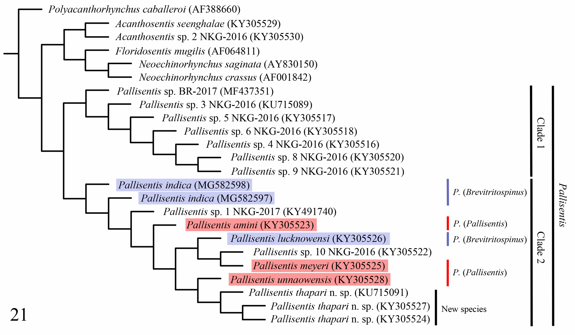
The amplification sizes of the rRNA of P. thapari ( KY305524 View Materials , KY305527 View Materials and KU715091 View Materials ) were 927, 909, 782 base pairs long and A. seenghale ( KY305529 View Materials ) was 913 base pairs long. The G+C contents were estimated as 45–46.1%. Sequences for the remaining taxa included in the analysis were downloaded from GenBank by referencing the accession numbers ( Table 1 View TABLE 1 ).
In the resultant cladogram ( Fig. 21 View FIGURE 21 ), three clades were identified: Pallisentis Clade 1, Pallisentis Clade 2, and a clade comprised of the two species of Acanthosentis as sister group to a clade of the other three species of Eoacanthocephala included in the analysis. Members of the genus Pallisentis were separated into the two clades mentioned above, one (Clade 1) comprised of sequences in GenBank that have not yet been identified to species and one clade (Clade 2) composed of nine identified species (including three sequences of P. thapari n. sp.) and two sequences from different putative species that have not yet been identified ( Fig. 21 View FIGURE 21 ).
| LU |
St. Petersburg University |
No known copyright restrictions apply. See Agosti, D., Egloff, W., 2009. Taxonomic information exchange and copyright: the Plazi approach. BMC Research Notes 2009, 2:53 for further explanation.
|
Kingdom |
|
|
Phylum |
|
|
Class |
|
|
Order |
|
|
Family |
|
|
Genus |