
Antecerococcus muntingi Hodgson & Williams
|
publication ID |
https://doi.org/10.11646/zootaxa.4091.1.1 |
|
publication LSID |
urn:lsid:zoobank.org:pub:76D13D36-682E-4E91-AC91-693CA9D3D465 |
|
DOI |
https://doi.org/10.5281/zenodo.6081588 |
|
persistent identifier |
https://treatment.plazi.org/id/03F2FF48-8179-0D6A-24B6-AAA2FDDAFCB6 |
|
treatment provided by |
Plazi |
|
scientific name |
Antecerococcus muntingi Hodgson & Williams |
| status |
sp. nov. |
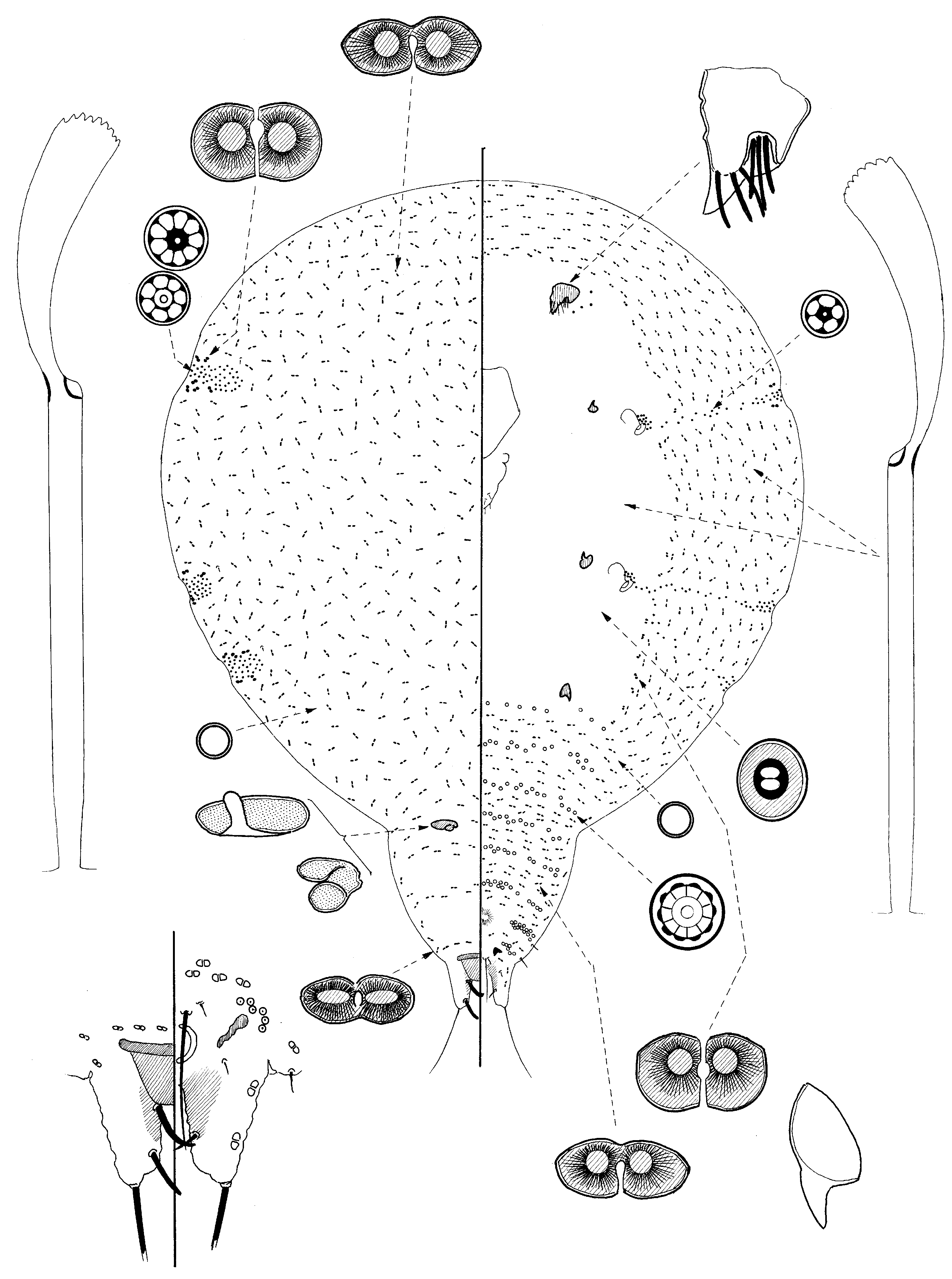
Antecerococcus muntingi Hodgson & Williams , sp. nov.
( Fig. 30 View FIGURE 30 )
Material studied. Holotype: SOUTH AFRICA, Cape Province, Tradouw Pass, on Erica sp. ( Ericaceae ), 11.i.1969, J. Munting (SANC): 1/1adf (good).
Mounted material. Body pear-shaped, small, 1.3 mm long and 1.05 mm wide. Margins of available specimen with shallow indentations in position of stigmatic pore bands.
Dorsum. Eight-shaped pores of 2 types: (i) a narrow, elongate pore, each 7–10 x 3–4 µm, quite evenly but fairly sparsely distributed throughout dorsum, but with smallest pores medially and posteriorly; and (ii) a slightly larger, more rounded pore, each 12–13 x 6.5–7.5 µm, restricted to 2–6 on each side of each stigmatic pore band. Simple pores, each about 3 µm wide, sparse, perhaps most abundant on posterior abdominal segments. Cribriform plates: 1 submedially on each side of abdominal segment IV, of unknown shape, that on one side elongate oval, about 60 x 20 µm, but with an odd finger-like process medially; other more unevenly shaped, 40 x 30 µm, possibly C-shaped; both with narrow margins and with very small micropores. Dorsal setae extremely few, each setose, and mainly 5 µm long. Tubular ducts with outer ductule 25–27 µm long, inner ductules quite long, each about 17 µm long; slightly broader than those on venter; abundant throughout. Anal lobes membranous, sclerotizations restricted to a small area on inner margins near their base, without obvious folding; most setae on lobes quite short; each lobe about 83 µm long with a long apical seta, each 145 µm long; more apical fleshy setae on dorsal surface each 20–26 µm long, more basal fleshy setae each 30–32 µm long; ventral surface seta near apex 14 µm long, exact position uncertain; medioventral setae probably absent; outer margin setae 8–10 µm long; each lobe with 2 small 8- shaped pores. Median anal plate bluntly quadrate, about 46 µm long, 65 µm wide at base. Anal ring with 4 pairs of setae, each about 100 µm long.
Venter. Eight-shaped pores of 2 types, as on dorsum; with elongate pore ( type i) in a broad submarginal band on head and thorax and in segmental bands across abdominal segments; and more rounded pore ( type ii) in a narrow band mesad to elongate pores, perhaps slightly smaller than those on dorsum, each 10 x 5 µm. Simple pores sparse. Small bilocular pores, each about 5.0 µm widest, frequent medially on head and thorax. Spiracular discpores each 4–6 µm wide with mainly 5 loculi but the number of loculi increasing (up to about 10) towards apex; posterior band bifurcated; each band in a group near peritreme and then in a sparse band initially, widening into a fairly broad apex; each anterior band with 70–90 pores, each posterior band with about 60 pores; each band without minute 8-shaped pores towards apex but some larger rounded 8-shaped pores sometimes intermingling with discpores; also with a small group of about 4 or 5 quinquelocular disc-pores just laterad to each antenna. Small convex closed pores absent; a few simple pores present near spiracles. Multilocular disc-pores, each about 7 µm wide, mainly with 10 loculi, distributed as follows: abdominal segment VIII about 6 on each side submarginally; VII 17– 20 on each side of vulva; VI 3 or 4 submarginally + 20 medially; V 2 or 4 submarginally + 19 medially; IV 2 or 3 submarginally + 20 medially; III 4 submarginally + 19 medially; II with 5 or 6 submarginally + 20 medially; plus a group of 3–7 just laterad to each hind-leg stub + 10 medially. Tubular ducts very slightly narrower than those on dorsum, present throughout. Ventral setae showing nothing distinctive; preanal setae each 60 µm long; smaller companion seta short. Leg stubs small. Antennae unusually large, unsegmented, each 60–62 µm long, 45–50 µm wide, with 6–8 fleshy setae, several in a wide, deep setal cavity; apex drawn out into a large cone-shaped triangular point. Clypeolabral shield 145 µm long. Spiracular peritreme each 23–27 µm wide.
Comment. In having rather elongate dorsal 8-shaped pores throughout the dorsum but more rounded pores on each side of the stigmatic pore bands, the adult female of A. muntingi is similar to A. asparagi and A. insleyae . However, it differs from both in the very large size of the antennae; also from A. asparagi in having many fewer 8- shaped pores on the dorsum and many fewer spiracular disc-pores in each pore band, and from A. insleyae in having multilocular disc-pores on all abdominal segments (only present on II and III in A. insleyae ) and in having almost all setae on anal lobes short, usually ≤30 µm long (usually ≥40 µm long on A. insleyae ).
A. muntingi can be diagnosed by the following combination of character-states: (i) dorsum with 8-shaped pores of two types; (ii) large 8-shaped pores entirely absent; (iii) intermediate-sized 8-shaped pores elongate, significantly longer than wide, randomly distributed; (iv) small 8-shaped pores absent from stigmatic bands; (v) one large cribriform plate on each side of abdominal segment IV; (vi) leg stubs present; (vii) posterior stigmatic band bifurcated; (viii) each stigmatic pore band with fewer than 100 pores, apex of each band lacking minute 8- shaped pores; (ix) multilocular disc-pores present across all abdominal segments, each band with approximately the same number of pores, and also across metathorax, and (x) each antenna exceptionally large, with a deep setal cavity and a strong cone-like point on apex.
The adult female of A. muntingi falls within Group B in the key to species of Antecerococcus , and is most similar to A. asparagi and A. insleyae , both also from South Africa.
Name derivation. A. muntingi is named after the collector, Jack (Jacobius) Munting, who not only collected this specimen but who, in the 1960s, published excellent papers on South African scale insects. He sadly died in 2013 (see Ben-Dov & Giliomee, 2014).
No known copyright restrictions apply. See Agosti, D., Egloff, W., 2009. Taxonomic information exchange and copyright: the Plazi approach. BMC Research Notes 2009, 2:53 for further explanation.
|
Kingdom |
|
|
Phylum |
|
|
Class |
|
|
Order |
|
|
Family |
|
|
Genus |