
Leptammina flavofusca
|
publication ID |
https://doi.org/ 10.5281/zenodo.187761 |
|
DOI |
https://doi.org/10.5281/zenodo.6217760 |
|
persistent identifier |
https://treatment.plazi.org/id/03F387BC-4840-FF83-EEFA-4C413BF9469B |
|
treatment provided by |
Plazi |
|
scientific name |
Leptammina flavofusca |
| status |
|
Leptammina flavofusca View in CoL gen. et sp. nov.
( Figs 3–5 View FIGURE 3 View FIGURE 4 View FIGURE 5 )
Material. Polarstern station 102#11 (Agassiz trawl) 125 specimens; Station 102#13 (Epibenthic sledge) 199 specimens; Station 80#9 (Epibenthic sledge) 11 specimens; Station 110#8 (Epibenthic sledge) 50 specimens; Station 81#9** (Agassiz trawl) 8 specimens.
Derivation of name: flavofusca is derived from the Latin words flavus, meaning yellow and fuscus, meaning brown, dusky or tawny. It refers to the colour of the test.
Diagnosis. Approximately spherical species of Leptammina , up to 1.2 mm in diameter, with test wall composed of very thin, finely agglutinated layer overlying inner organic lining. Single prominent circular aperture. Peduncular sheath well developed. Cell body brownish-yellow in colour, visible through semitransparent test wall, with single large nucleus.
Deposition of type material: The holotype from Station 102#11 and paratypes, from Stations 102#11, 102#13, 80#9, 110#8, and 81#9** are deposited in the Forschungsinstitut Senckenberg, Frankfurt am Main, Germany, under reg. no. SMF XXVII 7536.
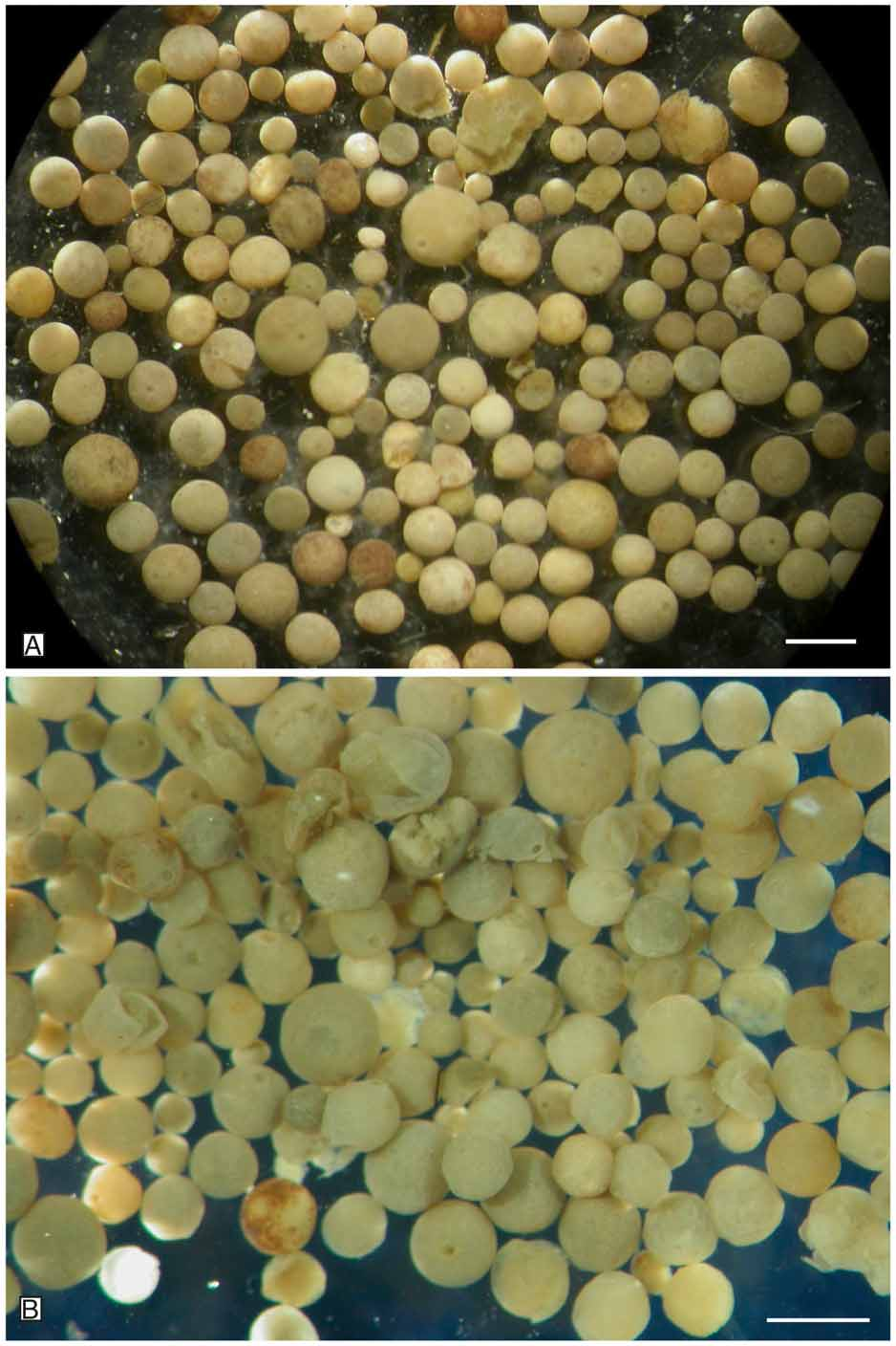
Description. General morphology. The test is free and brownish-yellow in colour. Unfixed specimens have a very even, approximately spherical shape ( Fig. 3 View FIGURE 3 ) and range in diameter from 345 to 1210 µm (mean=690 µm; median=702 µm; n=180). When dried for examination in the SEM, some distortion of the shape usually occurs. There is a single circular aperture surrounded by a thin collar at the end of a very short neck ( Figs 4 View FIGURE 4 B–C).
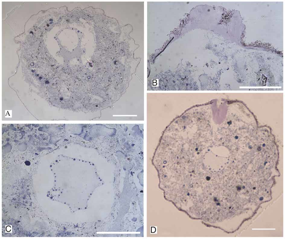
Test wall. The wall is <10 µm thick, semitransparent, and comprises a very thin and delicate outer agglutinated layer and an inner organic lining ( Fig. 5 View FIGURE 5 A–D). The surface is smooth, dull and non-reflective. The agglutinated particles that comprise the outer layer are <10 µm in size and generally angular, with larger particles embedded in a matrix of finer particles, one to a few µm in size ( Fig. 4 View FIGURE 4 E–F).
Cell body. The brownish-yellow cell body completely fills the test interior but may shrink after fixation. A distinct organic peduncular sheath (stomostyle) is located immediately inside the aperture ( Fig. 5 View FIGURE 5 B); it may extend into the apertural neck and onto the rim of the aperture. A projection from the peduncular sheath forms the inner organic lining along the entire inside of the test. Some vacuoles are visible in sectioned individuals. There is one large nucleus, up to 165 µm diameter, of the granular type, with few scattered nucleoli along its periphery ( Fig. 5 View FIGURE 5 A). The nucleus is a folded or approximately round in shape and is located within an exonuclear vacuole. A vacuole, up to 100 µm wide; is sometimes located in the center of the nucleus. Pseudopodia have not been observed but a thin string of cytoplasm projects through the centre of the peduncular sheath, along its central axis. However, examination of the cell body of critical-point dried individuals by SEM reveals a dense network of cytoplasm organized like fine pseudopodia (reticulopodia), in which are embedded stercomata, mineral particles, and other foreign inclusions ( Fig. 4 View FIGURE 4 D).
Remarks: Leptammina flavofusca gen. et sp. nov. differs from L. grisea gen. et sp. nov. in the smoother and more regular shape of the test, which is yellowish-brown rather than greyish in colour. Both species were most abundant at Station 102. It might be argued that the species look different merely due to the available inorganic particles available at any given site but the fact that they co-occurred argues against this possibility. Leptammina flavofusca gen. et sp. nov. is also clearly distinguished from Saccammina alba and Pilulina argentea by the distinctive colour of the test.
Distribution: Weddell Sea, central part at 4795 m depth; off Kapp Norvegia at 3138 and 4385 m depth.
Molecular characterization. Seven sequences of L. flavofusca gen. et sp. nov. and six sequences of L. grisea gen. et sp. nov. were obtained from four and three isolates of each species, respectively. No particular structural features (introns, insertions) were observed. The sequenced fragments ranged from 1110 to 1112 nucleotides in L. grisea gen. et sp. nov. and from 1132 to 1137 nucleotides in L. flavofusca gen. et sp. nov. The GC content was 39.0–39.5% in L. flavofusca gen. et sp. nov. and 40.8–41.2 % in L. grisea gen. et sp. nov. The sequences of both species differed by 15.4 to 16.2 %, while the divergence within each species was below 1 %.
Thirteen sequences of Leptammina gen. nov. were aligned to 41 sequences including the major groups of monothalamous foraminiferans ( Fig. 6 View FIGURE 6 ). Maximum likelihood phylogenetic analysis showed that both species group together with an undescribed Antarctic shallow water species, called “silver saccamminid” (Gooday et al. 1996) in a very strongly supported clade (100 %). The relationships between the two Leptammina species were relatively weakly supported (79 %) and in some trees the “silver saccammminid A26” branched among them. The three species clustered together with several other monothalamous species of the lineage C, a heterogeneous assemblage including, among others, genera such as Hippocrepinella Heron-Allen & Earland, 1932 , Rhizammina Brady, 1879 , Toxisarcon Cedhagen & Pawlowski, 2002 , Gloiogullmia Nyholm, 1974 , Cylindrogullmia Nyholm, 1974 , Bathyallogromia Gooday, Holzmann, Guiard, Cornelius & Pawlowski, 2004 , as well as the Xenophyophorea (Aranda da Silva, unpublished).


| SMF |
Forschungsinstitut und Natur-Museum Senckenberg |
No known copyright restrictions apply. See Agosti, D., Egloff, W., 2009. Taxonomic information exchange and copyright: the Plazi approach. BMC Research Notes 2009, 2:53 for further explanation.
|
Kingdom |
|
|
Phylum |
|
|
Class |
|
|
Order |
|
|
Family |
|
|
Genus |