
Australopacifica atrata ( Steel, 1897 )
|
publication ID |
https://doi.org/ 10.11646/zootaxa.4604.3.12 |
|
publication LSID |
lsid:zoobank.org:pub:73E8A9F4-EEDF-42FF-A2B0-2E55A04AC6CA |
|
DOI |
https://doi.org/10.5281/zenodo.5925041 |
|
persistent identifier |
https://treatment.plazi.org/id/03F387BF-FFE2-D57C-FF18-D9E3FF11F9BF |
|
treatment provided by |
Plazi |
|
scientific name |
Australopacifica atrata ( Steel, 1897 ) |
| status |
|
Australopacifica atrata ( Steel, 1897)
Synonymy:
Geoplana atrata Steel, 1897, p. 105 , Plate VII, Fig 10.
Geoplana atrata: Winsor, 1973, p. 101 , Fig 1 View FIGURE 1 .
Geoplana atrata: Winsor, 1977, p. 140 .
Geoplana atrata: Winsor, 1979, p. 156 , Fig 2 View FIGURE 2 .
Australopacifica atrata: Ogren & Kawakatsu, 1991, p. 42 .
Parakontikia atrata: Winsor, 1991, p. 45 .
Parakontikia atrata: Winsor, 1997, p. 577 .
Kontikia atrata: Jones, Johns & Winsor, 1998, p. 95 .
Parakontikia atrata: Johns, Boag and Yeates, 1998, p. 473 .
Material examined. NHMUK (18)97.11.1.47, Geoplana atrata , labelled “?co-type, Manning River, NSW, Australia, Mr. W.T. Steel”. One specimen preserved in ethanol.
NHMUK 2018.12.7.1-7. Specimens from Pontardawe, Glamorgan, 18 July 2015, D. R. James: three specimens preserved in 100% ethanol, separate vials; four sectioned specimens on slides: DJ1, anterior portion, 7 mm long, longitudinal sections at 15 µm, 5 slides; mid-portion about 2 mm long, transverse sections at 10 µm, 2 slides; posterior portion about 9mm long, longitudinal sections at 15 µm, 5 slides. DJ3, anterior portion, 8 mm long, longitudinal sections at 10 µm, 6 slides; mid-portion about 2 mm long, transverse sections at 10 µm, 2 slides; posterior portion about 12 mm long, longitudinal sections at 15 µm, 11 slides. DJ4, anterior portion, 9 mm long, longitudinal sections at 15 µm, 5 slides; posterior portion about 13 mm long, longitudinal sections at 15 µm, 8 slides. DJ9, whole specimen (apparently the anterior end of a specimen after fission), 9 mm long, longitudinal sections at 15 µm, 7 slides.
Other specimens have been deposited in NHMUK:
NHMUK 2018.12.7.8-11. Four specimens in 100% ethanol collected from Crediton , Devon, 30 June 2016, Dr Paul Chanin
NHMUK 2018.12.7.12-19. Eight specimens in 100% ethanol; collected Crediton , Devon, 14 July 2016, Dr Paul Chanin .
NHMUK 2018.12.7.20-22. Three specimens in 100% ethanol from RHS Wisley, 14 November 2018, coll. Dr Fryni Drizou.
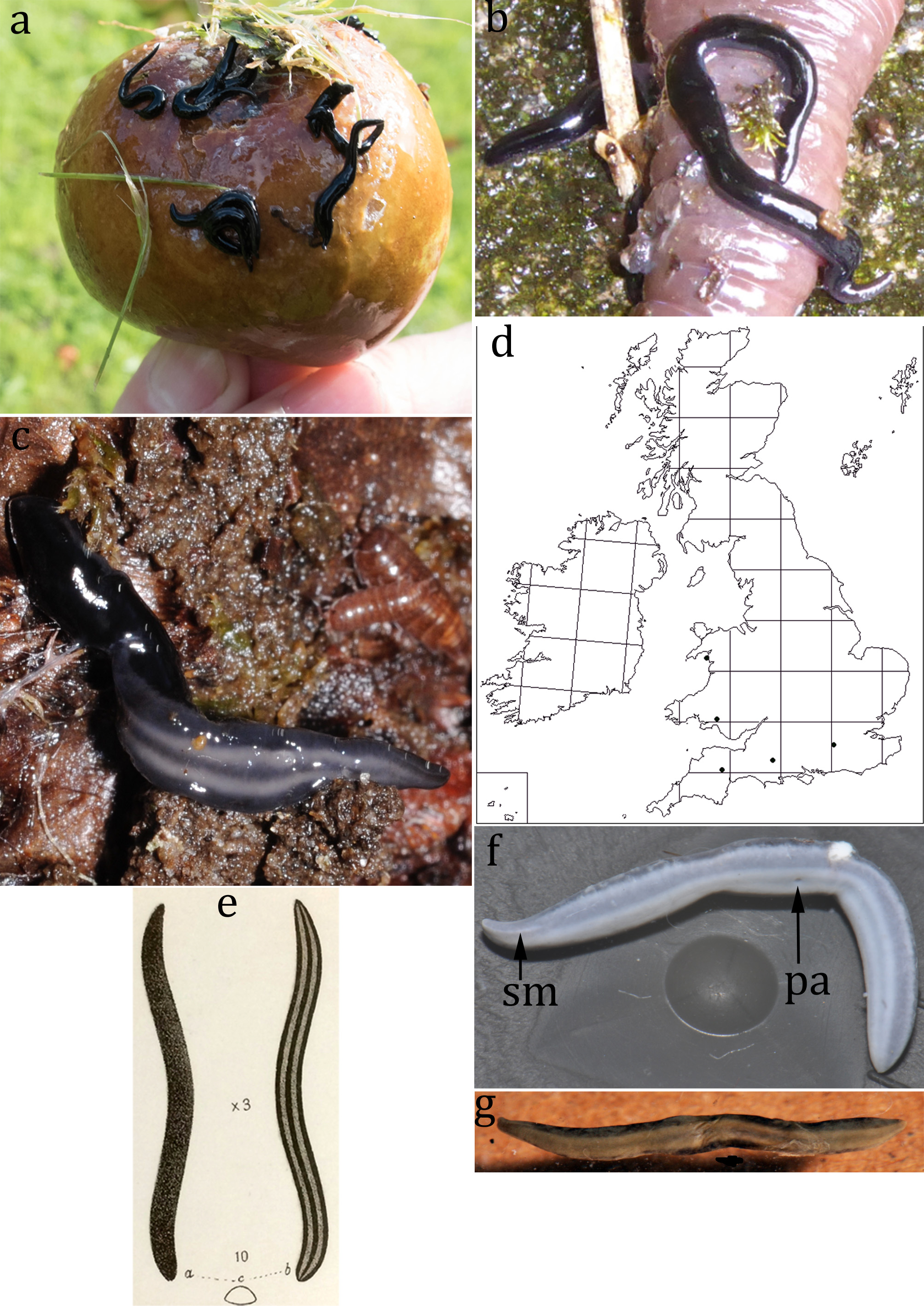
Description. The length of individuals varies considerably due to fragmentation and regeneration; the longest was about 3 cm long. They are slender; all are less than 2 mm wide and almost circular in cross section when crawling.
Living flatworms are uniform intense black dorsally and laterally ( Figs 1 View FIGURE 1 , 2 View FIGURE 2 ), the intense black colour terminates sharply at the lateral margin ( Fig 1g, h View FIGURE 1 ). Ventrally there is a median dark grey line, two slightly wider pale grey lines and two still wider dark grey lines ( Figs 1b View FIGURE 1 , 2c, e, f, g View FIGURE 2 ). At the lateral margin of the worm, the wider dark ventral lines are separated from the margin of the dorsal intense black colour by a narrow pale grey line, only obvious in live specimens under magnification and strong illumination ( Fig 1g, h View FIGURE 1 ) but visible in preserved specimens ( Fig 2f View FIGURE 2 ).
Eyes (black spots) are not readily visible in living worms, or in preserved worms. Under strong illumination and magnification uniserial eyes can be seen ( Fig 1h View FIGURE 1 ), sparsely distributed laterally in the grey line just ventral to the sharp margin of the intense black dorsal colour. In longitudinal sections eyes can be seen regularly spaced 400-500 µm apart in the anterior ( Fig 3d View FIGURE 3 ) and about 600 µm apart posteriorly. Each eye is about 35 µm in diameter.
Anteriorly there is a sensory margin about 1 mm long on either side, clearly visible only in preserved specimens ( Fig 2f View FIGURE 2 ).
The mouth (pharyngeal opening) is visible on the ventral surface ( Fig 2f View FIGURE 2 ) of larger specimens. Its position varies considerably between specimens, from 47% to 74% of body length (median 54.5%, n = 10) and it cannot be seen on small (<6 mm long) specimens. There is no sign of a gonopore in any examined specimen.
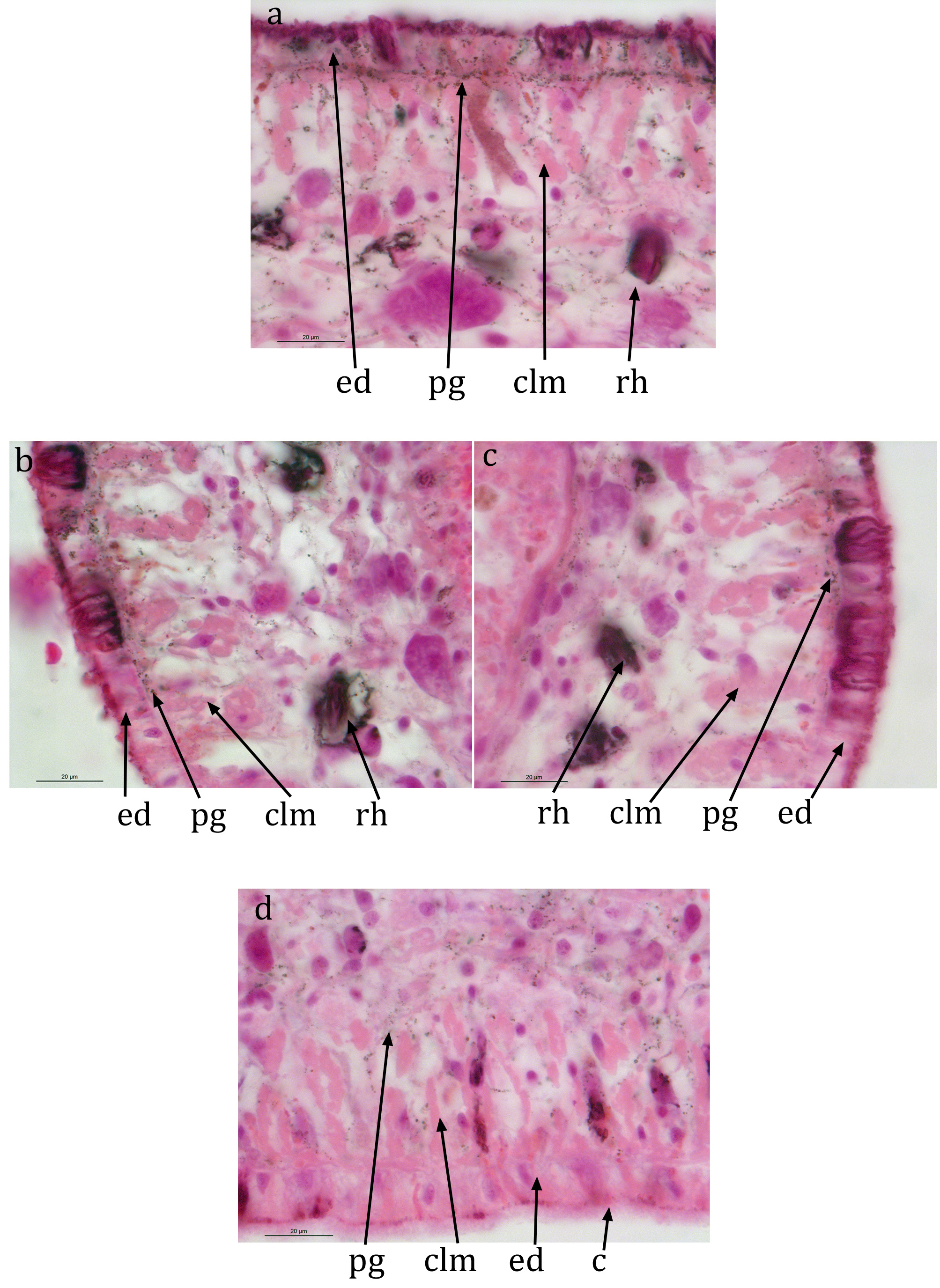
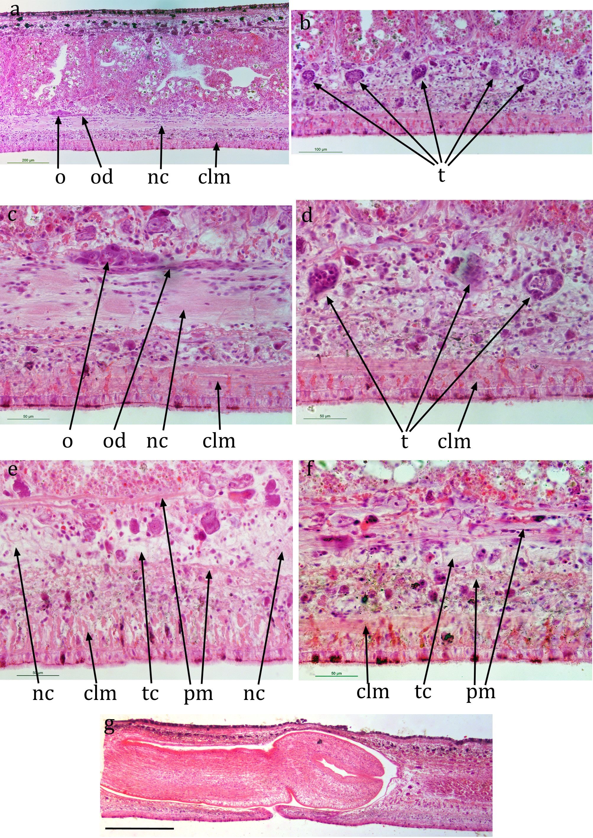
The ciliated creeping sole is about half the width of the sections, a little wider than the paired ventral nerve cords ( Fig 3a View FIGURE 3 ). The ventral cilia are about 5 µm long ( Figs 3c View FIGURE 3 ; 4d View FIGURE 4 ). The ventral epidermis is a columnar monolayer, 10-12 µm thick ( Figs 3c View FIGURE 3 ; 4d View FIGURE 4 ). Subcutaneous ventral circular muscle is about 5 µm thick, longitudinal muscle is in bundles about 35 µm thick ( Figs 3c View FIGURE 3 ; 4d View FIGURE 4 ). Dorsal and lateral epidermis is a non-ciliated, columnar monolayer ( Figs 3a, b View FIGURE 3 ; 4a, b, c View FIGURE 4 ), 15 µm thick; dorsal subcutaneous musculature 25-30 µm in total, mostly longitudinal muscle in bundles ( Fig 3b View FIGURE 3 ; 4a View FIGURE 4 ).
Small (<1 µm) black pigment granules are present scattered in the epidermis, subepidermal muscle layer and parenchyma, but their distribution round the circumference of the body varies ( Fig 4 View FIGURE 4 ). Dorsally, pigment granules are present scattered in the epidermis, most numerous immediately ental to the epidermis, and scattered in the subepidermal muscle and parenchyma ( Fig 4a View FIGURE 4 ). The pigment granules become less numerous at the lateral margin ( Fig 4b, c View FIGURE 4 ). Ventrally, pigment granules are present within the sub-epidermal muscle and the parenchyma as far dorsally as the nerve cords ( Fig 4d View FIGURE 4 ), but more numerous in the mid-line and towards the margins. The distribution of the granules corresponds with the dark pigment in the live animals. The more numerous and superficial pigment granules dorsally are presumed to account for the intense black colour, the less intense ventral dark lines being the result of fewer and less superficial pigment granules and the ventral pale lines by even fewer and less superficial pigment granules.
There is a ventral nerve plexus, about the same width as the ciliated sole, immediately dorsal to the longitudinal muscle bundles. A pair of ventral nerve cords, with transverse commissures, is present, about 360 µm apart (centre to centre) ( Fig 3a View FIGURE 3 ). Eyes are ental to the cutaneous longitudinal muscle ( Fig 3a View FIGURE 3 ).
Some parenchymal longitudinal and transverse muscle fibres are present, a few ventrally and dorsally to the ventral nerve cords ( Figs 3a, c View FIGURE 3 ; 5e, f View FIGURE 5 )). There is no ring zone of parenchymal longitudinal muscle ( Fig 3a View FIGURE 3 ).
The pharynx is cylindrical, about 2 mm long, 0.5 mm in diameter ( Fig 5g View FIGURE 5 ). The pharyngeal opening is about 1.25 mm from the anterior end of the pharyngeal pouch (63% of its length). The gut is typically triclad.
A pair of presumed early stage ovaries is present in each of the four sectioned specimens ( Fig 5a, c View FIGURE 5 ) immediately dorsal to the ventral nerve cord on either side. In DJ1 and DJ3 they are respectively about 3 mm and 4 mm behind the anterior end, in DJ3 they are about 320 µm apart. Each is about 100 µm long, 20 µm high and 30 µm wide. There is partial development of the ovovitelline duct from the postero-ventral surface of each ovary running posteriorly for a short distance along the dorsal surface of the ventral nerve cord on each side ( Fig 5a, c View FIGURE 5 ). However, ovovitelline ducts are not evident in more posterior transverse sections ( Fig 3a View FIGURE 3 ) or in posterior longitudinal sections (in fully mature specimens of land flatworms they are distinctive).
Numerous presumed early-stage testes are present lateral to the ventral nerve cord on either side from just behind the presumed ovaries to behind the pharyngeal pouch ( Fig 5b, d View FIGURE 5 ). They are ovoid, about 40 µm maximum dimension. Maturing spermatozoa are clearly visible in some. Sperm ducts cannot be distinguished with certainty. There is no sign of any development of the copulatory apparatus or of a gonopore in any of the sectioned specimens.


No known copyright restrictions apply. See Agosti, D., Egloff, W., 2009. Taxonomic information exchange and copyright: the Plazi approach. BMC Research Notes 2009, 2:53 for further explanation.
|
Kingdom |
|
|
Phylum |
|
|
Class |
|
|
Order |
|
|
Family |
|
|
Genus |
Australopacifica atrata ( Steel, 1897 )
| Jones, Hugh D. 2019 |
Kontikia atrata:
| Jones, Johns & Winsor 1998: 95 |
Parakontikia atrata
| : Johns, Boag and Yeates 1998: 473 |
Parakontikia atrata:
| Winsor 1997: 577 |
Australopacifica atrata
| : Ogren & Kawakatsu 1991: 42 |
Parakontikia atrata:
| Winsor 1991: 45 |
Geoplana atrata
| : Winsor 1979: 156 |
Geoplana atrata:
| Winsor 1977: 140 |
Geoplana atrata:
| Winsor 1973: 101 |
Geoplana atrata
| Steel 1897: 105 |