
Amnifleckia guttata
|
publication ID |
https://doi.org/10.5281/zenodo.174364 |
|
DOI |
https://doi.org/10.5281/zenodo.6261211 |
|
persistent identifier |
https://treatment.plazi.org/id/03F387EE-5A04-FFFB-D05D-FA04F793FA88 |
|
treatment provided by |
Plazi |
|
scientific name |
Amnifleckia guttata |
| status |
|
Amnifleckia guttata nov. sp. Zhang, Ren, and Cheng
Figs. 1–4 View FIGURES 1 View FIGURES 2 View FIGURES 3 View FIGURES 4
Material. Holotype D 2834 , part. Fossilized with abundance of conchostracans and a coleopterous larva, collection of Dalian Natural History Museum, PR China . Paratype. Specimen coll. NIGPAS 133711 , deposited in the Nanjing Institute of Geology and Palaeontology, Chinese Academy of Sciences .
Etymology. From Latin word guttatus (meaning “gutta, the shape of dripping”) for the distinct guttula in the third and fourth segments.
Species diagnosis. Amnifleckia guttata n. sp. differs from Amnifleckia splendida n. sp. in its MA and MP with two rows of cells between them, and IR2 and RP3/4 with two rows of cells between them.
Age and layer. Middle Jurassic, Jiulongshan Formation, Daohugou Village.
Description. Holotype. This fossil is a rather well-preserved adult specimen with outspread wings connected to body; parts of left wings, anal part of right hind wings and the tip of abdomen are missing. Strong tendency towards coloured wings. The gender is uncertain because of the lack of preservation of the hind wing anal part and the tip of abdomen.
Fore wing 68.7 mm long, 12.8 mm wide at nodus; distance between base and arculus, 7.9 mm, between arculus and nodus, 24.8 mm, between nodus and pterostigma, 19.6 mm, between pterostigma and apex, 11.6 mm; wing not petiolate, two rows of cells and a longitudinal secondary vein more or less parallel to posterior wing margin in the anal area; AA distally strongly bent towards posterior wing margin and parallel with CuA, distally fused with the secondary longitudinal vein into a strong vein parallel to posterior wing margin and distally reaching CuA; median space free; none (left wing) or only one crossvein (right wing) in submedian space; a curved strong vein Cup-crossing separates the submedian space and subdiscoidal space; discoidal space basally opened; subdiscoidal space free, transverse, 2.6 mm long and 1.1 mm wide; RP and MA separate at arculus; MAb 1.4 mm long, well aligned with distal free part of CuA; CuA divided into a very short CuAb (0.8 mm long) directed towards posterior wing margin and a CuAa (9.5 mm long) more or less parallel to posterior wing margin; area between CuA and MP with only one or two rows of large, traverse cells; distal of the end of CuA, up to seven rows of cells in area between MP and posterior wing margin; MP rather straight, reaching posterior margin at the level about the basal of pterostigma, 50.3 mm from wing base, at 73 percent of the total wing length; MAa slightly curved, more or less parallel with MP; postdiscoidal area, 1.9 mm wide, with only one row of cells, distal part much narrower than basal part; Ax0 visible, very near to the wing base; two primary antenodal veins very strong, Ax1 0.8 mm basal of arculus and Ax2 about 6.6 mm distal of arculus; no secondary antenodal cross-vein between C and ScP but seven antenodal cross-veins between ScP and RA; about fourteen antesubnodal cross-veins between arculus and subnodus; base of RP3/4 9.9 mm distal of arculus, closer to arculus than to nodus; base of IR2 very close to that of RP3/ 4, 2.6 mm (left wing) or 2.4 mm (right wing) distally, originating distinctly on RP; vein CP in the nodus bent towards ScP but not reaching it, with small empty space between it and ScP, this structure being very original, apparently unique among the Odonata ( Nel et al. 1993); subnodus oblique; ten postnodal cross-veins between C and RA, not aligned with the eleven postsubnodal cross-veins between RA and RP1; no pterostigmal brace vein; three cross-veins below the peterostigma; pterostigma strong, 4.8 mm long and 1.1 mm wide; C and RA thickened along the pterostigma; area between C and RA distal of pterostigma long, with about fifteen cross-veins; RP2 aligned with subnodus; eleven bridge-cross-veins; one oblique vein “O”, seven cells and 7.6 mm distal of base of RP2; RP2 nearly straight; base of IR1 five (left wing) or four (right wing) cells and 5.2 mm or 5.8 mm distal of base of RP2; IR1 basally zigzagged but distally nearly straight, more or less parallel to RP1; area between MA and RP3/4 strongly widened distally, with about fifteen rows of cells along posterior wing margin; area between RP3/4 and IR2 with one row of cells (two rows of cells near the posterior margin), narrowed distally; area between IR2 and RP2 with one row of cells from their bases to level of the middle of pterostigma, distally widened, with eight rows of cells along posterior wing margin; area between RP2 and IR1 progressively widened, with two distinct and several weaker, shorter intercalary longitudinal veins and about eight rows of cells along posterior wing margin; area between IR1 and RP1 not distally widened, with up to five rows of cells and one or two weak longitudinal intercalary veins in between.
Hind wing distinctly shorter (by about 9 percent) than fore wing, 62.6 mm long, 13.0 mm wide at nodus; distance between base and arculus, 6.2 mm, between arculus and nodus, 20.6 mm, between nodus and pterostigma, 18.6 mm, between pterostigma and apex, 10.8 mm; wing not petiolate; cells in anal area irregular, some being very small and others being very large, with several large, approximate rectangle cells arranged tidily along the basal side of subdiscoidal space; median space free; submedian space with one strong cross-vein, 6.6 mm distal of wing base; a curved vein Cup-crossing separating submedian and subdiscoidal spaces; subdiscoidal space not preserved well, but strong tendency towards being free and transverse; discoidal cell free, basally closed, 2.1 mm long and 0.95 mm wide; RP and MA separate at arculus; MAb 1.8 mm long, well aligned with distal free part of CuA; CuA divided into CuAa and CuAb; CuAa slightly zigzagged, very short (shorter than that of forewing), vanishing in area between MP and posterior margin about 6.3 mm from its base, and more or less parallel to posterior wing margin with two rows of cells and a secondary parallel vein between them; area between CuAa and MP with two to four rows of large cells; MP nearly straight, reaching posterior margin at the level about the basal of pterostigma, 43.8 mm from wing base, at 63.8 percent of the total wing length; MAa nearly straight, parallel to MP, with one row of cells in the postdiscoidal area, 1.7 mm wide, this area being narrower near posterior wing margin with two rows of small cells present; Ax0 visible, very near to wing base; two primary antenodal veins very strong, Ax1 0.7 mm basal of arculus and Ax2 5 mm distal of arculus, distance between them smaller than that of forewing; Ax1 nearly perpendicular to ScP and R+MA, Ax2 distinctly oblique; no secondary antenodal cross-vein between C and ScP but seven antenodal cross-veins between ScP and RA; ten antesubnodal cross-veins between arculus and subnodus; base of RP3/4 7 mm distal of arculus, much closer to arculus than to nodus; base of IR2 very close to that of RP3/4, 2.0 mm (left wing) or 2.4 mm (right wing) distally, originating distinctly from RP; nodal structure identical to that of the forewing; subnodus oblique; nine postnodal cross-veins between C and RA not strictly aligned with the thirteen postsubnodal cross-veins between RA and RP1; no pterostigmal brace vein; three cross-veins below the peterostigma; pterostigma long and strong, distinctly longer than the forewing one, 6.4 mm long and 1.1 mm wide; C and RA thickened along the pterostigma; area between C and RA distal of pterostigma long, with about ten cross-veins; RP2 aligned with subnodus; seven bridge cross-veins; one oblique vein “O”, four cells and 4.5 mm distal of base of RP2; RP2 nearly straight; base of IR1, five (left wing) or four (right wing) cells and 5.2 mm or 5.3 mm distal of base of RP2; IR1 basally zigzagged but distally nearly straight, more or less parallel to RP1; area between MA and RP3/4 strongly widened distally; area between RP3/4 and IR2 with one row of cells (two rows of cells near the posterior margin), narrowed distally; area between IR2 and RP2 with one row of cells from their bases to level of the middle of pterostigma, distally widened, with five rows of cells along posterior wing margin; area between RP2 and IR1 progressively widened, with one distinct and several weaker intercalary longitudinal veins and about ten rows of cells along posterior wing margin; area between IR1 and RP1 not distally widened, with up to five rows of cells and one weak longitudinal intercalary veins in between.
Abdomen parallel-sided, stout and rather slippery (no setae or spines appear), 67.5 mm long, 7.2 mm wide; dorsal carina, transverse carina and ventral carina visible (sensu Asahina 1954); no distinct strips but a pair of markedly guttula aside the dorsal carina in the middle of the third and fourth segments separately; the eighth and ninth segments weakly sclerotized; the end segments (sensu Tillyard 1917) is fragmentary because of missing the tip of abdomen.
Thorax stout, about 11.8 mm long and 10.7 mm wide; Dorsal, ventral and internal parts are fossilized together and difficult to interpret, but anepisternum (AES2), dorsal carina (DCR), collar (COL), antealar sinus (AAS) (sensu Zhao 1990) and mesopleural suture (MPS) (sensu Asahina 1954) can be easily distinguished; humeral plate 2 (HP2), axillary plate 2 (AP2), humeral plate 3 (HP3) and postnotum 3 (PN3) (sensu Asahina 1954) are well preserved and large.
Legs strong and long; coxa (CX) of all legs were covered and invisible; part of trochanter (TR) visible, pretarsus (PTAR) well preserved, claw (CL) strong and sharp; prothoracic leg and mesothoracic leg originated very close to each other, with the metathoracic leg originated a great distance from them. Femur (FE) of prothoracic leg 8.0 mm long, tibia (TI) 9.4 mm long; tarsus (TA) 4.3 mm long, with basitarsus (BTA) 1.9 mm long and distitarsus (DTA) 1.3 mm long; claw 0.8 mm long. Femur of mesothoracic leg 10.2 mm long; tibia 10.5 mm long; tarsus 4.7 mm long, with basitarsus 2.0 mm long and distitarsus 1.3 mm long; claw 0.9 mm long. Femur (FE) of metathoracic leg 14.1 mm long; tibia (TI) 13.8 mm long; tarsus (TA) 5.5 mm long, with basitarsus (BTA) 2.7 mm long and distitarsus (DTA) 1.4 mm long; claw 0.9 mm long; all femurs are about two times wider than tibias; only the spines of the prothoracic left leg tibia and the metathoracic left leg femur are preserved, stout and small, about 0.3 mm long.
Head is preserved, but jumbled and rather useless, about 7.3 mm long and 8.2 mm wide, seemingly of anisopteroid-type (after Fleck & Nel 2002).
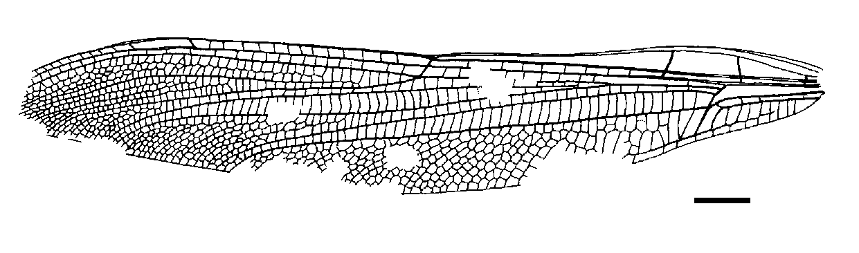
Paratype. A nearly complete fore wing, with few small zones missing toward apex; infuscate; wing 71.5 mm long, 12.2 mm wide at nodus level; distance between base and arculus, 8.8 mm, between arculus and nodus, 25.1 mm, between nodus and pterostigma, 20.3 mm, between pterostigma and apex, 11.8 mm; anal area 11.5 mm long and 3.0 mm wide, elongate, with two rows of large irregular cells between AA and AP; AA distally strongly bent towards posterior wing margin and nearly parallel with CuA, distally reaching CuAb; median area free of cross-vein; submedian area with a cross-vein and a curved vein CuP separating submedian and subdiscoidal areas; subdiscoidal space free, 4.0 mm long and 0.5 mm wide; discoidal cell basally opened, elongate, circa 10 mm long and 1 mm wide, free of cross-vein; RP + MA separated at nearly right angle from RA, strongly curved; RP separated from MA 0.5 mm distally; just distal of arculus, MA basally very strong, MA divided into MAa and MAb 1.2 mm distally; MAb short, 1.2 mm long, well aligned with distal free part of CuA; free part of CuA very strong, separating from MP 10 mm from wing base and directed towards posterior wing margin for 2.7 mm; CuA distally divided into CuAa and CuAb, CuAb rather short, directed towards wing base, meeting main branch of AA and posteriorly closing subdiscoidal space; CuAa basally more or less parallel to posterior wing margin, with one row of cells between them, and distally weakly zigzagged; postdiscoidal area about 3 mm wide just distal of discoidal cell; area between CuAa and MP basally 3.8 mm wide, with 1–2 rows of large cells; distal of end of CuAa, about 7–8 rows of cells in area between MP and posterior wing margin; MP nearly straight but with a curve close to its apex, reaching posterior margin well distal of nodus level, 55 mm from wing base, at 77% of total wing length; MAa nearly straight; one row of cells in postdiscoidal area, except at posterior wing margin, this area being only slightly widened near posterior wing margin; Ax0 visible very near wing base; two very strong primary antenodal cross-veins, Ax1 1.2 mm basal of arculus and Ax2 5.5 mm distal of arculus, Ax1 nearly perpendicular to ScP and R + MA but Ax 2 oblique; no secondary antenodal cross-vein of first row between C and ScP but eight antenodal cross-veins of second row between ScP and RA, all distal of Ax 2; more than nine cross-veins in area between RA and RP, between arculus and nodus; four cross-veins between RP and MAa, basal of midfork; base of RP 3/4 12 mm distal of arculus, closer to arculus than to nodus; base of IR2 two cells distal to that of RP3/4; IR2 apparently emerging from RP3/4; nodal Cr and subnodus aligned but not very oblique; 13 postnodal cross-veins not corresponding to the 14 postsubnodal cross-veins; no pterostigmal brace; three cross-veins below pterostigma; pterostigma sclerotized, very long and narrow, 5 mm long and 0.86 mm wide, basally recessed but after midway between nodus and apex; vein C strongly widened along pterostigma; area between C and RA distal of pterostigma long, with 14 cross-veins; RP2 aligned with subnodus; first oblique cross-vein ‘O’ four cells and 6 mm distal of subnodus, second oblique cross-vein five cells and 12.5 mm distally; RP2 nearly straight; base of IR1 five cells distal of base of RP2; IR1 basally slightly zigzagged but distally nearly straight, more or less parallel to RP1; area between MA and RP3/4 widened distally with 16 rows of cells along posterior wing margin; area between RP3/4 and IR2 only slightly widened at wing margin, with two rows of cells between them; area between IR2 and RP2 with ten rows of cells between them along posterior wing margin; area between RP2 and IR1 progressively widened, with three strong intercalary longitudinal veins and eight rows of cells along posterior wing margin; one row of cells between RP1 and RA distal of pterostigma.
Discussion. Amnifleckia guttata n. sp. is rather similar to Bellabrunetia catherinae Fleck et Nel, 2002, the type species of Bellabrunetia: long gaff, short CuAa, fore wing pterostigma distinctly shorter than that of the hind wing, pterostigma basally recessed, similar nodus structure, discoidal space and subdiscoidal space, and the similar pattern of the main longitudinal veins ( Fleck & Nel 2002). As for B. catherinae, it differs from the poorly known genus Pternopteron Pritykina, 1970 in the fore wing Ax1 distinctly basal of arculus, instead of being aligned with it ( Pritykina 1970: fig. 6).
The main differences with Bellabrunetia are: (1) at least two cross-veins below the pterostigma instead of a maximum of one cross-vein below the pterostigma in Bellabrunetia; (2) hind wing shorter than fore wing only by 9 percent, not like that of 15 percent in Bellabrunetia; (3) hind wing Ax1 and Ax2 not parallel to each other, unlike in Bellabrunetia; (4) the position of the oblique vein “O” much more distal of the subnodus than in Bellabrunetia; (5) cells in hind wing anal area more numerous than in Bellabrunetia, and distinctly denser; (6) a longitudinal row of spines along Costa, which is visible in ventral view in Bellabrunetia, is not present in Amnifleckia ; (7) Amnifleckia has distinctly shorter wings, around 68-70 mm long, instead of around 89 mm in Bellabrunetia; (8) the area between MA and RP3/4 in Amnifleckia with only about 15 rows of cells along posterior margin, much smaller than that of Bellabrunetia (with more than 20 rows); (9) without the very hairy body, unlike that of Bellabrunetia; wings infuscate instead of being hyaline; area between the veins RP and MA without any constriction distal of arculus, but with a basal cross-vein; IR2 apparently branching on RP3/4; MA and MP not parallel along posterior wing margin, with two rows of cells between them; IR2 and RP3/4 not parallel along posterior wing margin, with two rows of cells between them.


No known copyright restrictions apply. See Agosti, D., Egloff, W., 2009. Taxonomic information exchange and copyright: the Plazi approach. BMC Research Notes 2009, 2:53 for further explanation.
|
Kingdom |
|
|
Phylum |
|
|
Class |
|
|
Order |
|
|
Family |
|
|
Genus |