
Amnifleckia splendida, Zhang, Bing-Lan, Fleck, Gunther, Huang, Di-Ying, Nel, André, Ren, Dong, Cheng, Xiao-Dong & Lin, Qi-Bin, 2006
|
publication ID |
https://doi.org/10.5281/zenodo.174364 |
|
DOI |
https://doi.org/10.5281/zenodo.6261213 |
|
persistent identifier |
https://treatment.plazi.org/id/03F387EE-5A0F-FFFE-D05D-FA50F2FEFE94 |
|
treatment provided by |
Plazi |
|
scientific name |
Amnifleckia splendida |
| status |
sp. nov. |
Amnifleckia splendida n. sp. Huang, Fleck, Nel & Lin
( Figs 5–6 View FIGURES 5 View FIGURES 6 )
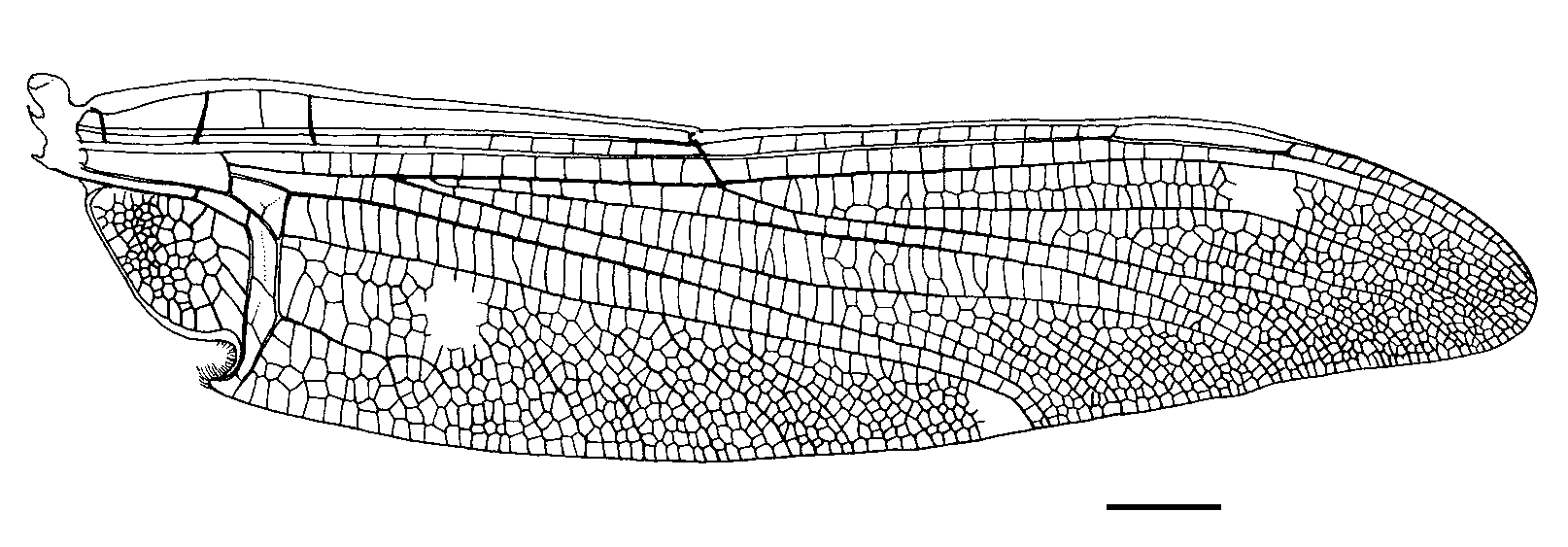
Diagnosis. This hind wing is also very close to that of Bellabrunetia catherinae and Amnifleckia guttata n. sp. Bellabrunetia was considered closely related to Pternopteron Pritykina, 1970. This new fossil shares with this latter the same highly specialized anal angle hook-like in its hind wing. The main difference between Pternopteron and Amnifleckia splendida n. sp. is its CuAa with a strong curved distal branch, instead of being straight as in Pternopteron. B. catherinae has also a CuAa forked at apex, but with a curved branch less distinct than in Amnifleckia splendida n. sp. Other differences with B. catherinae are as follows: hind wing distinctly shorter (65.4 mm long versus 76.0 mm for B. catherinae); wing with infuscate spots in cubito-anal area instead of being hyaline; area between the veins RP and MA without any constriction distal of arculus; five cross-veins below the pterostigma.
The fore wing of Amnifleckia guttata n. sp. has a length compatible with that of the hind wing of Amnifleckia splendida n. sp. (hind wing shorter than fore wings). Amnifleckia splendida n. sp. differs from Amnifleckia guttata n. sp. in its MA and MP parallel along posterior wing margin, with only one row of cells between them, and IR2 and RP3/4 parallel along posterior wing margin, with one row of cells between them. These differences only justify a specific separation.
Description. A nearly complete hind wing of a male, with few small zones missing toward apex; hyaline with two rather large infuscate spots in the cubito-anal area; wing 65.4 mm long, 14.7 mm wide at nodus level; distance between base and arculus, 7.2 mm, between arculus and nodus, 20.6 mm, between nodus and pterostigma, 18.3 mm, between pterostigma and apex, 12.9 mm; anal area 9.7 mm long and 5.8 mm wide, rounded and broad, with numerous small irregular cells between AA and AP; a strong tornus, hooklike, covered with hairs at apex of AA; AA distally strongly bent towards posterior wing margin and nearly parallel with CuA, but not distally reaching CuAb; median area free of cross-vein; submedian area free; a curved vein CuP separating submedian and subdiscoidal areas; subdiscoidal space crossed by three transverse veins, elongate and narrow, 6 mm long and 1 mm wide; discoidal cell closed, elongate, circa 2.1 mm long and 2.4 mm wide, free of cross-vein; RP + MA separated at nearly right angle from RA, strongly curved; RP separated from MA 0.7 mm distally; just distal of arculus, MA basally very strong, MA divided into MAa and MAb 2.6 mm distally; MAb short, 1.6 mm long, well aligned with distal free part of CuA; free part of CuA very strong, separating from MP 10 mm from wing base and directed towards posterior wing margin for 3.2 mm; CuA distally divided into CuAa and CuAb, CuAb also very strong, 4 mm long, straight and directed towards posterior wing margin, not meeting main branch of AA; CuAa short, basally more or less parallel to posterior wing margin, with 3–4 rows of cells between them, and distally forked into a curved branch and a weakly zigzagged branch, reaching posterior wing margin about 3.2 mm from its base; postdiscoidal area 1.8 mm wide just distal of discoidal cell; area between CuAa and MP basally 4.2 mm wide, with three rows of large cells; distal of end of CuAa, about 9–12 rows of cells in area between MP and posterior wing margin; MP nearly straight but with a slight curve close to its apex, reaching posterior margin well distal of nodus level, 42.7 mm from wing base, at 65% of total wing length; MAa nearly straight; one row of cells in postdiscoidal area, this area being narrowed near posterior wing margin; Ax0 visible very near wing base; two very strong primary antenodal cross-veins, Ax1 1.1 mm basal of arculus and Ax2 3.8 mm distal of arculus; maybe a weak secondary antenodal cross-vein of first row between C and ScP, between Ax1 and Ax2, but seven antenodal cross-veins of second row between ScP and RA, all distal of Ax2; 14 cross-veins in area between RA and RP, between arculus and nodus; three cross-veins between RP and MAa, basal of midfork; base of RP3/4 8.1 mm distal of arculus, closer to arculus than to nodus; base of IR2 one cell distal to that of RP3/ 4, 7 mm distally; IR2 apparently not emerging from RP3/4; nodal Cr and subnodus aligned but not very oblique; 12 postnodal cross-veins not corresponding to the 14 postsubnodal cross-veins; no pterostigmal brace; five weak cross-veins below pterostigma; pterostigma sclerotized, very long and narrow, 7.8 mm long and 0.7 mm wide, basally recessed but after midway between nodus and apex; vein C strongly widened along pterostigma; area between C and RA distal of pterostigma long, with 19 cross-veins; RP2 aligned with subnodus; first oblique cross-vein ‘O’ four cells and 3.5 mm distal of subnodus, no second oblique cross-vein; RP2 nearly straight; base of IR1 five cells and 5.8 mm distal of base of RP2; IR1 basally zigzagged but distally nearly straight, more or less parallel to RP1; area between MA and RP3/4 widened distally with about 13 rows of cells along posterior wing margin; area between RP3/4 and IR2 broadened in its mid part, with two rows of cells, but narrowed at wing margin; area between IR2 and RP2 strongly broadened at apex; area between RP2 and IR1 progressively widened, with two strong intercalary longitudinal veins and seven rows of cells along posterior wing margin; one row of cells between RP1 and RA distal of pterostigma.
Holotype. Specimen coll. NIGPAS 133712 , deposited in the Nanjing Institute of Geology and Palaeontology, Chinese Academy of Sciences.
Type locality. Daohugou Village , Ningcheng County, Inner Mongolia, NE China.
Type strata. Middle Jurassic, Jiulongshan Formation.
Etymology. After the wonderful state of preservation of the type material.

No known copyright restrictions apply. See Agosti, D., Egloff, W., 2009. Taxonomic information exchange and copyright: the Plazi approach. BMC Research Notes 2009, 2:53 for further explanation.
|
Kingdom |
|
|
Phylum |
|
|
Class |
|
|
Order |
|
|
Family |
|
|
Genus |