
Epidaus wangi Chen, Zhu, Wang & Cai
|
publication ID |
https://doi.org/ 10.11646/zootaxa.4154.1.6 |
|
publication LSID |
lsid:zoobank.org:pub:88CB9162-1494-4788-8EED-E31768CE1EBE |
|
DOI |
https://doi.org/10.5281/zenodo.6068510 |
|
persistent identifier |
https://treatment.plazi.org/id/03F3C958-FFC1-FFA2-E28F-B0AFEF68D61A |
|
treatment provided by |
Plazi |
|
scientific name |
Epidaus wangi Chen, Zhu, Wang & Cai |
| status |
sp. nov. |
Epidaus wangi Chen, Zhu, Wang & Cai View in CoL , sp. nov.
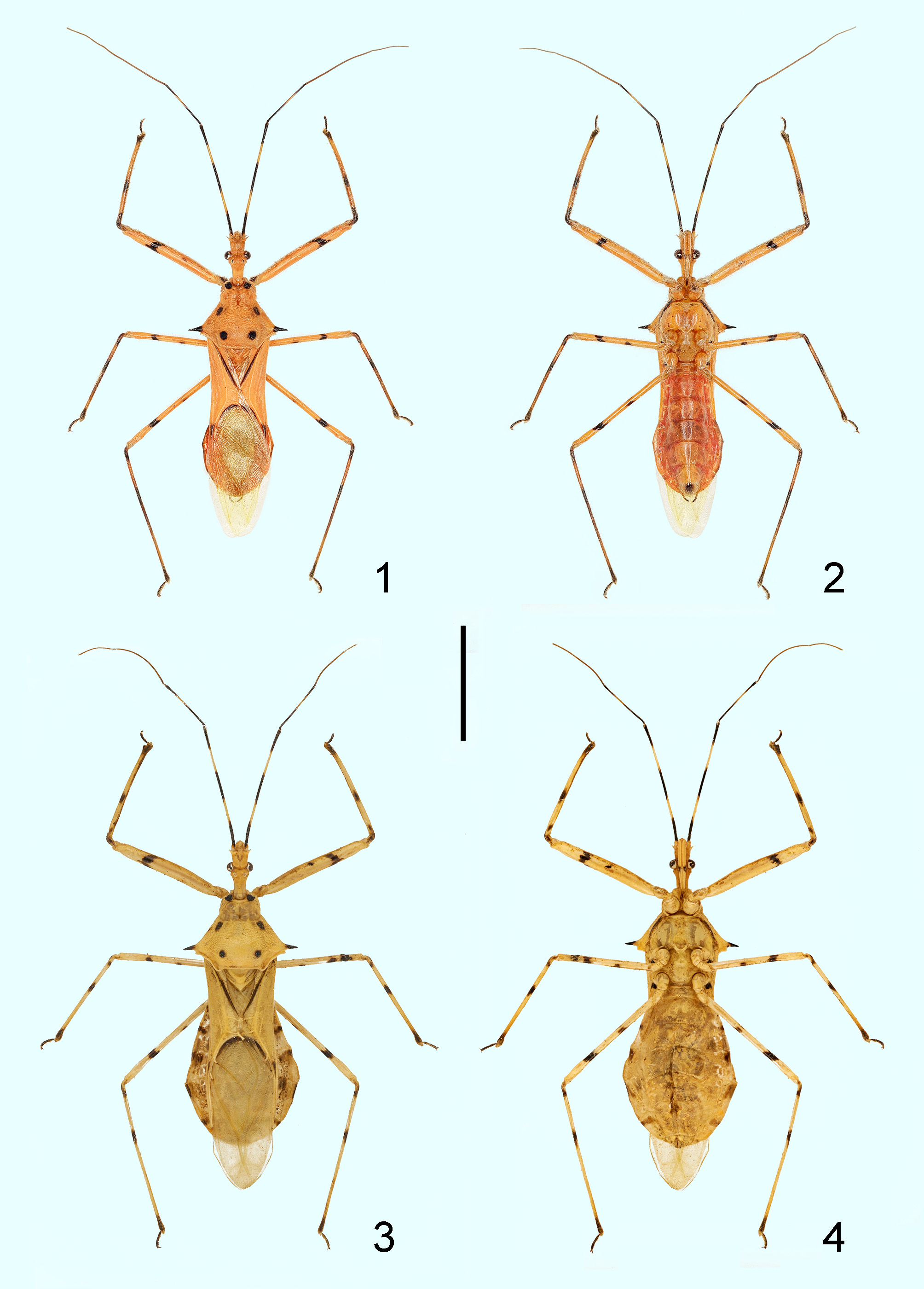
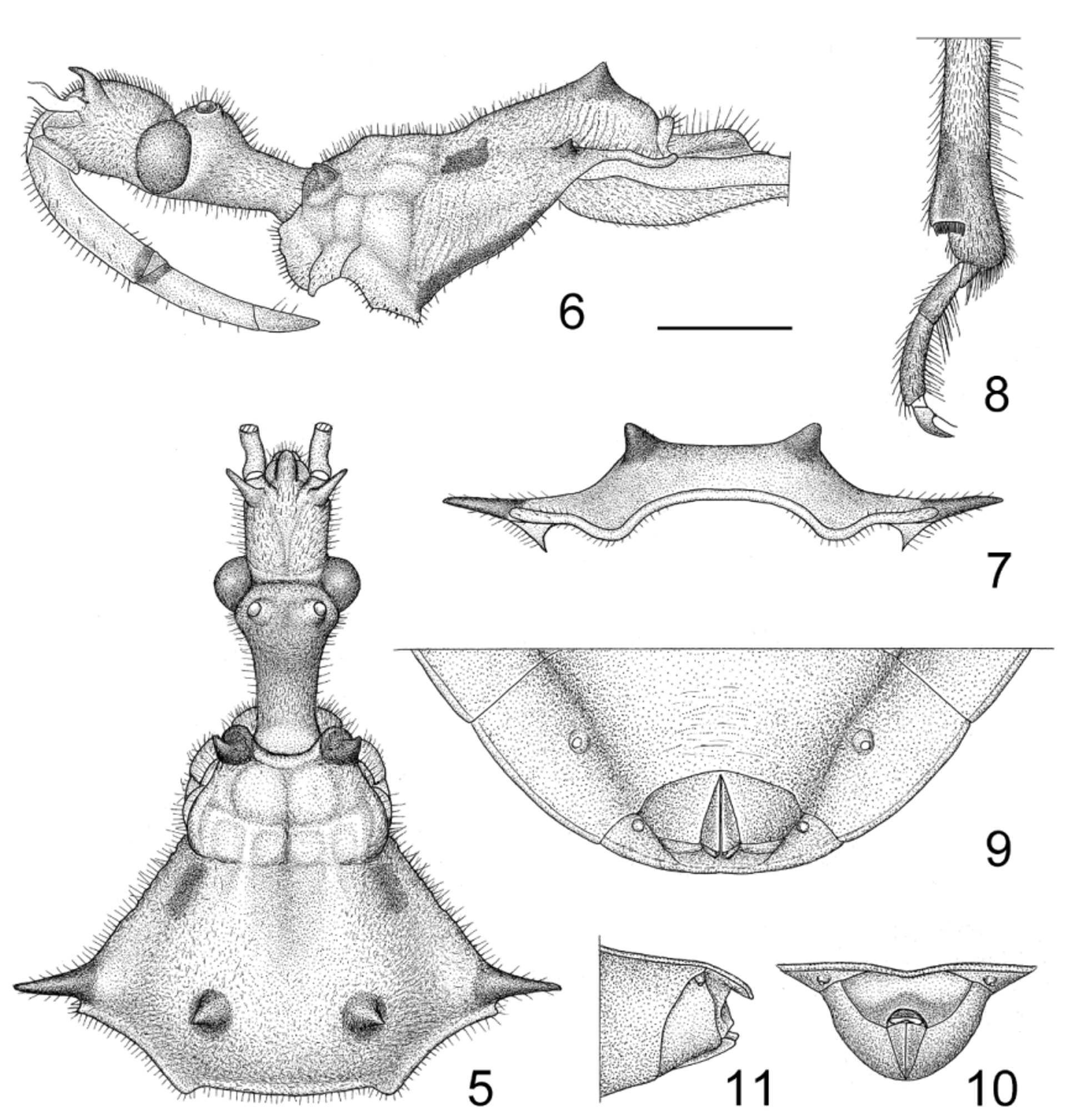
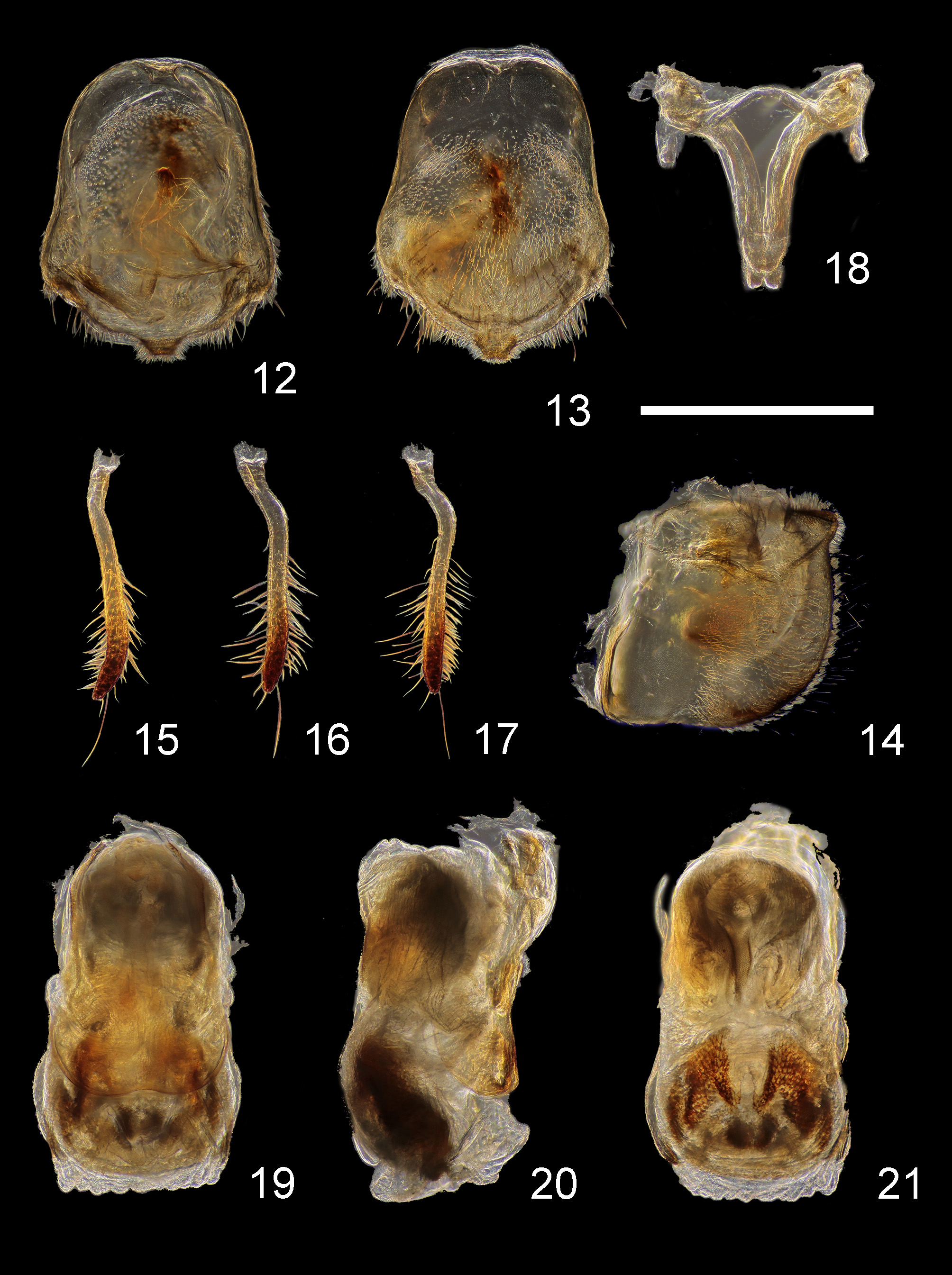
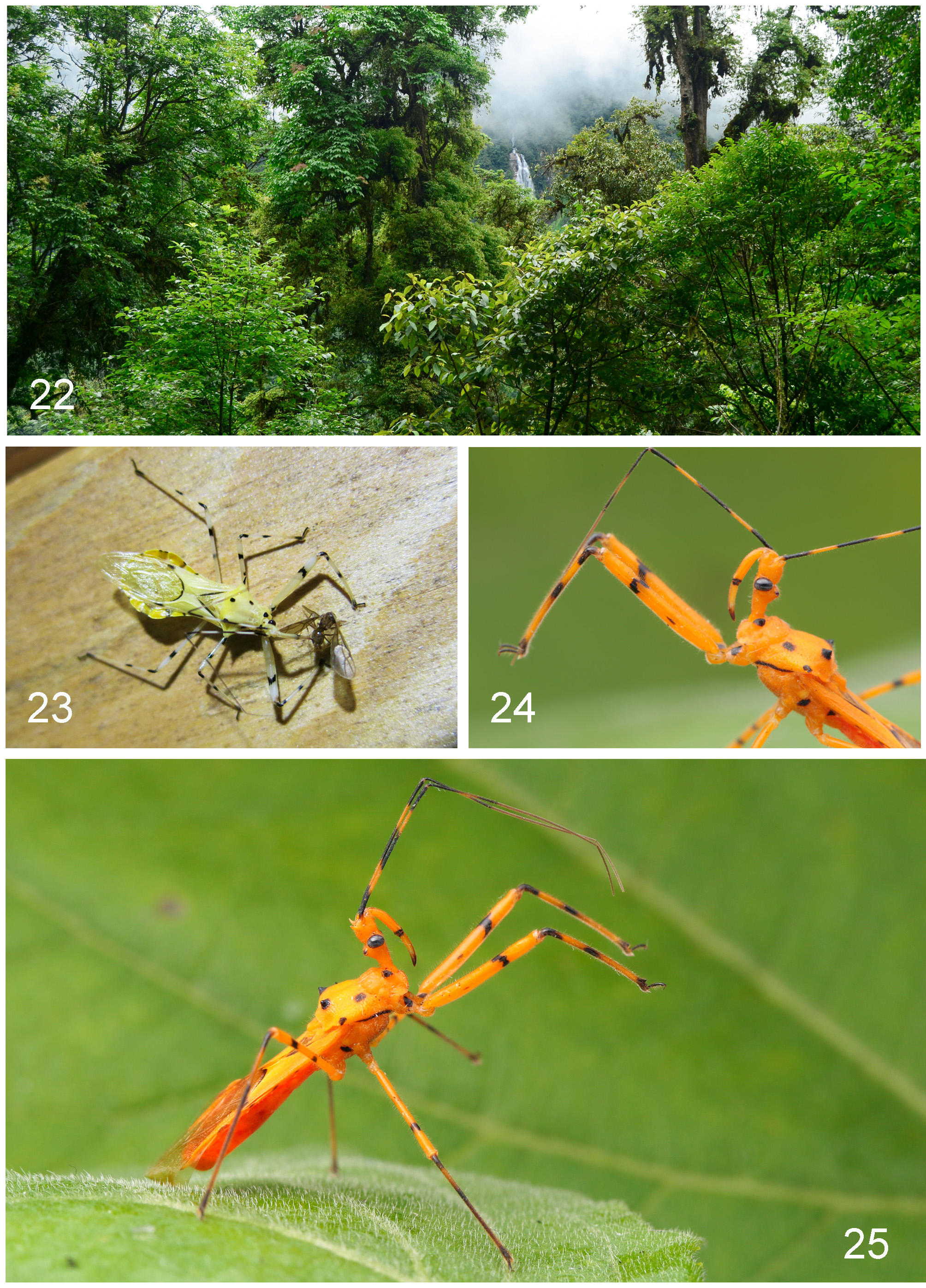
( Figs 1–21 View FIGURES 1 – 4 View FIGURES 5 – 11 View FIGURES 12 – 21 , 23–25 View FIGURES 22 - 25 )
Diagnosis. Body length 21.81 to 26.84 mm; pale yellow, antennae and legs with black annulations and spots, pronotum with six spots ( Figs 1–4 View FIGURES 1 – 4 , 23–25 View FIGURES 22 - 25 ), eyes, darker strips along clavus, and apex of corium blackish; two tubercules at middle of posterior part of pronotal lobe small, lateral pronotal angles spine-shaped and black ( Figs 1–7 View FIGURES 1 – 4 View FIGURES 5 – 11 ); apical portion of fifth abdominal segment of female roundly expanded and widest ( Figs 3, 4 View FIGURES 1 – 4 ).
Description. Colouration: Body generally lemon yellow to yolk yellow, with black stripes and spots, slightly shining ( Figs 1–4 View FIGURES 1 – 4 , 23–25 View FIGURES 22 - 25 ). Eyes, apical portion of first visible rostral segment, base of second visible rostral segment, third visible rostral segment, basal, middle, and apical portions of first antennal segment, basal and apical portions of second antennal segment, collar tubercles, two spots behind transverse sulcus of pronotum, two tubercles on disc of posterior pronotal lobe, spines of lateral pronotal angles, ventral margin of propleuron, markings on anterior margin of metapleuron, markings on fore coxa and trochanter, annular markings of femora, basal, subbasal and apical portions of tibiae, tarsi, claval suture, apical angle of corium, basal margin of membrane, lateral markings of second abdominal segment blackish brown to black; ocelli brown, shining; middle of second antennal segments, apical two antennal segments, tibiae (except basal, subbasal, and apical portions) yellowish brown; basal parts of each connexival segments in female and of fifth and sixth segment of connexivum in male slightly dark brown; abdomen of male tinged with red beneath ( Figs 2 View FIGURES 1 – 4 , 25 View FIGURES 22 - 25 ).
Structure: Body medium-sized, elongate. Body clothed with pale yellow short pubescence; first antennal segment with sparse, long, yellowish brown setae and short, yellow pubescence, second antennal segment with dense thick setae of various length, third and fourth antennal segments with short pubescence and sparse short setae; ventral surface of fore trochanter and each femora, inner surface of each tibiae with abundant thick setae; fossula spongiosa of fore and mid tibiae weakly developed; apex of fore tibiae with comb, slightly expanded ( Fig. 8 View FIGURES 5 – 11 ).
Head cylindrical, postocular portion slightly longer than anteocular portion; process behind antennal insertion spine-shaped, acute at apex, slightly bent; first antennal segment longest, subequal to combined length of head and pronotum, fourth segment shortest; eyes protruding laterally; ocelli widely separated, protruding dorsally; first visible rostral segment subequal to combined length of second and third segments, exceeding posterior margin of eye, third segment shortest ( Fig. 6 View FIGURES 5 – 11 ). Collar processes developed, coniform; pronotum slightly shorter than its width; middle longitudinal sulcus of anterior lobe deep at base, anterior lobe with groove; posterior lobe longer than anterior lobe, rugose; anterior margin concave, posterior margin nearly straight, posterior angles rounded and slightly protruding; two tubercles at middle of posterior part of pronotal lobe obtuse, coniform ( Figs 1–7 View FIGURES 1 – 4 View FIGURES 5 – 11 ); lateral pronotal angles spine-shaped, produced laterally, pointed outward ( Figs 1–5, 7 View FIGURES 1 – 4 View FIGURES 5 – 11 ); scutellum distinctly wider than its width, triangular; fore femora thickest, longer than mid or hind femora; hemelytra significantly exceeding apex of abdomen, corial cell longer than its width, base of inner cell more than twice as wide as base of outer cell, apical 3/ 4 of clavus membranous. Fifth and sixth abdominal segments slightly (Ƌ) or distinctly (♀) expanded laterally.
Male genitalia: Pygophore oblong, simple in form, with an extended, wide, simple process ( Figs 12–14 View FIGURES 12 – 21 ); paramere club-shaped, bent dear base, rounded at apex, with several long setae on apical half ( Figs 15–17 View FIGURES 12 – 21 ); basal plate of phallus thin, basal plate bridge very thin and arched; pedicel very short and wide ( Fig. 18 View FIGURES 12 – 21 ); phallosoma ovate in dorsal and ventral views ( Figs 19, 21 View FIGURES 12 – 21 ); dorsal phallothecal sclerite slightly warped upwards at apical part ( Fig. 20 View FIGURES 12 – 21 : arrow); struts porrect, horizontal, shorter than half of phallus in resting condition ( Figs 19, 20 View FIGURES 12 – 21 ).
Female genitalia: Eighth abdominal tergite short, anterior and posterior margins slightly emarginated; anterior margin of ninth abdominal tergite nearly straight, posterior margin broadly emarginated; first valvifer subtriangular; first valvula very thin ( Figs 9–11 View FIGURES 5 – 11 ).
Measurements [in mm, Ƌ (n=1) / ♀ (n=1)]. Length of body: to apex of fore wings 21.81/26.84; to apex of abdomen 18.81/23.71; length of head 3.79/4.33; length of anteocular part 1.32/1.64; length of postocular part 1.38/ 2.01; synthlipsis 0.79/1.12; interocellar space 0.49/0.62; length of antennal segments I–IV = 7.61/9.86, 3.54/4.05, 4.43/4.91, 2.04/2.57; length of anterior pronotal lobe 1.39/1.48; length of posterior pronotal lobe 2.72/3.58; width of thorax 6.76/8.50; length of scutellum 1.19/1.65; length of hemelytron 15.25/18.30.
Type material. Holotype: Ƌ, CHINA, Tibet, Bolonggong , light trap, 31.vii.2015, leg. Z. Chen, J.Y. Wang & Y.S. Zhao ( CAU) . Paratype: 1 ♀, CHINA, Tibet, Chayu , 6.vii.2011, leg. Y. Liu ( CAU) .
Etymology. This species is named after Prof. Wang Baohai for his excellent contribution to the exploration of the entomofauna of Tibet.
Distribution. China (Tibet).
Remarks. The new species is similar to E. tuberosus in morphology and colouration, but it is easy to separate it from the latter by the presence of four distinct black spots on posterior lobe of pronotum and dark annuli on legs, as well as much longer lateral pronotal spines (vs. two small black spots on the posterior lobe of pronotum, without annulation on legs, and short lateral pronotal spines in E. tuberosus ). The new species resembles E. bicolor in pale coloration, but it differs from the latter in antennae, legs, and pronotum with black markings, apical part of female abdomen nearly rhombic (vs. antennae, legs, and pronotum without black markings, apical part of female abdomen somewhat oval in E. bicolor ).
Biology. This species can be attracted to artificial light.
| CAU |
China Agricultural University |
No known copyright restrictions apply. See Agosti, D., Egloff, W., 2009. Taxonomic information exchange and copyright: the Plazi approach. BMC Research Notes 2009, 2:53 for further explanation.
|
Kingdom |
|
|
Phylum |
|
|
Class |
|
|
Order |
|
|
Family |
|
|
Genus |