
Kalasha Distant, 1907
|
publication ID |
https://doi.org/ 10.11646/zootaxa.4545.3.5 |
|
publication LSID |
lsid:zoobank.org:pub:0929C032-C0C1-43B6-AB1C-E3F351D7D046 |
|
DOI |
https://doi.org/10.5281/zenodo.5936319 |
|
persistent identifier |
https://treatment.plazi.org/id/03F3F531-4172-FFD3-FF4B-1A9CFB52FEDC |
|
treatment provided by |
Plazi |
|
scientific name |
Kalasha Distant |
| status |
|
Genus Kalasha Distant View in CoL
Kalasha Distant, 1908: 254 View in CoL ; Jacobi, 1914: 379; Evans, 1946: 47; Metcalf, 1962: 9; Oman et al., 1990: 222; Shen & Zhang, 1995: 185; Knight, 2010: 143.
Type Species: Kalasha nativa Distant, 1908 , by original designation and monotypy.
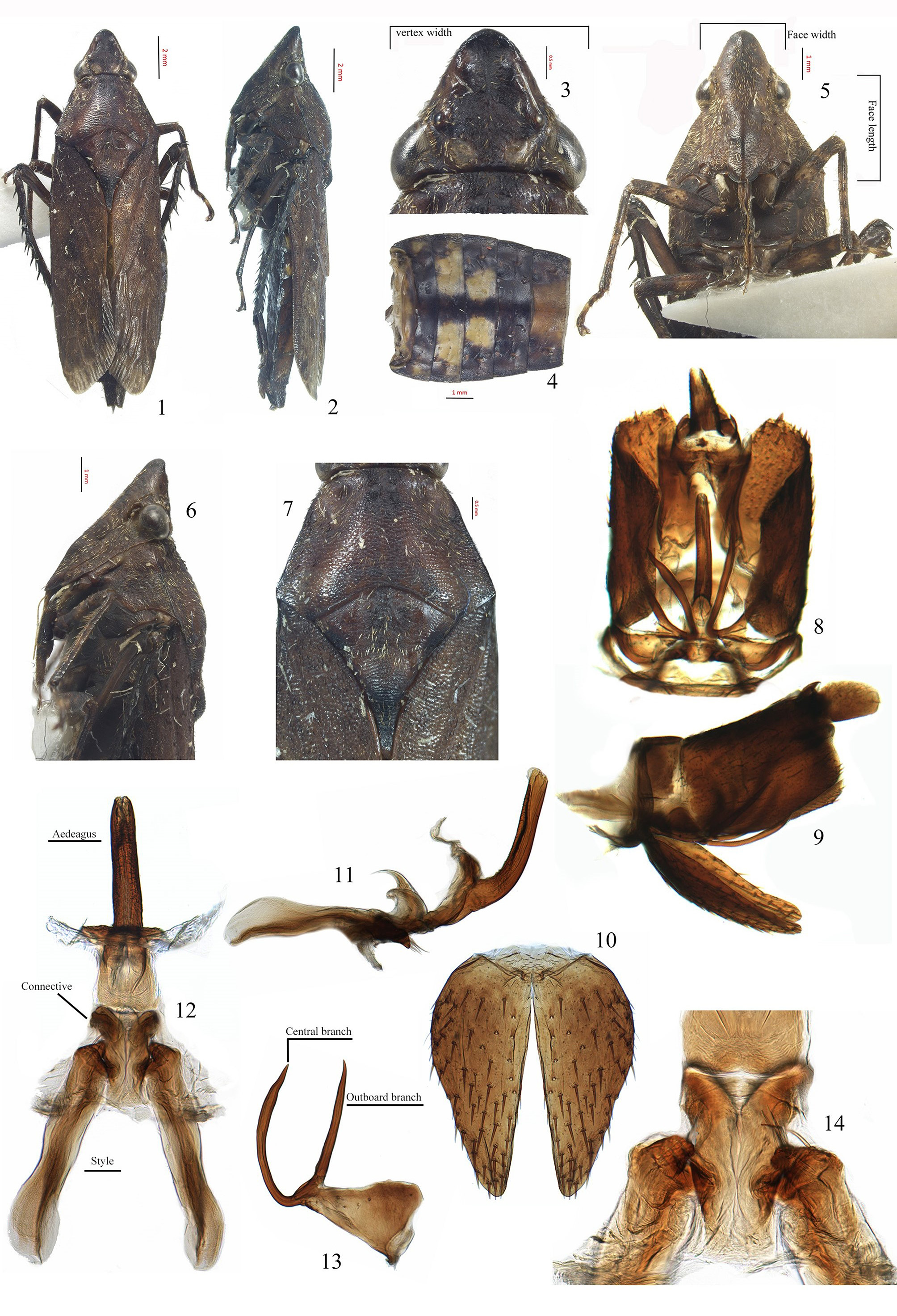
Diagnosis. Crown subtriangularly produced in front of eyes, longer or slightly shorter than basal width, rostrum reaching mesothoracic coxae, front tibiae slender, male ventral appendages with apex bifurcated and pointing laterad.
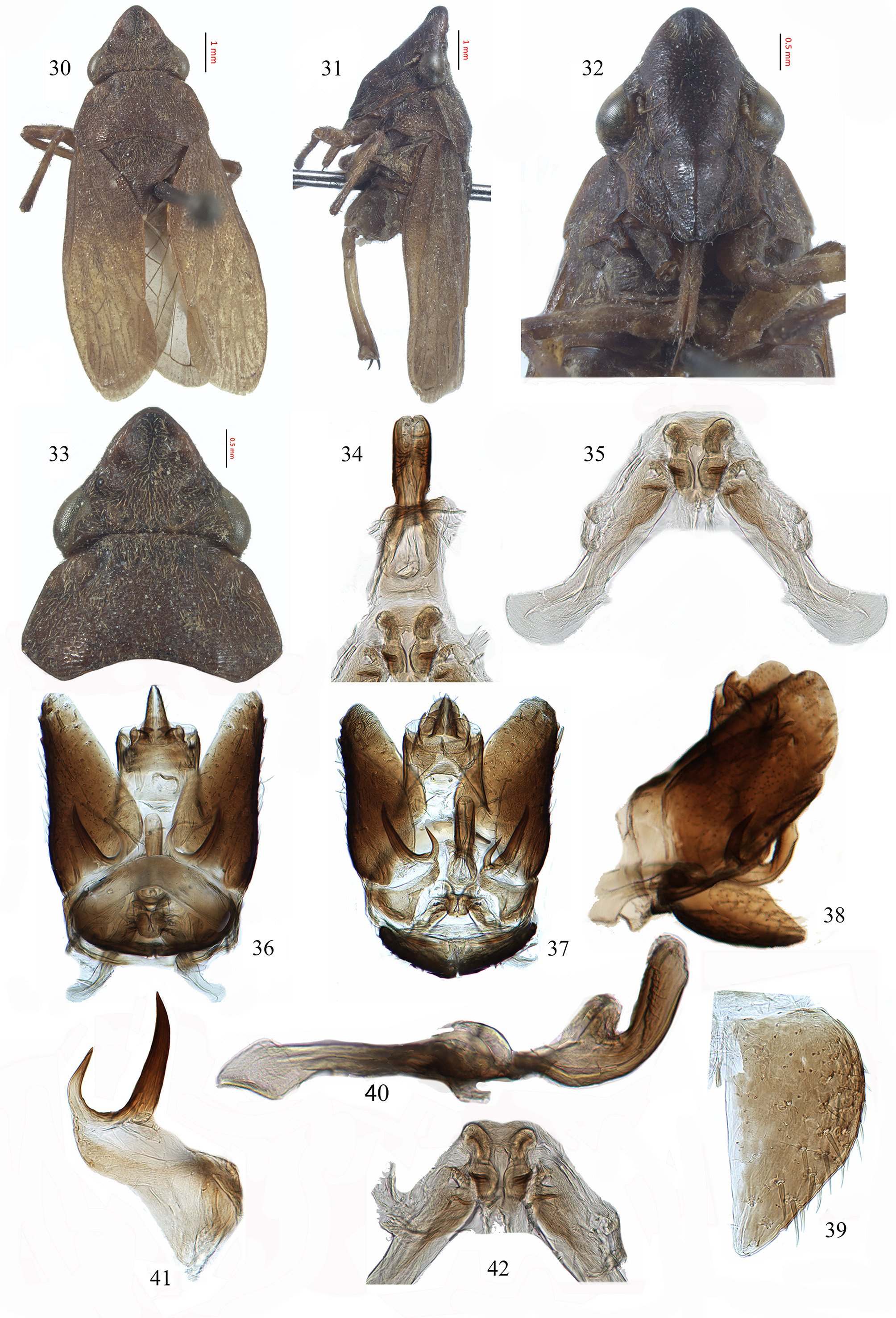
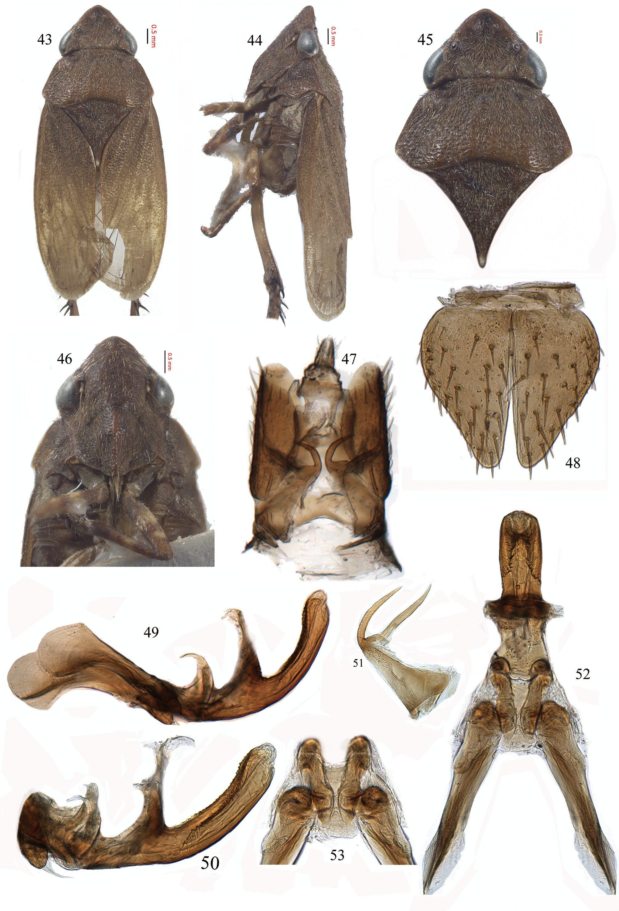
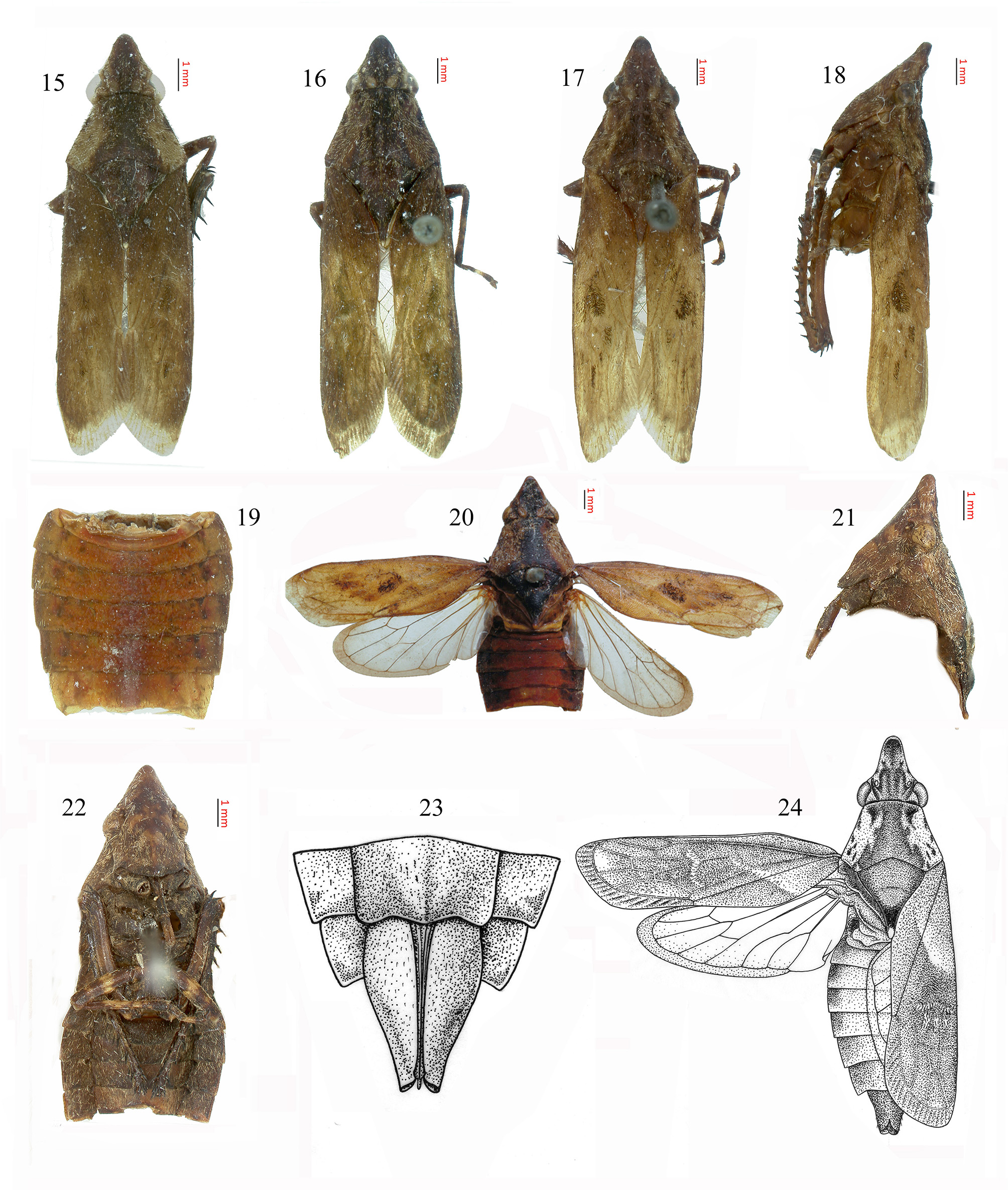
Description. Medium to large leafhoppers (body length 10.0–17.3 mm including forewing). Body yellow brown to dark brown and densely covered with tiny dark and light setae. Crown ( Figs. 3 View FIGURES 1–14 , 33 View FIGURES 30–42 , 45 View FIGURES 43–53 , 56 View FIGURES 54–68 ) triangular, produced anteriorly in front of eyes, as long as or longer than width between eyes, depressed medially, with lateral submarginal ridge, apex slightly upturned, one pair of short tubercles located subapically mesad of lateral ridge, one pair of submedial ovoid depressions without setae between eyes near posterior margin, posterior margin somewhat concave; eyes reniform, ocelli located slightly anteromesad of eyes on small tubercule near crown margin. Face ( Figs. 5 View FIGURES 1–14 , 22 View FIGURES 15–24 , 32 View FIGURES 30–42 , 46 View FIGURES 43–53 , 60 View FIGURES 54–68 ) with antennal ridge moderately well developed, curved, not concealing antennal base in anterior view; base of antennal flagellum paler than rest of antenna, lateral frontal suture extended onto crown in front of ocelli, frontoclypeus in profile slightly bent forward and with short longitudinal ridge at apex, clypeal sulcus obvious; anteclypeus convex and gradually narrowed apically with indistinct median longitudinal ridge, apex extended slightly beyond lower margin of gena; lorum narrow, well separated from gena, dorsal end even with clypeal sulcus, gena exposing long, narrow proepisternum; rostrum reaching mesothoracic coxae. Pronotum ( Figs. 6, 7 View FIGURES 1–14 , 31, 33 View FIGURES 30–42 , 44, 45 View FIGURES 43–53 , 58, 59 View FIGURES 54–68 ) trapezoidal with lateral margins straight, carinate and strongly divergent posterad, elevated posterad and sloping laterally, anterior margin slightly produced, two pairs of dimples located anterolaterally, posterior margin concave, surface granulose and transversely rugulose, lateral surface rugose, narrowed downward and concealing most of mesepisternum. Exposed part of mesonotum and scutellum ( Figs. 6, 7 View FIGURES 1–14 , 30, 31 View FIGURES 30–42 , 44, 45 View FIGURES 43–53 , 55, 59 View FIGURES 54–68 ) granulose and as long as or slightly longer than pronotum, sides of mesonotum slightly protuberant, scutellum swollen distad of scutoscutellar sulcus in lateral view and attenuate in dorsal view. Forewing granulose, claval suture obviously down folded, venation largely obscured by opaque sclerotization in basal two-thirds, veins R, M fused basally, vein RA1 very weak or absent, veins Pcu and A1 separated from each other; central anteapical cell slightly longer than outer and inner anteapical cells; 4–5 apical cells, 3rd apical cell usually shortest; appendix well developed, extended around wing apex from claval suture to 4th apical cell. Abdomen ( Figs. 1, 4 View FIGURES 1–14 , 19 View FIGURES 15–24 , 54, 57 View FIGURES 54–68 ) extending beyond wings at rest, yellow brown to dark red, lateral margins not or only slightly extended laterad of forewings at rest, nearly parallel-sided and gradually narrowed posteriorly; abdominal tergites of male with irregular spots and most of tergites with paired foveae. Foreleg and midleg with pale round spots, front tibia slender, not dilated. Hind femur macrosetal formula 3+1+0; tibia row AD with spinelike setal bases, with ca. 10–11, 8–11 setae in rows AD, PD, respectively.
Male genitalia: Pygofer ( Figs. 8, 9 View FIGURES 1–14 , 36–38 View FIGURES 30–42 , 47 View FIGURES 43–53 , 61–63 View FIGURES 54–68 ) base short, separated from lobe by vertical membranous cleft, with bifurcate ventral appendages partly separated from base by membrane and extended dorsolaterad on inner surface of lobe ( Figs. 13 View FIGURES 1–14 , 41 View FIGURES 30–42 , 51 View FIGURES 43–53 , 68 View FIGURES 54–68 ); lobe with many macrosetae, parallelogram-shaped in lateral view. Segment X with pair of long, slender apodemes. Valve ( Figs. 10 View FIGURES 1–14 , 48 View FIGURES 43–53 , 65 View FIGURES 54–68 ) stongly reduced and fused with subgenital plate. Subgenital plate ( Figs. 10 View FIGURES 1–14 , 39 View FIGURES 30–42 , 48 View FIGURES 43–53 , 65 View FIGURES 54–68 ) depressed with lateral margin evenly convex, shorter than pygofer side, gradually narrowing posteriorly and terminating in blunt angle, with scattered macrosetae on ventral surface. Connective ( Figs. 11, 14 View FIGURES 1–14 , 40, 42 View FIGURES 30–42 , 49, 53 View FIGURES 43–53 , 64 View FIGURES 54–68 ) subtrapezoidal with lateral margins vertically expanded to keeled process, anterior part of vertical process posteriorly curved and gradually acute. Style ( Figs. 11, 12 View FIGURES 1–14 , 35, 40 View FIGURES 30–42 , 49, 52 View FIGURES 43–53 , 64, 66 View FIGURES 54–68 ) with apodeme long, apophysis reduced, lobelike, with several slender setae on ventral surface. Aedeagus ( Figs. 11, 12 View FIGURES 1–14 , 34, 40 View FIGURES 30–42 , 49, 50, 52 View FIGURES 43–53 , 64, 66, 67 View FIGURES 54–68 ) symmetrical and curved dorsad, tubular, dorsal surface flat or slightly concave and with sculpture, preatrium and dorsal atrium not strongly developed.
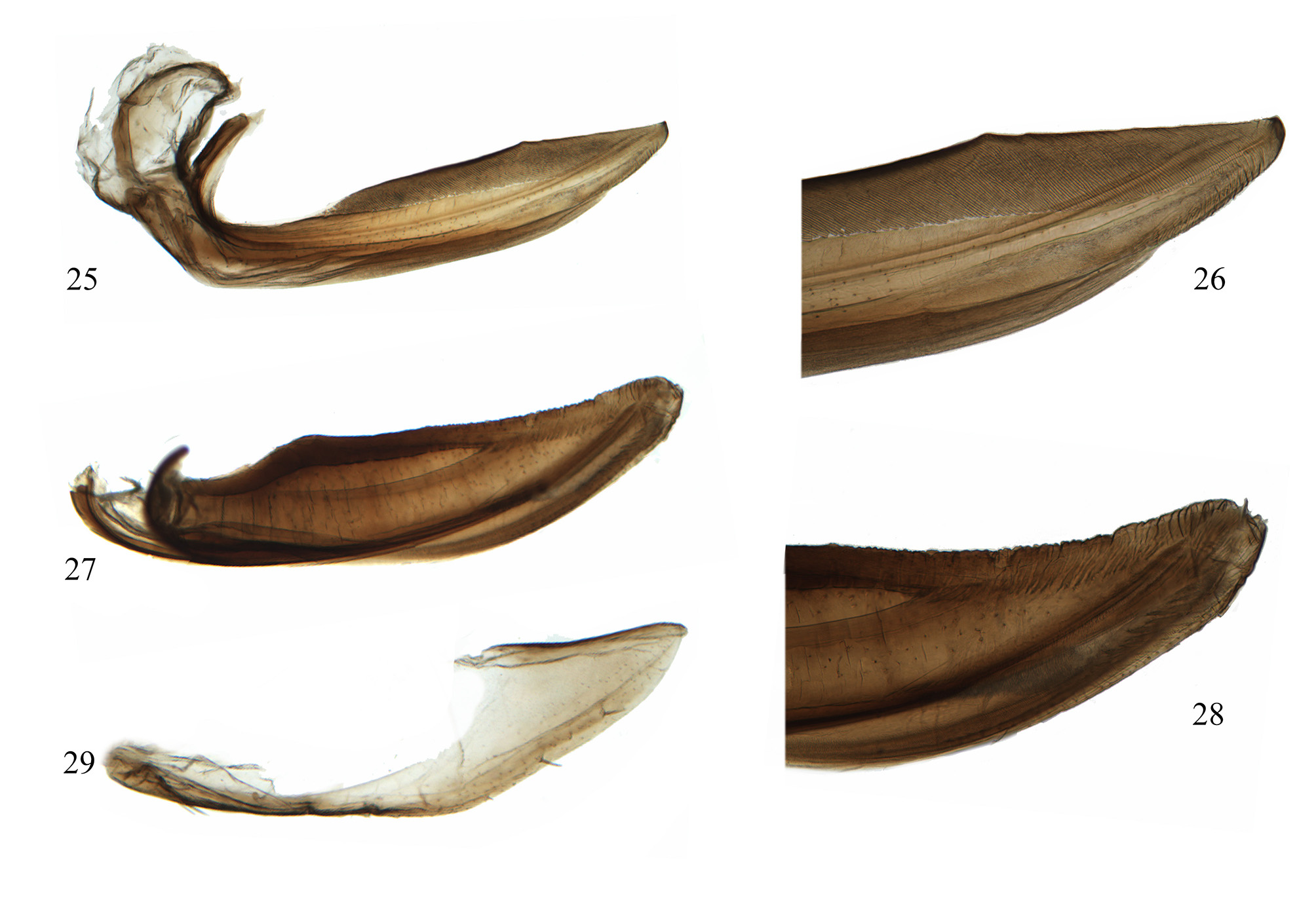
Female. Pygofer ( Fig. 23 View FIGURES 15–24 ) slightly emarginate apically rather than deeply bifid. Sternite VII ( Fig. 23 View FIGURES 15–24 ) with middle convex and both sides protruded backward in ventral view. Ovipositor not reaching pygofer apex. First valvula ( Figs. 25, 26 View FIGURES 25–29 ) sword-like with many irregular punctations along medial ridge, blade narrowest at base, dorsal sculpture densely strigate. Second valvula ( Figs. 27, 28 View FIGURES 25–29 ) knife-shaped, narrow at base, broadened in distal two-thirds, dorsal margin slightly concave with numerous small teeth on distal half, ventral margin convex, apex narrowly rounded. Third valvula sheath-shaped, connected with pygofer by membrane, with several small setae on ventral margin.
Ecology. This genus is arboreal and usually collected in forests..
Distribution. China, Malaysia (new record), Thailand (new record), Viet Nam, West Indonesia.

No known copyright restrictions apply. See Agosti, D., Egloff, W., 2009. Taxonomic information exchange and copyright: the Plazi approach. BMC Research Notes 2009, 2:53 for further explanation.
|
Kingdom |
|
|
Phylum |
|
|
Class |
|
|
Order |
|
|
Family |
Kalasha Distant
| Tang, Jiu & Zhang, Yalin 2019 |
Kalasha
| Knight, W. J. 2010: 143 |
| Shen, L. & Zhang, Y. - L. 1995: 185 |
| Oman, P. W. & Knight, W. J. & Nielson, M. W. 1990: 222 |
| Metcalf, Z. P. 1962: 9 |
| Evans, J. 1946: 47 |
| Jacobi, A. 1914: 379 |
| Distant, W. 1908: 254 |