
Microdon analis (Macquart, 1842)
|
publication ID |
https://doi.org/10.1080/00222933.2015.1010314 |
|
DOI |
https://doi.org/10.5281/zenodo.4330134 |
|
persistent identifier |
https://treatment.plazi.org/id/03F4879D-FF83-507D-9259-B666FDC35E66 |
|
treatment provided by |
Carolina |
|
scientific name |
Microdon analis |
| status |
|
Zoophagy: Microdon analis View in CoL and Melangyna cincta (Syrphidae)
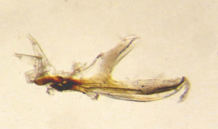
The larva of M. analis lives in galleries excavated by Formica ants ( Hymenoptera , Formicidae ) under bark of fallen pine trees and stumps ( Pinus sylvestris L., Pinaceae ) where access is restricted+ and feeds on their early stages ( Table 1). The larva of M. cincta feeds mainly on the aphid, Phyllaphis fagi (L.) ( Hemiptera , Aphididae ) which forms colonies on the undersides of leaves of beech, Fagus sylvatica L. ( Fagaceae ). Infested leaves often curl up and access is unrestricted to restricted+ ( Table 1). Aphids are surrounded and covered in varying amounts of secreted, white flocculence and honeydew is present, some of which is in the form of wax-coated droplets. Uniquely, these two species have antennomaxillary organs borne on elongate, cylindrical projections at the apex of the pseudocephalon. Between the projections is a shallow groove, along which the head skeleton protracts and retracts.
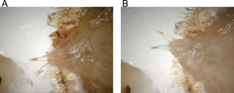
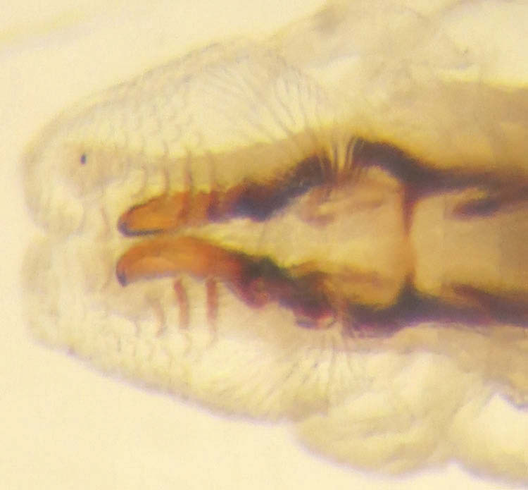
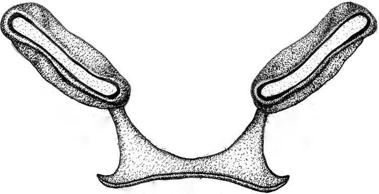
Microdon analis : rear compartment, anal segment to the metathorax, is hemispherical or domed in shape and rigid, although not sclerotised, and ventrally coated in fine setae. The metathorax is divided into an upper, basal section and a lower apical one, the latter being concealed along with the front compartments, under the basal section with the border between them being a continuation of a band of setae that circumvents the entire ventral margin of the body. The middle compartment, mesothorax to the pseudocephalon, is narrow in relation to the rear section and highly retractile. The relatively large mandibles are at the front of the head skeleton ( Figure 8 View Figure 8 ) and are thin and blade-like with serrated ventral margins, the eight or so posterior teeth being longer and rotated backwards and inwards ( Figure 9 View Figure 9 ). Posteriorly, the mandible is inflated and the rear face has an oval cavity which articulates with a peg-like, labial projection ( Figure 9 View Figure 9 ). A conspicuous, curved, tapering apodeme extends from the postero-ventral margin, round which attaches a tendon for the mandibular muscles ( Figure 9 View Figure 9 ). The apex of the labium is separate and comprises a pair of flat, elongate-oval sclerites, the labial sclerites, and between them, a triangular sclerite, the labial plate, which has a rounded apex and curves posteriorly to articulate with the basal section of the labium. The rear, inside margin of the labial plate bears upright spines.
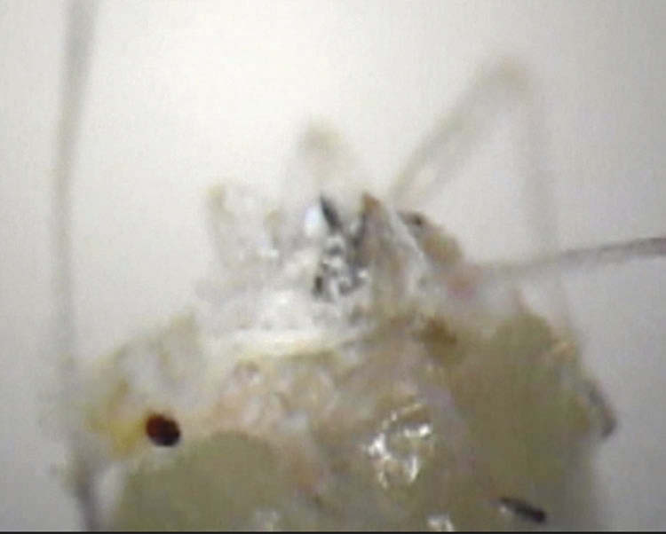
During movement, the middle compartment extends to slightly beyond the marginal band of setae and sweeps up to 70 degrees either side of the longitudinal midline ( Figures 10A and 10B View Figures 10 ). Locomotion is slow and, if a barrier or obstacle is encountered, the larva contracts the body at the point of contact and by shuffling backwards and sideways, attempts to bypass it. The larva can also rotate the entire body on a point, but does not grip the substrate with mandibles. Feeding consists of repeated lunges into the prey via protraction and retraction of the head skeleton. When protracting, the mandibles are depressed to a shallow 25–40 degrees angle relative to the horizontal. At the limit of protraction, the labial sclerites and plate depress to almost a right angle and the mandibles rise to the horizontal and may separate slightly, these actions expose the opening of the pharynx ready for food to be sucked in (Film 5, Figure 11 View Figure 11 ). The pump operates independently of lunging and 3–8 pumping actions occur per lunge. By altering the direction of protraction, the head skeleton can reach different areas inside the prey, but no seal is made between it and the larva and prey fluids spill from the wound.
Melangyna cincta : at rest, the larva of this species is similar to M. analis in that the front margin is the metathorax with the mesothorax and prothorax contracted underneath, but the metathorax is not fixed into two sections and when feeding or moving, these segments unfold. The larva is subcylindrical, highly flexible and the entire body has a complex, repeated, segmental pattern of integumental grooves and lines along which the integument collapses during movement. Rear compartment segments, anal segment to the prothorax posterior to the anterior spiracles, lack vestiture and parts of the ventral surface are smooth. The apex of the anal segment has a smooth-surfaced, transverse, grasping bar. The prothorax overlaps the mesothorax. Antero-laterally the prothorax has a pair of black, triangular-shaped sclerites that are basally attached to the integument ( Figure 12A and 12B View Figures 12 ). The labrum and labial plate are sclerotised and taper together at the front of the head skeleton ( Figures 12A and 12B View Figures 12 ). The labial plate articulates with the labial sclerites which are embedded in the sides of the head skeleton with muscles inserting on a postero-ventral apodeme ( Figure 12C View Figures 12 ). The mandibles are bar-shaped, taper posteriorly, lack muscles and are embedded into the lateral margins of the head skeleton ( Figure 12C View Figures 12 ).
This larva is the most prehensile of those studied here. It sweeps by holding on with the rear end and moving the front of the body up to about 75 degrees to either side. Unlike other species, the border between the rear and front compartments is not fixed and, depending on the extent of sweeping, the body bends at any point from about the sixth abdominal segment to the mesothorax. To initiate forward locomotion, muscles contract in front of the anus and a crease forms across the body. This pulls the grasping bar on to the substrate and a peristaltic wave moves forward. The sides of the body bulge with body fluids which forces the ventral surface against the substrate and gripping power moves along the body. When the peristaltic wave reaches the prothorax, it inclines and either holds on using a prominent, mid-ventral pad or, the tip of the head skeleton is pressed against the substrate and held there, presumably by suction pressure from the pump. Simultaneously, the crease at the rear end fills out and the grasping bar lifts to release the anal segment which is pulled forward and another peristaltic wave begins. When it reaches the prothorax, it lifts and extends forward and ahead of the previous position and so, forward locomotion occurs. The maximum rate recorded was one body length in 2.4 seconds.
The larva can also raise the body up as far as the fourth and fifth abdominal segments, and move forward with just the rear three or four segments while the front end remains elevated (Film 6). If flocculence is encountered when lunging for food, it probes it with the head skeleton and if it sticks, the larva wipes it off on the substrate. If a wax-coated droplet of honeydew is encountered, it pierces it and imbibes or spills the contents. If it encounters an aphid with any part of the ventral surface of the anterior compartments, the larva shuffles backwards until it can bend the prothorax and capture it. Capture involves holding it in place with sticky saliva, contraction of the prothoracic apex to form a cup-shape into which the prey is drawn and simultaneously exposes the triangular, lateral hooks to further grip the prey. Feeding occurs by protraction of the head skeleton co-ordinated with contraction of the prothorax which presses the sharp apices of the labrum and labial plate against the prey until it is pierced and, usually, the prey is lifted from the substrate. To extract food, the head skeleton protracts and retracts repeatedly and the labium lowers so that prey fluids enter the head skeleton and are sucked in by the pump (Film 7, Figure 13 View Figure 13 ). Recorded lunge rates were fairly constant at one lunge per 0.4–0.5 second. From capture to wiping off the remains at the end of feeding, handling time in a filmed sequence was 2 minutes 20 seconds, during which about 311 lunges were made.
Higher Cyclorrhapha
Compared with lower Cyclorrhapha , the labium is inactive in food gathering. The upper arm articulates with the mandibles, while the lower arm comprising labial plate and labial sclerites is incorporated into a new structure, the atrium. Also new is an oral cavity and an anal lobe, and food gathering includes a functional partnership between the pseudocephalon and mandibles. Four compartments are usually present: anal segment to abdominal segment 2; abdominal segment 1 to the anterior spiracles; anterior spiracles to the apex of the prothorax and, the head skeleton.
Saprophagy: Silba fumosa (Egger) , Lonchaea hackmani Kovalev (Lonchaeidae) ; Palloptera trimacula (Meigen) (Pallopteridae) ; Meiosimyza platycephala (Loew) (Lauxaniidae) ; Coelopa frigida (Fabricius) (Coelophidae) ; Clusia flava (Meigen) (Clusiidae) ; Neophyllomyza acyglossa (Villeneuve) (Milichiidae) ; Chymomyza costata (Zetterstedt) (Drosophilidae) ; Calliphora vomitoria (Linnaeus) (Calliphoridae) .
These larva feed on watery to soft-solid biofilm in narrow, concealed spaces where access is restricted + to ++ ( Table 1). However, C. frigida and C. vomitoria also feed in less constrained places where volumes of decay can be many times larval heights ( Table 1). Rear compartment segments relatively uniform and locomotory spicules straddle the borders between segments, always with fewer spicules on the segment in front; those spicules may incline forward and those behind incline backwards. Similar spicules coat the anal lobe and the anterior face of the prothorax, and reduced numbers are usually present at the border between abdominal segment 1 and the metathorax. The pseudocephalon comprises a pair of lobes each of which ensheaths a mandible with a gap ventro-apically through which the mandible protrudes ( Figure 14 View Figure 14 ). Anastomising cirri cover the lateral and ventral margins of the lobes and lead towards the pharynx between the mandible bases ( Figure 14 View Figure 14 ). Mandibles comprise two parts or sections: a subrectangular base and a curved, distal mandibular hook ( Figures 15 View Figure 15 and 16 View Figure 16 ). The mandibular base bears two apodemes to which elevator muscles attach at the upper, inner point and depressor muscles underneath the lower, outer point ( Figure 16 View Figure 16 ). The inside face of the mandibular hook is often flattened or scalloped ( Figure 14 View Figure 14 ).The mandibular base is often indented medially ( Figure 16 View Figure 16 ). A hinge joint exists between the mandible and the intermediate sclerite, with a ridge on the rear face of the mandible ( Figure 17 View Figure 17 ) fitting into a groove on the front margin of the intermediate sclerite ( Figure 18 View Figure 18 ). The anterior end of the intermediate sclerite is inflated to accommodate the groove and greatly more sclerotized relative to the base and the ventral bridge at this end of the intermediate sclerite ( Figure 15 View Figure 15 ). Dental sclerites extend under the base of each mandible and are enveloped in depressor muscle tendon ( Figure 16 View Figure 16 ). The basal sclerite is variably sclerotised but usually more in the upper half and the cornua are more or less parallel ( Figure 15 View Figure 15 ).
Feeding and movement consist of sweeping and lunging (Film 1, Figure 19A–C View Figures 19 ). Lunging is inclination of the prothorax and except for C. flava , in which the head skeleton is fixed in the thorax, involves protraction and retraction of the head skeleton and depression of the mandibles (Films 8–11, Figure 20A–E View Figures 20 ).
As one lunge ends:
● abdominal segments 8–2 are extended and stationary;
● abdominal segment 1, the metathorax, mesothorax and the prothorax to the level of the anterior spiracles are contracted;
● the prothorax beyond the spiracles is inclined;
● the pseudocephalon and head skeleton are retracted and the mandibles depressed ( Figure 20A and 20B View Figures 20 ).
A new lunge begins when:
● the head skeleton pivots and protracts, the mandibles elevate and the prothorax beyond the anterior spiracles rises and extends; these actions bring the mandibles together which closes the oral cavity, tapers the pseudocephalon and moves these compartments towards food or a substrate ( Figure 20C View Figures 20 );
● at the limit of extension, the prothorax beyond the anterior spiracles inclines down and the mandibles depress and separate and the labial lobe retracts; these actions extend the mandibular hooks from their sheaths, opens the oral cavity and exposes the pharynx ( Figure 20D View Figures 20 );
● on reaching a firm substrate, the entire ventral length of the mandibles are briefly parallel with it ( Figure 20E View Figures 20 ), i.e. the mandibular hooks are in the food, the oral cavity is open and food is gathered into it by retraction of the head skeleton and depression of the mandibles ( Figures 20B View Figures 20 and 21 View Figure 21 );
● the mandibles depress to their limit which lifts the oral cavity ( Figure 20B View Figures 20 ) and biofilm within the oral cavity is sucked into the head skeleton by the pump in the basal sclerite; from inward movement of the dorsal cornua, there is more than one pumping action per depression.
In deep quantities of biofilm, food may be gathered by repeated elevation and depression of the mandibles alone. Extra-oral digestion was not observed in these species, i.e. saliva secreted prior to ingestion. Lunge times were within the range of one lunge per 0.4–0.9 sec ( Table 2). The distances head skeletons protract from the thorax were 29–100% of head skeleton length ( Table 2). The range of angles over which mandibles elevate and depress was greater below than above the horizontal axis ( Table 2). During depression, the mandibles separate to about three times their distance apart at elevation ( Figure 21 View Figure 21 ).
Forward locomotion starts with contraction across the ventral and lower lateral margins of the seventh abdominal segment anterior to the anus, which presses the anal lobe against the substrate, and is followed by a peristaltic wave passing along the body. When the wave reaches the prothorax, a lunge takes place and the mandibles depress and grip the substrate. This momentarily blocks the next wave and, at the segment border with the prothorax, the mesothorax folds and slides forward over the prothorax as far as the anterior spiracles ( Figure 22 View Figure 22 ) and the front of the prothorax slides over the pseudocephalon ( Figure 23A and 23B View Figures 23 ). At the next wave, the mandibles elevate and a lunge occurs which moves the anterior end ahead of the previous position. The main difference between feeding and locomotory lunges is that, in locomotion, the mandibles partially depress to grip the substrate, whereas in feeding, the mandibles depress to their limit.


No known copyright restrictions apply. See Agosti, D., Egloff, W., 2009. Taxonomic information exchange and copyright: the Plazi approach. BMC Research Notes 2009, 2:53 for further explanation.