
Hymedesmia (Hymedesmia) peruana, Salani & Willenz & Fernandez & Hajdu, 2022
|
publication ID |
https://doi.org/ 10.11646/zootaxa.5165.2.4 |
|
publication LSID |
lsid:zoobank.org:pub:B0D96EEA-B3FA-4607-8C28-4A19B3027BBF |
|
DOI |
https://doi.org/10.5281/zenodo.6831781 |
|
persistent identifier |
https://treatment.plazi.org/id/03F487E0-FFB9-9B5A-21FA-FB0ECAC7D0E0 |
|
treatment provided by |
Plazi |
|
scientific name |
Hymedesmia (Hymedesmia) peruana |
| status |
sp. nov. |
Hymedesmia (Hymedesmia) peruana View in CoL sp. nov.
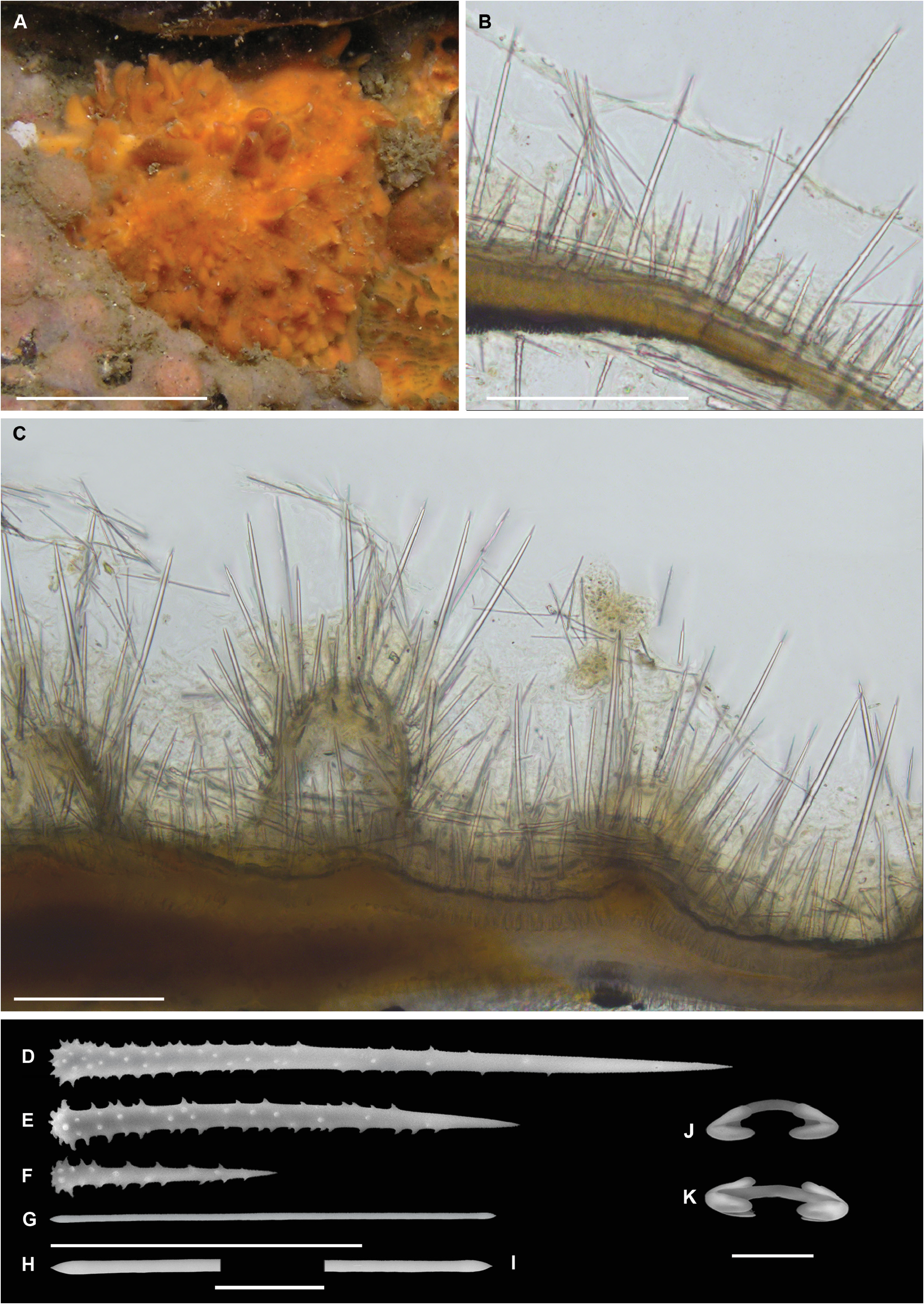
( Figures 3–4 View FIGURE 3 View FIGURE 4 ; Table 1 View TABLE 1 )
Material examined. Holotype ― MNRJ 13694 View Materials (Vouchers: RBINS – IG 32841 –POR 13694, MHNG 85931 View Materials ), Isla Foca , Piura Region (5.19529S, 81.2160W), depth 26 m, coll. Y. Hooker & M. Rios (13/XII/2009). GoogleMaps
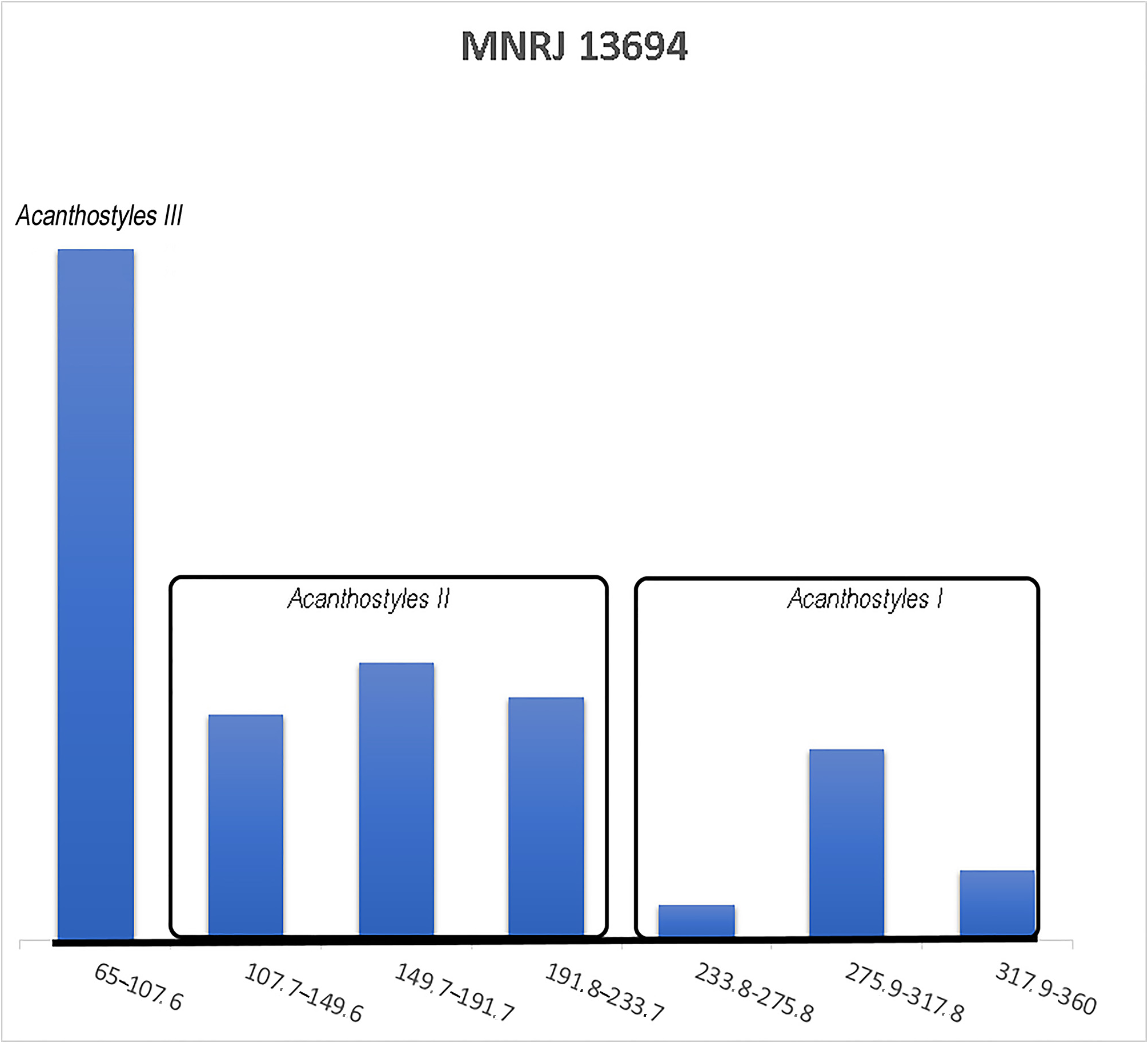
Diagnosis. Hymedesmia (H.) orange in situ. Acanthostyles I echinating and acanthostyles II forming microconules at surface. Three categories of acanthostyles, bigger and intermediate ones partially spined, and smaller entirely spined (I. 262–360 × 7–10 µm; II. 118–231 × 3.5–10 µm; III. 65–106 x 2.6–6.1), tornotes (117–146 ×1.6–3µm), arcuate chelae (14.7–21.3 µm).
Description ( Fig. 3A View FIGURE 3 ). Thinly encrusting sponge (8.8 cm 2), less than 1 mm thick (0.16 to 0.6 mm). Consistency very soft and fragile. Surface smooth. Color in life orange ( Fig. 3A View FIGURE 3 ), turning to beige when preserved in ethanol.
Skeleton ( Figs 3B–C View FIGURE 3 ). Ectosomal skeleton with ectosomal strongyles forming bundles (4 to 7 spicules) extending up from the substrate. Choanosomal acanthostyles I may pierce the surface occasionally ( Fig. 3B View FIGURE 3 ). Subectosomal and choanosomal skeletons overlapping, with subectosomal canals (45–265 µm diameter). Choanosomal skeleton composed of typical hymedesmioid structure, consisting of a basal layer of spongin with acanthostyles I and II erect on the substrate ( Fig. 3B–C View FIGURE 3 ). Acanthostyles I pierce the ectosome and the acanthostyles II support the surface, forming microconules. Isochelae appear scattered in the ectosome, and some tylostyles lay parallel to, or flat on the substratum.
Spicules ( Figs 3D–K View FIGURE 3 , 4 View FIGURE 4 ; Table 1 View TABLE 1 ). Ectosomal diactines: tornotes, straight, isodiametric ( Fig. 3G View FIGURE 3 ), with conical extremities, 117– 132.3 –146 (± 11.8) × 1.6– 2.2 –2.9 (± 0.4) µm ( Figs 3H–I View FIGURE 3 ). Choanosomal acanthostyles I ( Figs 3D View FIGURE 3 , 4 View FIGURE 4 ): hastate to acerate tip tapering gradually, rounded base; shaft straight, with spines occupying the basal two thirds of its length; spines mostly slightly curved, those of the base larger than those of the shaft, 262– 300.7 –360 (± 34.3) × 7.7– 8.3 –9.8 (± 0.8) µm. Choanosomal acanthostyles II ( Figs 3E View FIGURE 3 , 4 View FIGURE 4 ): similar to acanthostyles I, but slightly smaller, 118– 182.7 –231 (± 40.3) × 3.5– 6.5 –9.6 (± 2.0). Choanosomal acanthostyles III ( Figs 3F View FIGURE 3 , 4 View FIGURE 4 ): straight, tapering gradually, acerate tip and rounded base, spined all over, 65.7– 78.2 –106 (± 11.1) × 2.6– 4.1 –6.1 (± 0.8) µm. Arcuate isochelae ( Figs 3J–K View FIGURE 3 ): one free spatulate ala and two lateral alae semi-fused with the shaft ( Fig. 3K View FIGURE 3 ), shaft characteristically curved, bow shaped, 14.7– 16.4 –21.3 (± 1.4) µm.
Ecology. This single specimen was epibiontic on a bivalve shell at 26 m depth.
Distribution. Known only from its type locality, Isla Foca.
Etymology. The species name “ peruana ” is used as a noun in apposition that refers to Peru.
Remarks. Hymedesmia (H.) peruana sp. nov. differs from H. (H.) santarositae sp. nov., by the combined absence of sigmas and microstrongyles ( Table 1 View TABLE 1 ). Hymedesmia (H.) croftsae Goodwin et al., 2012 has similarly small ectosomal tornotes, but these are strongylote, and acanthostyles, albeit exhibiting similar overall dimensions, seemingly cannot be separated in three categories. Hymedesmia (H.) humboldti sp. nov. also bears some similarities, which are further elaborated in its remarks (see below). Other species of Hymedesmia (H.) considered here (reported from the Antarctic, Sub-antarctic, SW Atlantic and SE Pacific) possess considerably larger spicules, none has three categories of acanthostyles, and many are only known from the deep sea.
| RBINS |
Royal Belgian Institute of Natural Sciences |
No known copyright restrictions apply. See Agosti, D., Egloff, W., 2009. Taxonomic information exchange and copyright: the Plazi approach. BMC Research Notes 2009, 2:53 for further explanation.
|
Kingdom |
|
|
Phylum |
|
|
Class |
|
|
Order |
|
|
Family |
|
|
Genus |
|
|
SubGenus |
Hymedesmia |