
Dysphaea vanida, Hämäläinen, Matti, Dow, Rory A. & Stokvis, Frank R., 2015
|
publication ID |
https://doi.org/ 10.11646/zootaxa.3949.4.1 |
|
publication LSID |
lsid:zoobank.org:pub:B3123099-882F-4C42-B83B-2BA1C2906F65 |
|
DOI |
https://doi.org/10.5281/zenodo.5691011 |
|
persistent identifier |
https://treatment.plazi.org/id/03F487E5-FFCD-FFC7-FF78-0C494C219E01 |
|
treatment provided by |
Plazi |
|
scientific name |
Dysphaea vanida |
| status |
sp. nov. |
Dysphaea vanida View in CoL spec. nov.
( Figs. 12 View FIGURES 9 – 12 , 16 View FIGURES 13 – 18 , 29–30 View FIGURES 25 – 30 , 34, 36 View FIGURES 31 – 36 , 40 View FIGURES 37 – 40 , 44 View FIGURES 41 – 44 , 48 View FIGURES 45 – 48 , 52 View FIGURES 49 – 52 , 56 View FIGURES 53 – 56 , 62 View FIGURES 57 – 62 , 68 View FIGURES 63 – 68 , 74 View FIGURES 69 – 74 , 81–82 View FIGURES 77 – 82 , 86 View FIGURE 86 )
Dysphaea dimidiata Selys forma (?);— Asahina (1985: 34, 36, Figs. 45–47 View FIGURES 45 – 48 , 67 View FIGURES 63 – 68 , ‘South Thailand’).
Dysphaea dimidiata View in CoL [nec Selys, 1853]; Asahina (1981: 6; same specimens as above);— Asahina (1990: 10–11, 17, Figs. 40–41 View FIGURES 37 – 40 View FIGURES 41 – 44 ; Ranong and Chumphon);— Hämäläinen & Pinratana (1999: 29, 123, part; records from Tak, Kanchanaburi and Ranong);— Michalski (1992: 61, Kanchanaburi);— Donnelly (1998: 140, Tak);— Donnelly (2000: 162, Kanchanaburi);— Khunwiset & Chanpaisaeng (2002: 193, Kanchanaburi);— Kitagawa & Katatani (2005: 26, 27, Figs. 9–10 View FIGURES 9 – 12 , Kanchanaburi). Dysphaea walli View in CoL [nec Fraser, 1927];— Hämäläinen (1988: 24, Kanchanaburi); Hämäläinen (1989: 34, Ranong).
Material studied: Holotype ♂: Thailand, Ranong province, Khlong Nakha, Khlong Bang Man, alt. 20–40 m, 12– 13 v 1999, leg. M. Hämäläinen. Deposited at RMHN, Leiden. Paratypes (30 ♂, 2 ♀, all from Thailand), deposited in Coll. Hämäläinen, if not otherwise stated; 4 ♂, Ranong, same data as for holotype; 3 ♂, as above, but date 8–10 ii 1988; 2 ♂, as above, but date 10–11 iv 1998; 1 ♂, as above, but date 9–11 iv 2000; 2 ♂ (in RMNH: RMNH.INS.505755, 505749), as above, but date 25–26 iii 2001; 2 ♂ (1 ♂ in RMNH, RMNH.INS.505799, as above, but date 8–12 v 2002; 1 ♂ (in RMNH: RMNH.INS.505742), as above, but date 5 iv 2003; 2 ♂, Kanchanaburi province, Thong Pha Phum District, Lam Khlong Ngu, alt. 490 m, 3 x 1986, leg. M. Hämäläinen; 3 ♂, as above but date 8–9 v 1999, leg. M. Hämäläinen; 2 ♂ (in Coll. Dow), as above, but date 4–6 v 2002; 2 ♂ (in RMNH, RMNH.INS.509984, 509985), as above but date 25 v 2003; 1 ♂, 1 ♀ (in tandem), Kanchanaburi, Thong Pha Phum District, Nang Kroan, alt. 600 m, 20 x 1999, leg. M. Hämäläinen; 1 ♂, Thong Pha Phum District (obviously Lam Khlong Gnu), 1990s, ex. coll. A. Pinratana; 1 ♂, 1 ♀, Tak province, Mae La Mao, 6 v 1998, ex coll. A. Pinratana; 1 ♂ (in Coll. André Günther), Surat Thani province, Khao Sok, Sok river, 22 ii 2001, leg. A. Günther; 1 ♂ (in Coll. André Günther), Surat Thani, Khao Sok, Bang Laen river, 21 ii 2001, leg. A. Günther; 1 ♂ (in Coll. André Günther), Phangnga province, Khao Lak, Bang Niang river, 9 iv 2009, leg. A. Günther. Other material. Earlier the first author has studied ca. 30 male specimens, collected in 1987–2003 by Bro. Amnuay Pinratana and his co-workers in the same sites as the paratypes from Ranong, Kanchanaburi and Tak provinces. These specimens are preserved at Insect Museum at St Gabriel’s College (Bangkok), which houses Pinratana’s collection.
Etymology. The species epithet is based on the common Thai girl name Vanida . In Thai the name means ‘girl’. The name is a noun in apposition and is not named after any particular person.
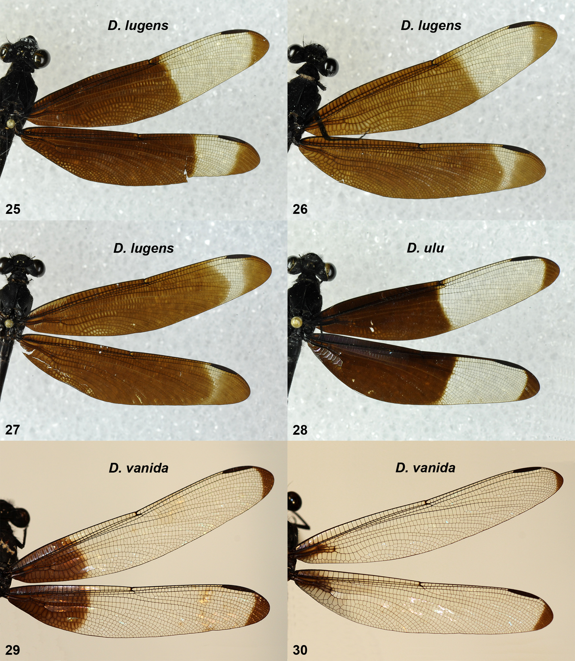
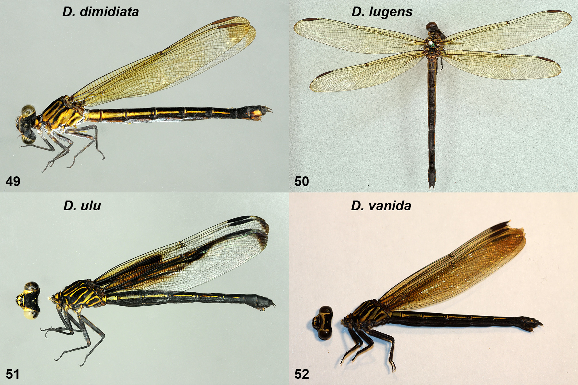
Diagnosis. A narrow winged Dysphaea species, males of which have only a small opaque patch at the wing base. Wing tips narrowly darkened.
Description of holotype male ( Fig. 12 View FIGURES 9 – 12 ). Head: Labium, labrum, base of mandibles and clypeus shining black, frons and vertex matt black.
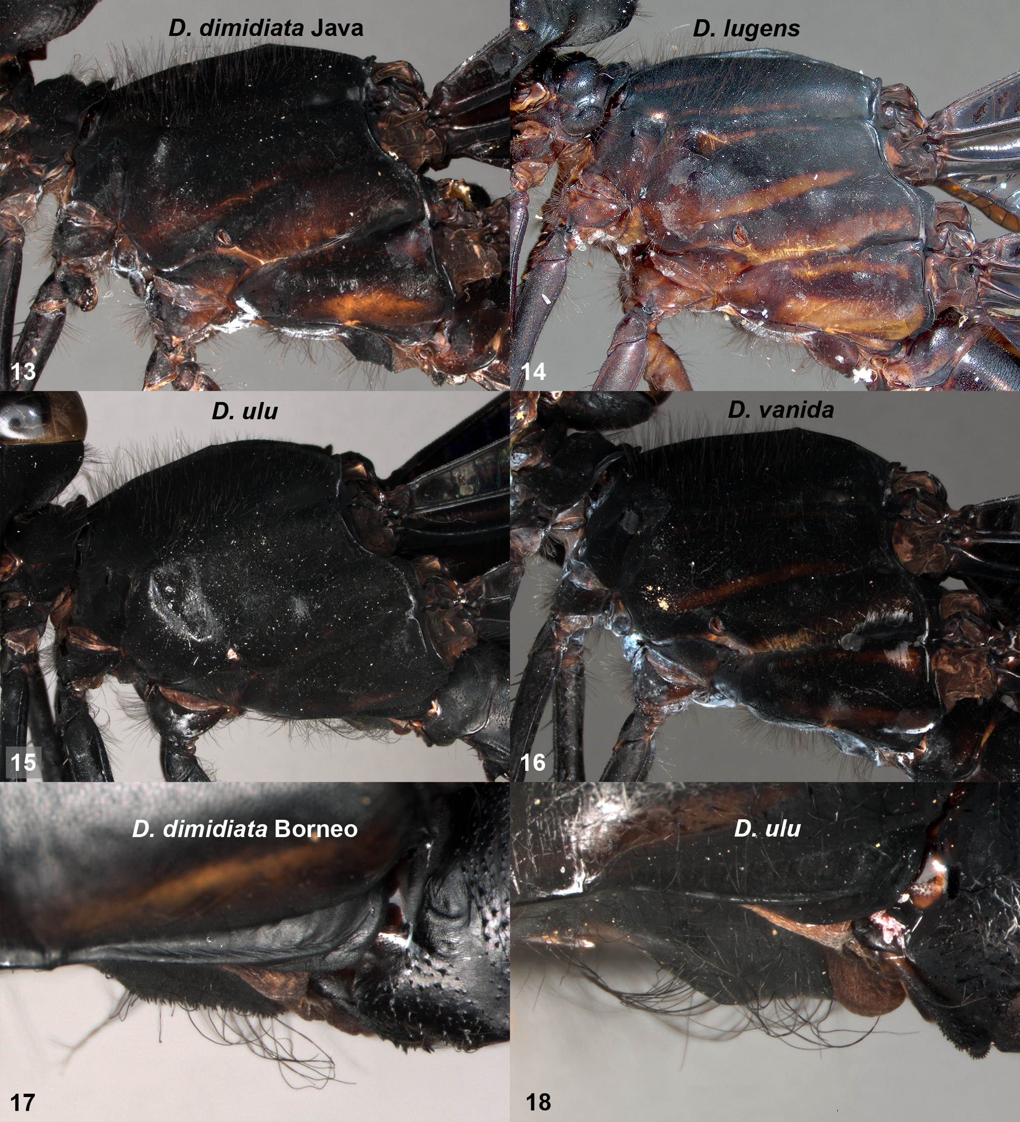
Thorax: Synthorax matt black with rather obscure, narrow, brownish stripes on sides as in Fig. 16 View FIGURES 13 – 18 . The upper stripe on mesepimeron, below the humeral suture, very narrow. Venter of thorax without tiny tubercles on poststernum. Legs wholly black, except posterior corner of hind coxa brownish.
Wings: Basal area of wings opaque black, with slight violet lustre. In Fw opaque area extends to half way between base and nodus. In Hw basal opaque area extends slightly more apicad, over half way between base and nodus. Tips of wings narrowly opaque, in Hw slightly more extensively than Fw (cf. Fig. 29 View FIGURES 25 – 30 ). Venation typical for the genus. Fw with 33–34 antenodals in first row; Hw correspondingly with 26–27 antenodals. Quadrangle with 0– 1 crossveins in Fw, 1–2 in Hw. Cubital field with 3 crossveins in Fw and Hw, all crossveins in the apical half of the field. Pterostigma long and narrow, broadest in middle; covering 9–11 underlying cells.
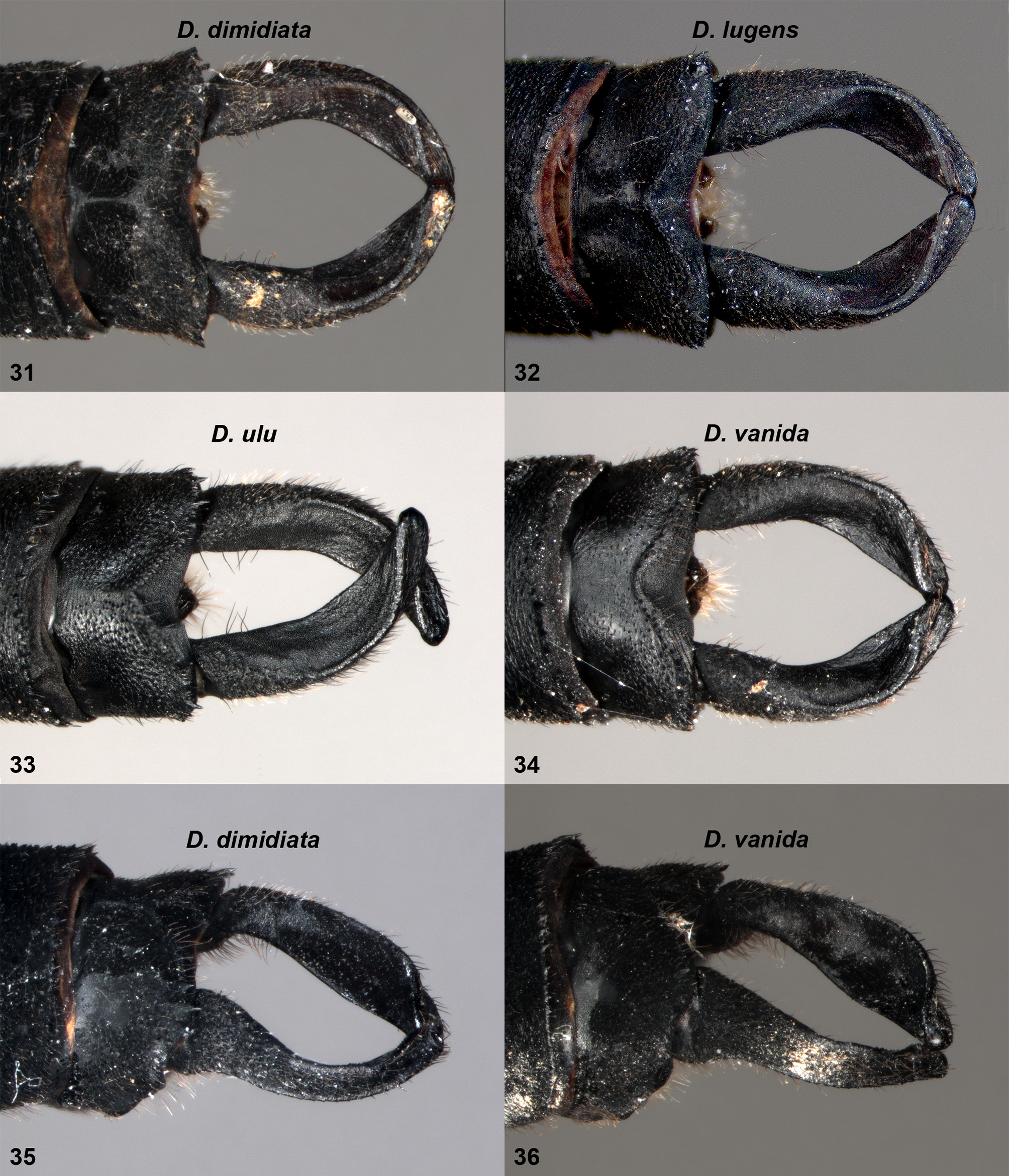
Abdomen: Matt black, with obscure remnants of brownish lateral stripes on S2–S4. Small pale dorsolateral markings at base of S3–S6. Appendages black; cerci in dorsal and ventral view of typical shape for genus; in lateral view ventral margin of cercus distinctly arched ( Figs. 34 View FIGURES 31 – 36 , 40 View FIGURES 37 – 40 ). In oblique dorsal view, cerci clearly broader in the middle part ( Fig. 36 View FIGURES 31 – 36 ). Paraprocts rudimentary.
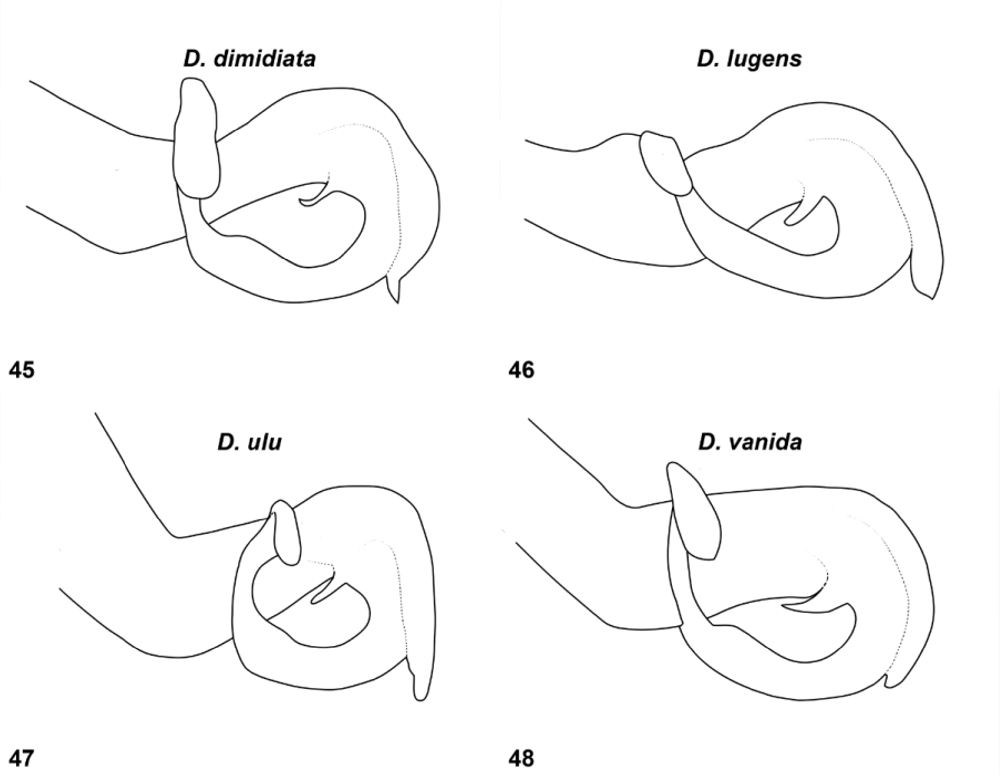
Penis: Terminal segment with two long, flat, rectangular, apical arms directed straight upwards on either side of shaft, then outwards and downwards ( Figs. 44 View FIGURES 41 – 44 , 48 View FIGURES 45 – 48 ).
Measurements (mm): Fw 35, Hw 33, abdomen (apps. excl.) 36, cerci 2.
Description of female. Based on a specimen collected in tandem with a male paratype at Nang Kroan in Kanchanaburi ( Fig. 52 View FIGURES 49 – 52 ).
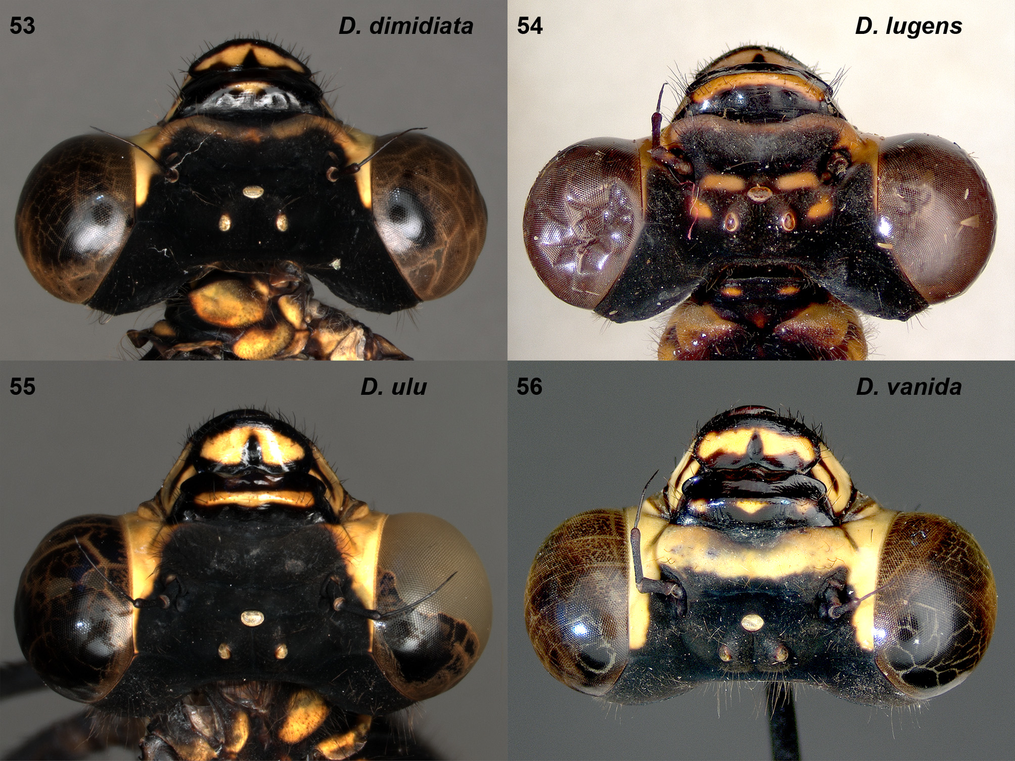
Head: Labium shining black with lateral lobes and sides of middle lobe yellow in basal half. Labrum shining black, with yellow marking in centre, indented with black basally. Base of mandibles largely yellow with black incomplete stripe medially. Clypeus shining black with three tiny yellow spots along ridge. Frons matt black with broad yellow band throughout antefrons, colour contiguous with yellow genae ( Fig. 56 View FIGURES 53 – 56 ). Antennae black. Yellow of genae extends almost to the level of lateral ocelli. Vertex and occiput matt black, with two tiny yellow spots on occiput. Eyes in life chestnut brown above, pale greenish below.
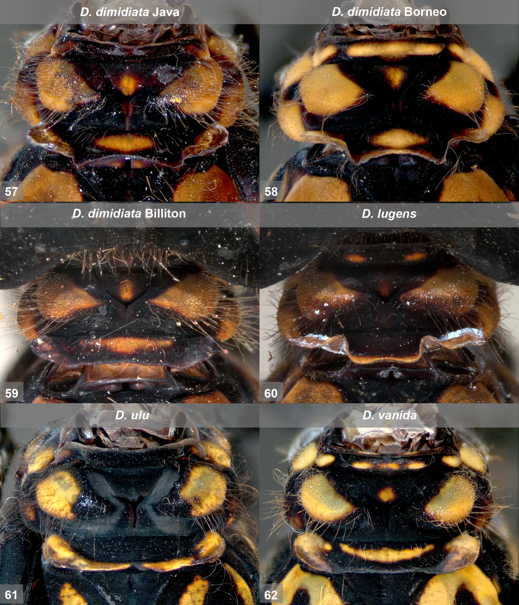
Thorax: Prothorax black with a row of tiny yellow spots on anterior lobe, rounded large yellow spots on either side of dorsum of middle lobe and a tiny spot medially. Posterior part of hind lobe raised obliquely upwards to form an elongate rectangular flap with prominent lateral ends ( Fig. 62 View FIGURES 57 – 62 ). Flap long, the median part slightly more than half length of entire hind lobe, with narrow yellow median stripe and with ends broadly yellow laterally. Synthorax matt black, with pale (pale greenish in life) stripes as in Figs. 68 View FIGURES 63 – 68 , 74 View FIGURES 69 – 74 . Stripes on mesepisternum and metepimeron forming a complete loop. Legs black, with yellow markings on posterior side of coxae.
Wings: Hyaline with slightly brownish tinge, especially at tips of wings. Fw with 31 antenodals in the first row; Hw with 26–27. Quadrangle with 1–2 cross veins in Fw, 2–3 in Hw. Pterostigma long, covering 8–9 underlying cells.
Abdomen: Matt black, with pale (pale greenish in life) markings as follows: S1 with large lateral spot, covering most of the side of segment. S2–7 with lateral stripes, broadest on S2–3 and gradually narrowing towards the apical segments. Obscure tiny lateral spot on S8 and a little larger, distinct lateral spot at S 9. Appendages black.
Measurements (mm): Hw 33, abdomen 31 (apps. excl.), cerci 1.
Variation in male paratypes. In some specimens the pale markings on synthorax and on sides of the basal abdominal segments are more distinct, clearly an age-dependent character. In younger specimens the small yellowish dorsolateral markings at the base of S3–6 are more distinct (cf. Figs. 81–82 View FIGURES 77 – 82 ). The extent of the opaque area in the wings is somewhat variable. In some specimens from the type locality the opaque basal area in the Fw extends only one third of the length of area between base and nodus and in the Hw less than half of this distance. In one specimen from the type locality the opaque patch on wings is very small, reaching only to the apical end of quadrangle ( Fig. 30 View FIGURES 25 – 30 ). A specimen from the same site with quite similarly reduced wing patches was illustrated by Asahina (1990, Fig. 40 View FIGURES 37 – 40 on p. 17). In a few specimens from Kanchanaburi the opaque area is slightly more extensive than in the holotype, in the Fw reaching over the half way of the distance between base and nodus, in the Hw reaching to the level of 7 cells before the nodus. There is also slight variability in venational details.
Measurements (mm): Hw 33–35, abdomen (excl. cerci) 38–39.5.
Variation in female paratypes. Specimen from Tak is slightly larger in size; Hw 34 mm, abdomen 32 (apps. excl.) mm, cerci 1 mm. Colour pattern of body and wings is quite similar. Fw with 33–35 antenodals in the first row; Hw with 25–27. Quadrangle with 1–2 crossveins in Fw, 2–3 in Hw. Pterostigma long, covering 8–11 underlying cells.
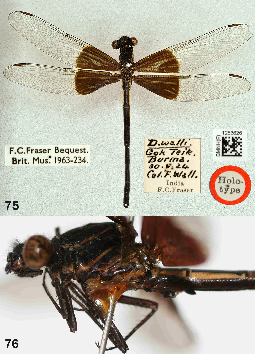
Distinguishing characters. Male: D. vanida is easy to separate from D. dimidiata by the less extensive basal opaque area in both wings. The shape of the cerci of these two species differs, clearly broader in the middle in D. vanida ( Fig. 36 View FIGURES 31 – 36 ) than in D. dimidiata ( Fig. 35 View FIGURES 31 – 36 ) in oblique dorsal view. The extent of the basal opaque patch on wings of D. vanida resembles that of the north Burmese species D. walli Fraser, 1927 , and the Indochinese and southern Chinese D. basitincta Martin, 1904 . However, in D. walli the wing tips are hyaline and the wings ( Fig. 75 View FIGURES 75 – 76 ) are proportionally considerably broader than in D. vanida ( Figs. 29–30 View FIGURES 25 – 30 ). D. walli has more distinct pale markings on the thorax and abdomen ( Fig. 75–76 View FIGURES 75 – 76 ); according to the description by Fraser (1927, 1934) the markings on the abdomen are pale blue. Fraser’s (1927) description of D. walli was based on four male specimens from ‘Maymyo, North Shan States, Upper Burma’. Only the holotype is available at BMNH ( Kimmins 1966); the three paratypes appear to be lost. D. basitincta is larger in size (Hw 37–41 mm), and the opaque area in wing tips is larger ( Fig. 77 View FIGURES 77 – 82 ). The penis structure of D. basitincta is entirely different, the apical arms not being extended laterally but curled downwards close to the stem (cf. Wilson & Reels 2001: 158, Figs. 8–9 View FIGURES 5 – 8 View FIGURES 9 – 12 ). Female: The female resembles that of D. dimidiata quite closely, but the wing tip is not as distinctly darkened as in D. dimidiata .


| RMNH |
National Museum of Natural History, Naturalis |
No known copyright restrictions apply. See Agosti, D., Egloff, W., 2009. Taxonomic information exchange and copyright: the Plazi approach. BMC Research Notes 2009, 2:53 for further explanation.
|
Kingdom |
|
|
Phylum |
|
|
Class |
|
|
Order |
|
|
Family |
|
|
Genus |