
Mecynostomella flinti, Johanson, 2003
|
publication ID |
https://doi.org/10.11646/zootaxa.270.1.1 |
|
DOI |
https://doi.org/10.5281/zenodo.5014385 |
|
persistent identifier |
https://treatment.plazi.org/id/03F5102F-656A-1641-FE9A-A94B145EFD2F |
|
treatment provided by |
Felipe |
|
scientific name |
Mecynostomella flinti |
| status |
sp. nov. |
Mecynostomella flinti sp.n.
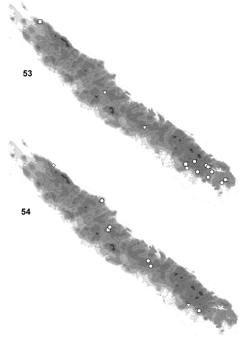
( Figs 1314 View FIGURES 614 , 4650 View FIGURES 4650 , 54 View FIGURES 5354 )
Material examined: Male holotype: Mt. Aoupinié, 700 m, 21°11'S, 165°18'E, 68.iii.1984 [ M. Pogue] ( NMNH, pinned) GoogleMaps .
Etymology: flinti , named after Oliver S. Flint, Jr. for his contribution in Trichoptera systematics. To be treated as a noun in genitive case.
Diagnosis: In flinti , the forewings have a dark area densely covered by coneshaped sensillae as in fusca . The species is easily separated from fusca and congeners by the presence of small spines on the dorsal part of segment X, and by the lateral branches originating distally on segment X. The genitalia are unique in the posterior margin of segment IX holding a triangular lobe over the gonocoxite, a very short segment IX, presence of lateral branches on segment X, and apex of segment X that is divided into two wide lobes.
Male.
Head: Frontal surface greybrownish, frons covered by short, dark brownish setae; dorsally divided into dark anterior and pale posterior half; few, long, golden brown setae along the eye margin and posterior margin of head. Antennal flagellum with 41 flagellom eres.
Thorax: Pronotum dark yellowishgrey, with few dorsolateral, stout, dark brownish setae on distinct warts. Mesonotum dark brownish with golden setae, setal warts absent.
Wings: Forewing length 10.2 mm (n=1), hind wing length 8.5 mm (n=1). Forewing dark brownish; distinct dark area between Cu and A as a in M. fusca made of dark, curving, coneshaped sensillae ( Figs 13, 14 View FIGURES 614 ) attached by large, oval tubercles ( Fig. 13 View FIGURES 614 ). Hind wing pale grey. Venation as described for genus, except R1 fading distally, fuses with Sc before wing margin. Approximately 14 long, straight hamuli along anterior margin.
Genitalia ( Figs 4650 View FIGURES 4650 ): Segment IX, lateral view ( Fig. 46 View FIGURES 4650 ), about 1.9x higher than long; anterior part substraight, anteroventral corner slightly produced anteriorly; dorsal margin rounded, with minute incision; posteroventral margin ( Fig. 48 View FIGURES 4650 ) with wide incision being nearly straight anteriorly; posterior margin with large, pointed dorsal marginal process above basal part of gonocoxite. Segment X nearly 5x longer than high, about parallelsided and with obtuse apex being slightly bent ventrally ( Fig. 46 View FIGURES 4650 ); setae confined to apex; row of spinous microtrichiae present at midlength; in dorsal view ( Fig. 47 View FIGURES 4650 ), apex bilobed, deeply incised centrally. Lateral branch smooth, about onesixth the length of central branch, originates at midlength on segment X; pointed, substraight ( Fig. 46 View FIGURES 4650 ), oriented posteriad ( Fig. 47 View FIGURES 4650 ). Median lobe, lateral view ( Fig. 46 View FIGURES 4650 ), small, partly hidden inside segment IX; with setae on posterior margin; not visible in dorsal view ( Fig. 47 View FIGURES 4650 ); in ventral view ( Fig. 48 View FIGURES 4650 ), with Ushaped incision, median corners slightly incised. Gonocoxite, lateral view ( Fig. 46 View FIGURES 4650 ), substraight, apex curving dorsad; dorsal and ventral margins nearly parallel; widening distally in ventral view ( Fig. 48 View FIGURES 4650 ), slightly converging, apex shallowly notched; gonocoxite covered by setae except at proximal part. Phallus simple; phallobase, lateral view ( Fig. 49 View FIGURES 4650 ), slightly longer than threequarts the height of segment IX; endotheca produced dorsally at midlength, without spines; in ventral view ( Fig. 50 View FIGURES 4650 ), lateral incisions at proximal part located anteriorly to anterior margin of phallus opening.
Female unknown.


| NMNH |
Smithsonian Institution, National Museum of Natural History |
No known copyright restrictions apply. See Agosti, D., Egloff, W., 2009. Taxonomic information exchange and copyright: the Plazi approach. BMC Research Notes 2009, 2:53 for further explanation.
|
Kingdom |
|
|
Phylum |
|
|
Class |
|
|
Order |
|
|
Family |
|
|
Genus |