
Endecous (Endecous) chape Souza-Dias & de Mello
|
publication ID |
https://doi.org/ 10.11646/zootaxa.4237.3.2 |
|
publication LSID |
lsid:zoobank.org:pub:BC7FE3FF-4B54-41FB-B74D-0AAAB784CD3B |
|
DOI |
https://doi.org/10.5281/zenodo.5690747 |
|
persistent identifier |
https://treatment.plazi.org/id/03F5167E-D109-3E76-FF75-FB3DC5EAEEA2 |
|
treatment provided by |
Plazi |
|
scientific name |
Endecous (Endecous) chape Souza-Dias & de Mello |
| status |
sp. nov. |
Endecous (Endecous) chape Souza-Dias & de Mello n. sp.
Figures 1 View FIGURE 1 , 2 View FIGURE 2 (A, C), 3.
Type locality. Brazil, Paraná State, Parque Nacional do Iguaçu (Iguaçu National Park).
Type material. Holotype male, 7 male paratypes, 10 female paratypes. Holotype: Brasil, Parque Nacional do Iguaçu. Trilha do Poço Preto. 20-30.i.2008. Dias, P.G.B.S. & de Mello, F.A.G. col. ( MZSP) . Paratypes: 1 male, 5 females, same data as the holotype ( MZSP) ; 1 male, same data as the holotype, with FWs and genitalia removed and kept with the specimen ( MZSP) ; 1 male, same data as the holotype (PSD33) ( MZSP) ; 1 male, same data as the holotype, with right FW removed and fixed in microscope slides ( MZSP) ; 1 male, labeled: Brazil, Parque Nacional do Iguaçu , Poço Preto. 22-28.xii.2010. Dias, P.G.B.S. col. Pitfall ( MZSP) ; 1 male, labeled: “BR [ Brazil], SC [Santa Catarina State], Concórdia. 28.xi-01.xii.2011. Dias, P.G.B.S. col.”, (PSD34) ( MZSP) ; 1 female, same data as the holotype (PSD35) ; 1 female, with the copulatory papilla removed and kept with the specimen, labeled: Brazil, Parque Nacional do Iguaçu , Trilha das Cataratas. 21.xii.2010. Dias, P.G.B.S. ( MZSP) ; 1 female, with the copulatory papilla removed and kept with the specimen, labeled: Brazil, PR [Paraná State], Foz do Iguaçu, Parque Nacional do Iguaçu , Trilha do Poço Preto. P 2—adultrap. 02-08.xi.2005. Szinwelski, N. col. ( MZSP) ; 1 female, labeled: Brazil, PR [Paraná State], Foz do Iguaçu, Parque Nacional do Iguaçu , Trilha do Poço Preto. 11.x.2010. Dias, P.G.B.S. col. ( MZSP) ; 1 male, 1 female, labeled: Brazil, Parque Nacional do Iguaçu , Trilha das Cataratas. 21.xii.2010. Dias, P.G.B.S. (Departamento de Zoologia, Instituto de Biociências, UNESP Botucatu).
Distribution. Southern Brazil, Atlantic Forest of western Paraná and Santa Catarina States. E. chape n. sp. is reported for the Iguaçu National Park, municipalities of Foz do Iguaçu and Céu Azul (Paraná), and the area of Concórdia municipality (Santa Catarina).
Etymology. Chape is the nickname of Associação Chapecoense de Futebol, a football team from the city of Chapecó, the largest city of the west of Santa Catarina State, which is part of the distribution of this new species. On 29th November 2016, a plane crash killed almost all the entire team, including the players, coaches, staff and journalists. E. chape n. sp. is a tribute to all people that made part of this team’s beautiful history, which was, unfortunately, interrupted by this terrible tragedy. The word “ chape ” is a noun in apposition.
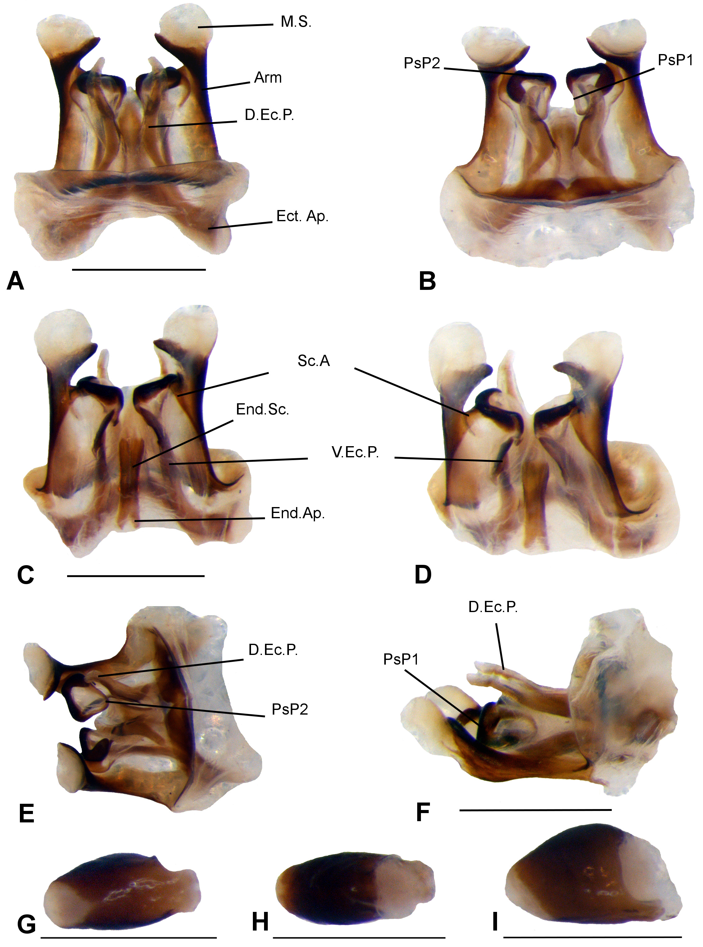
Diagnosis. Within the genus, E. chape n. sp. can be recognized by the following characters: males FWs short, rounded, not trespassing the third abdominal tergite; mirror irregular, with six cells, the distal weak and irregular; harp with three weak veins; lateral field with veins irregular, weak. Median part of the pseudepiphallic sclerite fused; pseudepiphallic arms lateral, hard, straight, apex pointed, curved inwards, with a conspicuous membranous sphere on the apex; sclerite A fused with the pseudepiphallic arm; PsP1 and PsP2 connected, forming a circular structure; PsP2 smaller and less sclerotized than PsP2; dorsal ectophallic projections (D.Ec.P.) long, almost reaching the apex of the pseudepiphallic arms, clearly separated and detached of the phallic complex; endophallic sclerite medial, long, grooved, forming a medio-dorsal crest; endophallic apodeme present, small, paired.
Description. General body coloration medium to reddish brown, almost uniform ( Figs 1 View FIGURE 1 A, B). Head. Dorsum with soft pubescence, medium brown, yellowish in the middle ( Fig. 1 View FIGURE 1 B). Vertex and fastigium medium brown ( Figs 1 View FIGURE 1 B, D). Fastigium little wider than long, narrower than scape, slightly narrower toward the apex, with a double row of small bristles, and a spot yellowish brown on apical portion, corresponding to the area of the median ocellus ( Figs 1 View FIGURE 1 B, D). Ocelli absent. Eyes obovate, prominent ( Figs 1 View FIGURE 1 B, C). Antennal scape medium brown, outer border yellowish brown. Antenna not annulated; antenomeres medium brown ( Figs 1 View FIGURE 1 B–D). Frons medium brown speckled with small, light brown spots, and a macula light brown below each antennal fossa. In frontal view, gena medium brown; line diagonal, thin, light brown that goes from gena toward frons. In lateral view, gena medium brown with diagonal band light brown, which surround the external and superior margin of the eye, and weak ascendant anastomosed lines medium brown ( Fig. 1 View FIGURE 1 C). Clypeus and labrum light brown. Maxillary palpi elongated, thin; joints 1–2 whitish, joints 3–5 elongate, same-sized, medium brown, speckled with very small light brown spots; apical third of joint 5 curved, apex rounded, whitish ( Figs 1 View FIGURE 1 C, D). Thorax. Pronotum DD wider than long, slightly pubescent, general coloration medium brown; two spots yellowish brown on anterior portion, with reticulated lines medium brown; two blotches large, yellowish brown on median portion, divided by stripe medium brown, which is divided by a line yellowish brown that goes from the anterior to posterior portion of DD; two spots yellowish brown on posterior portion; DD cephalic and caudal margins sub-straight; LL ventro-cephalic angle slightly salient, rounded, ventro-caudal angle almost straight, ventral margin gradually ascendant ( Figs. 1 View FIGURE 1 C, D). Legs. Legs I and II yellowish to medium brown, not annulated ( Fig. 1 View FIGURE 1 A). Auditory tympanum present on both sides of TI; internal tympanum oval, small; external rounded. TI and TII with two same-sized ventral apical spurs. FIII light to medium brown, with diagonal medium brown stripes, distal half medium brown. TIII medium brown; subapical spurs 4/4, the first smaller and associated to the apical; serrulation between and above subapical spurs 1 and 2; apical spurs 3/3, more developed on inner face; dorsal and median apical spurs same-sized and the longest on both faces ( Fig. 1 View FIGURE 1 A, I). Basitarsus I, II and III medium brown; basitarsus III with double row of spines ( Fig. 1 View FIGURE 1 I). Abdomen. Medium to yellowish brown, pubescent ( Fig. 1 View FIGURE 1 A). Subgenital plate wider than long; anterior margin sub-straight, posterior margin broad, rounded, with short distal projections ( Fig. 1 View FIGURE 1 E). Supra anal plate medium brown, with a central macula yellowish brown; anterior margin slightly concave, posterior margin sub-straight, without distal projections ( Fig. 1 View FIGURE 1 F). Cerci as long as FIII, medium brown. Male. FWs membranous, short, rounded, not trespassing the third abdominal tergite ( Figs 1 View FIGURE 1 A, B); right FW hard, medium brown ( Figs. 1 View FIGURE 1 B, 2A, C); left FW membranous, transparent; stridulatory vein present, with ca. 82 - 94 teeth (n=09) ( Figs 1 View FIGURE 1 B, 2A, C); chord with two elongated cells; mirror irregular, with six cells, the distal weak and irregular ( Figs. 1 View FIGURE 1 B, 2A, C); harp with three weak veins, one incomplete ( Figs. 1 View FIGURE 1 B, 2B, D); lateral field with veins irregular, weak ( Figs 2 View FIGURE 2 A, C). Male genitalia. Pseudepiphallus: median part of the pseudepiphallic sclerite fused ( Figs 3 View FIGURE 3 A, B); pseudepiphallic arms lateral, hard, straight, apex pointed, curved inwards ( Figs 3 View FIGURE 3 A–D); presence of a conspicuous membranous sphere on the apex of the pseudepiphallic arms ( Figs 3 View FIGURE 3 A–E); sclerite A fused with the pseudepiphallic arm, the apical part of the sclerite is visible in ventral view ( Figs 3 View FIGURE 3 C, D). PsP1 and PsP2 connected, forming a circular structure, visible in superior and lateral views ( Figs. 3 View FIGURE 3 B, E, F); presence of a membrane between the contact points of PsP1 and PsP2 ( Figs. 3 View FIGURE 3 B, E); PsP1, hard, visible in ventral and dorsal views ( Figs. 3 View FIGURE 3 B–F); PsP2 small, less sclerotized than PsP1, curved inwards ( Figs 3 View FIGURE 3 B, E, F). Ectophallic invagination: ectophallic apodeme short, robust ( Figs 3 View FIGURE 3 A, B); ectophallic arc below the median part of the pseudepiphallic sclerite ( Figs 3 View FIGURE 3 A, B); dorsal ectophallic projections long, almost reaching the apex of the pseudepiphallic arms ( Fig. 3 View FIGURE 3 A), clearly separated and detached of the phallic complex ( Figs 3 View FIGURE 3 A, F), apex curved inwards; apex of the ventral ectophallic projections pointed, curved inwards ( Figs 3 View FIGURE 3 C, D); ectophallic fold membranous, with a weak sclerotization visible in dorsal view ( Figs 3 View FIGURE 3 A, B). Endophallus: endophallic sclerite medial, long, grooved, forming a medio-dorsal crest ( Figs 3 View FIGURE 3 C, D); endophallic apodeme present, small, paired ( Figs 3 View FIGURE 3 C, D).
Female. Larger than male, general coloration similar. Apterous. Supra anal plate medium brown, similar to male. Subgenital plate medium brown, short, wider than long, anterior margin slightly convex, posterior margin rounded, with a central concavity. Female genitalia: c opulatory papilla laterally flattened, apex rounded, basis with a dorsal projection, as in Figs 3 View FIGURE 3 G–I.
Remarks on paratype. One male paratype had the FWs different from the holotype showing chord with two cells, one elongated and other reduced ( Figs 2 View FIGURE 2 A, C).
Measurements (mm). Males (n=8): HW—3.53 (3.2–3.9); IOD—1.88 (1.5–2.3); PL—2.96 (2.5–3.9); AWP—3.31 (2.6–3.9); PWP—4.57 (4.2–5.1); PW—5.22 (4.5–5.6); FWL—5.66 (5.1–6.2); FWW—4.9 (4.5–5.2); LFIII—16.25 (14–17.5); WFIII—3.9 (3.5–4.1); LTIII—17.2 (15.2–19.2); LBt–III—5.35 (4.1–6). Females (n=11): HW—3.42 (3.2–3.6); IOD—1.8 (1.5–1.9); PL—2.84 (2.2–3.5); AWP—3.16 (2.7–3.4); PWP— 4.57 (4.2–5.1); PW—5.02 (4.9–5.2); LFIII—15.6 (12.2–16.9); WFIII—3.86 (3.5–4.1); LTIII—17 (14.9–18.9); LBt-III—5.25 (4.5–5.9); OL—13.6 (10.1–15.1).


| MZSP |
Sao Paulo, Museu de Zoologia da Universidade de Sao Paulo |
No known copyright restrictions apply. See Agosti, D., Egloff, W., 2009. Taxonomic information exchange and copyright: the Plazi approach. BMC Research Notes 2009, 2:53 for further explanation.