
Palaeoproteus cf. miocenicus (Vasilyan and Yanenko, 2020)
|
publication ID |
https://doi.org/ 10.26879/1323 |
|
DOI |
https://doi.org/10.5281/zenodo.11033511 |
|
persistent identifier |
https://treatment.plazi.org/id/03F52665-D050-FF8F-FC60-9E34FB8C74CD |
|
treatment provided by |
Felipe |
|
scientific name |
Palaeoproteus cf. miocenicus |
| status |
|
Figures 3–4 View FIGURE 3 View FIGURE 4
Material. Hambach 6C: three atlases (IPB-HaH 2117, IPB-HaH 2175, IPB-HaH 2164); one trunk vertebra (IPB-HaH 2162). Hambach 11: two dentaries (IPB-HaR 2043, IPB-HaR 2071); one anterior trunk vertebra (IPB-HaR 2183); one trunk vertebra (IPB-HaR 2008). Hambach 11C: five trunk vertebrae (IPB-HaR 2404/2408).
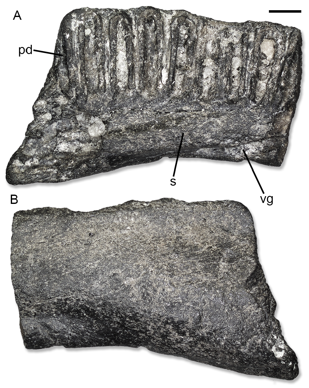
Description. IPB-HaR 2071 ( Figure 3 View FIGURE 3 ) represents part of the posterior end of a very large dentary. It is very fragmentary. IPB-HaR 2043 is also very large and robust. It represents a portion originally located somewhere in the middle of the dentary. The pars dentalis of the dentary is composed by a very high dental lamina and a very low subdental lamina. On the medial side, nine tooth positions are visible, hosting the poorly-preserved remains of pleurodont, very high, rather narrow and closely spaced teeth provided with thick walls. Ventral to the pars dentalis, there is a robust shelf, which is not strongly developed medially. The preservation of this shelf is rather poor, but a narrow and shallow groove is visible ventrally. The groove moves towards the medial surface near the anterior end of the fragment. The lateral surface is smooth. In lateral/medial view, the ventral margin is somehow concave, suggesting ventral development of the posterior part of the dentary.
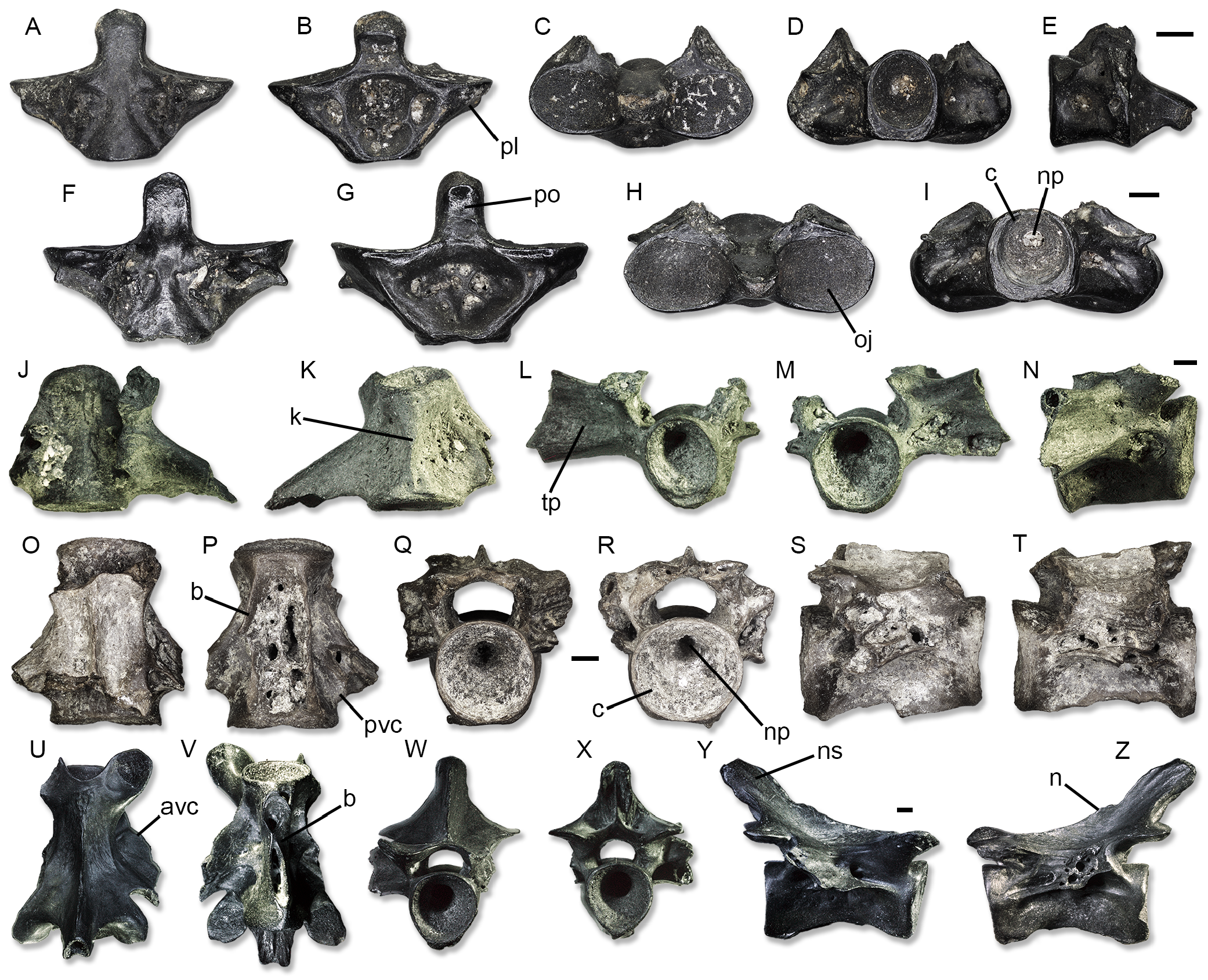
IPB-HaH 2164 is a very small atlas, but the other ones are larger. The length of the largest atlas, IPB-HaH 2175 ( Figure 4 View FIGURE 4 F-I), is 6 mm. All atlases miss almost completely the neural arch, preserving only the centrum. Anteriorly, the occipital joints are wide and mediolaterally elongated, with a suboval/subelliptical shape (i.e., slightly dorsoventrally compressed) in anterior view. They are very shallowly concave and do not coalesce in the middle, being separated by a thin and long processus odontoideus. The processus is slightly slenderer in IPB-HaH 2175 than in the other two specimens. It has a flat dorsal surface and an anteroventrally-directed and strip-like articular surface that is not separated into two distinct lateral areas. There is no postodontoideus foramen at the base of the process. The posterior end of the centrum is represented by a posterior cotyle with a notochordal pit in the middle. The cotyle is circular in IPB-HaH 2175 ( Figure 4I View FIGURE 4 ) and more mediolaterally compressed in IPB-HaH 2117 ( Figure 4D View FIGURE 4 ). In both specimens, it is larger than the processus odontoideus. This feature cannot be evaluated in IPB-HaH 2164 because the area is damaged. In IPB-HaH 2715, the ventral surface of the centrum shows a concave area with a number of large foramina ( Figure 4G View FIGURE 4 ). On the other hand, IPB-HaH 2117 bears a very deep fossa in the middle of the centrum, which is flanked by two smaller symmetrical foramina by the sides and by other even smaller foramina posteriorly ( Figure 4B View FIGURE 4 ). The ventral surface of IPB-HaH 2164 displays a depressed area by each side of the centrum. Foramina also cover the lateral surface of each processus lateralis in all specimens.
Both IPB-HaR 2008 ( Figure 4 View FIGURE 4 O-T) and IPB-HaR 2408 ( Figure 4 View FIGURE 4 U-Z) are amphicoelous and large sized (the centrum length reaches 6.5 mm and 9.5 mm, respectively). A notochordal pit is present in the middle of the large and hourglassshaped centrum. The cotyles are circular in both anterior and posterior views. The ventral surface of the centrum bears two high and sharp longitudinal basapophyses, which run parallel along the entire length of the vertebra. Only in IPB-HaR 2408, the basapophyses contact in the middle at about one third of the centrum length ( Figure 4V View FIGURE 4 ). Between the basapophyses, various central foramina are present. At least three large ones are visible on IPB-HaR 2008 ( Figure 4P View FIGURE 4 ), surrounded by other smaller ones, whereas only a number of small ones are visible on IPB-HaR 2408 ( Figure 4V View FIGURE 4 ). The neural arch is robust and dorsally flattened, being better preserved in IPB-HaR 2408. A neurapophysis is present. It is low anteriorly, but rises to a moderate degree posteriorly. A robust spine is present at the posterior end of the arch, strongly projecting posterodorsally beyond the postzygapophyses. The posterior end of the spine is truncated. A wide and deep, U-shaped anterior notch on the neural arch is visible on IPB-HaR 2408 ( Figure 4U View FIGURE 4 ), allowing the anterior cotyle to be visible in dorsal view. The deepest part of the notch reaches the posterior margin of the prezygapophyses. The zygapophyses are more or less horizontal in both anterior and posterior view. The zygapophyseal facets are suboval. In posterior view, two shallow depressions are visible on the posteroventral surface of the neural arch, flanking a low longitudinal ridge running along the ventral side of the neural spine. The transverse processes are moderately developed and posterolaterally directed. Ventrally, they are connected to the centrum by moderately-(IPB-HaR 2008) or well-developed (IPB-HaR 2408) anterior ventral crests (anterior alar process in Vasilyan and Yanenko, 2020) and little-developed posterior ventral crests. On the other hand, the zygapophyseal crests are not developed. The other vertebrae only preserve isolated centra or fragments of centra, some of them being much smaller than the two previously described (IPB-HaR 2406 is about 4 mm long). Nevertheless, they share the same morphology.
IPB-HaR 2183 ( Figure 4 View FIGURE 4 J-N) is the only trunk vertebra displaying some differences from the other ones. This large vertebra has a very massive, almost 6 mm long centrum, which is amphicoelous, hour-glass-shaped and notochordal. The overall aspect is relatively shorter compared to the trunk vertebra IPB-HaR 2008, coming from the same level. The anterior cotyle is moderately mediolaterally compressed, appearing subelliptical in anterior view. The posterior cotyle, on the other hand, is subcircular in posterior view. The ventral surface of the centrum bears a narrow keel and no basapophyses. A number of small foramina are present by the ventrolateral sides of the centrum, in place of real subcentral foramina. Most of the neural arch is missing, preserving only the right lateral wall and the base of the left one. The transverse processes are long and moderately robust; they are directed posterolaterally. The anterior ventral crest is very low, whereas the posterior one is slightly more developed. Zygapophyseal crests are not developed. The base of the right prezygapophysis is also preserved, but not the zygapophysis itself. The shortness of this vertebra, together with its ventral keel, identify it as an anterior trunk vertebra ( Estes et al., 1967).
Remarks. The diagnostic features recently reported by Vasilyan and Yanenko (2020) allow a rather straightforward identification for these remains as a batrachosauroidid salamander. The dentaries of these urodeles have thick-walled teeth and a ventrally-projecting posterior part. Trunk vertebrae are amphicoelous, with subcircular cotyles, basapophyses, and a posterodorsally-projecting neural spine. The atlases are provided with both anterior and posterior cotyles with a rounded outline, among which the former are large and concave. The well-developed paired ventral basapophyses, as well as maybe the flat neural arch and developed anterior ventral crest, suggest that the batrachosauroidid from Hambach is a member of the European genus Palaeoproteus ( Vasilyan and Yanenko, 2020) . In particular, the following combination of features observed in the studied material is diagnostic of P. miocenicus following Vasilyan and Yanenko (2020): overall large size; poorly-developed subdental shelf of dentary; vertebrae with a long neural spine; weakly-concave and slightly dorsoventrally-compressed anterior cotyles of the atlas; external surface of the atlas pierced by foramina of different sizes; strongly pronounced, lip-shaped odontoid process of the atlas ( Vasilyan and Yanenko, 2020: p. 8, stated that “In P. miocenicus , the odontoid process is very large in comparison to all known batrachosauroidids”). However, the atlases from Hambach 6C show no postodontoid foramen, in contrast with the type and referred material of P. miocenicus ( Vasilyan and Yanenko, 2020) . All four P. miocenicus atlases share the presence of this foramen, whereas it is absent in Palaeoproteus klatti Herre, 1935 , and Palaeoproteus gallicus Estes et al., 1967 . Despite this difference, we refrain to name a new species here and attribute the fossils from Hambach 6C to Palaeoproteus cf. miocenicus , due to the overall similarity between bones of P. miocenicus and the German taxon as well as the low sample of atlases that hinders a complete understanding of the real variation of this feature in these animals. Material coming from the youngest level in Hambach also shares the same attribution, given that atlases from this level are currently unknown, and so it is not possible to verify the presence or absence of a postodontoid foramen.
No known copyright restrictions apply. See Agosti, D., Egloff, W., 2009. Taxonomic information exchange and copyright: the Plazi approach. BMC Research Notes 2009, 2:53 for further explanation.