
Stygarctus ayatori, Fujimoto, Shinta, 2014
|
publication ID |
https://doi.org/ 10.11646/zootaxa.3784.2.8 |
|
publication LSID |
lsid:zoobank.org:pub:B0B44D7B-32A4-4749-86D0-BE09EC1D31E5 |
|
DOI |
https://doi.org/10.5281/zenodo.5690711 |
|
persistent identifier |
https://treatment.plazi.org/id/433EB653-9E59-4691-A44F-83718D9C0C1B |
|
taxon LSID |
lsid:zoobank.org:act:433EB653-9E59-4691-A44F-83718D9C0C1B |
|
treatment provided by |
Plazi |
|
scientific name |
Stygarctus ayatori |
| status |
sp. nov. |
Stygarctus ayatori View in CoL sp. nov.
(Figs. 2–6, Table 1 View TABLE 1 )
Diagnosis. Stygarctus with club shaped primary clavae and bent sausage-like secondary clavae; funnel shaped lateral processes without micro-spines; pair of dorsal spines with terminal bifurcation; caudal plate with pair of antero-lateral lobes, pair of round lateral processes with minute spike and pair of caudal spikes; ventral surface without plates; female genital structure with seminal receptacle ducts entangled near exterior opening and internal thickening situated peripheral to female gonopore and between female gonopore and anus.
Materials examined. Holotype: KUZ Z677: adult female mounted in Fluoromount-G TM.
Paratypes: KUZ Z678: adult female mounted in glycerol; KUZ Z679, Z680, Z681: three adult females mounted in Hoyer’s medium; KUZ Z682: adult male mounted in Fluoromount-G TM; KUZ Z683: adult male mounted in Hoyer’s medium.
Type Locality: Okinoshima, Tateyama Bay, Boso Peninsula, Honshu, Japan (34°59′27″N, 139°49′24″E). Coll. Shinta Fujimoto on 18th April 2013.
Type depositories: The type series is deposited in the Zoological Collection of Kyoto University ( KUZ). The two specimens mounted in distilled water and the four specimens on a SEM stub are deposited in the author’s personal collection.
Description of holotype. Adult female with body length of 112 Μm. Dorsal surface with sculptured dorsal plates (Figs. 2, 5A); ventral surface smooth without plates or cuticle folds (Fig. 3). Cephalic plate with medial lobe, pair of medio-lateral lobes and pair of lateral lobes. Ventral lobes on each side of anteriorly protruded mouth cone.
FIGURE 2. Drawing of Stygarctus ayatori sp. nov., holotype female [KUZ Z677] (dorsal view). cE, cirrus E; cs, caudal spike; ds, dorsal spine; ec, external cirrus; ic, internal cirrus; lc, lateral cirrus; mc, median cirrus; ms, minute spike; pcl, primary clava; scl, secondary clava; s 4, leg IV sensory organ.
FIGURE 3. Drawing of Stygarctus ayatori sp. nov., holotype female [KUZ Z677] (ventral view). a, anus; cE, cirrus E; cs, caudal spike; ds, dorsal spine; ec, external cirrus; fg, female gonopore; ic, internal cirrus; it, internal thickening; lc, lateral cirrus; ms, minute spike; pcl, primary clava; scl, secondary clava; sr, seminal receptacle; s 4, leg IV sensory organ.
Bucco-pharyngeal apparatus dissolved before observation. Unpaired median cirrus (12 Μm) with short scapus (2 Μm) and flagellum (10 Μm) inserted medially at level of incision between medio-lateral and lateral lobes. Medial lobe bears pair of internal cirri (12 Μm) each with short scapus (3 Μm) and flagellum (9 Μm) arising from cirrophore. Lateral lobe bears lateral cirrus (14 Μm) with short scapus (3 Μm) and flagellum (11 Μm) and club shaped primary clava (6 Μm) inserted dorso-posterior to lateral cirrus. Thin sheet present exterior to lateral lobe. Ventral lobe bears external cirrus (11 Μm) with short scapus (2 Μm) and flagellum (9 Μm) and sausage-like secondary clava (16 Μm) inserted interior to external cirrus.
Three dorsal body plates with funnel shaped lateral processes, reduced to thin sheets at lateral margin and micro-spines absent. Two intersegmental plates present. Pair of dorsal spines (23 Μm) with terminal bifurcation fringed with thin cuticle inserted on posterior ridge of second dorsal body plate. Caudal plate with pair of anterolateral lobes, pair of round lateral processes each with minute spike and pair of caudal spikes. Round lateral process reduced to thin sheet at lateral margin. Pair of Cirri E (24 Μm) each with proximal portion (6 Μm) and flagellum (18 Μm) inserted between antero-lateral lobe and round lateral process. Papilla shaped leg IV sense organ (2 Μm) inserted at base of leg IV.
Rosette-like female gonopore opens ventrally 8 Μm anterior to anus. Pair of seminal receptacles each consists of sinuous duct opening 2 Μm posterior from level of gonopore and small vesicle positioned 11 Μm laterally from the gonopore. Seminal receptacle duct forming a three-dimensional nodule of entanglement near duct opening. Terminal anus consists of two cuticle lobes. Region peripheral to gonopore and between gonopore and anus with internal thickening.
Four claws inserted directly on each leg with dorsal, long, thin filaments on median claws. Claws of leg IV (7 Μm) longer than those of leg I–III (5 Μm).
Etymology. The specific epithet, ayatori , is a Japanese word for ‘string figure’ referring to the seminal receptacle ducts of the new species.
Remarks on paratypes and additional material. The bucco-pharyngeal apparatus was observed in specimens mounted in distilled water ( Figs. 4 View FIGURE 4 A, 5B). It consists of a buccal tube (22 Μm), two stylets with furca, three apophyses (3 Μm) and three placoids (3 Μm) for a specimen with body length of 111 Μm. The pharyngeal bulb was not observed.
The female genital structure was identical in all material observed with no variation in the number of turns or loops in the ducts or in the shape of the internal thickening ( Figs. 4 View FIGURE 4 B, 5C). The male gonopore, situated immediately anterior to the anus, is a simple round opening of a posteriorly directed 4 Μm long tube ( Fig. 4 View FIGURE 4 C). The difference in the genital structures of the female and male was the only sexual dimorphism recognized in the new species.
Results of electron scanning microscopy are described as follows. The dorsal plates are strongly sculptured and each plate is partitioned only by the elevation of the cuticle ( Figs. 6 View FIGURE 6 A, B). The three-dimensional appearance of the cephalic region is better observed than under DIC microscopy ( Fig. 6 View FIGURE 6 C). The female gonopore opens on a triangular button shaped lobe, which is not visible with DIC ( Fig. 6 View FIGURE 6 D). The smooth surface posterior to the gonopore indicates the internal thickening observed with DIC is an internal structure ( Fig. 6 View FIGURE 6 D).
Differential diagnosis. Stygarctus ayatori sp. nov. is distinguished from S. arbornatus (for which there is no information on the genital structure) by the lack of dorsal spines, which are present in the new species. Stygarctus bradypus is most similar to S. ayatori sp. nov. in the morphology of sensory organs, dorsal plates, lateral processes, dorsal spines and caudal spikes but the new species is distinguished from S. bradypus , and the other congeners, by the entangled seminal receptacle ducts and internal thickening in the female genital region. In contrast to the new species, these congeners have simple ducts with loops or spirals and no internal thickening reported with the exception of a v-shaped thickening situated between the pair of seminal receptacles in S. gourbaultae Renaud- Mornant, 1981 ( Pollock 1970a, b; Renaud-Mornant 1970, 1981; Grimaldi de Zio et al. 1987; Hansen et al. 2012).
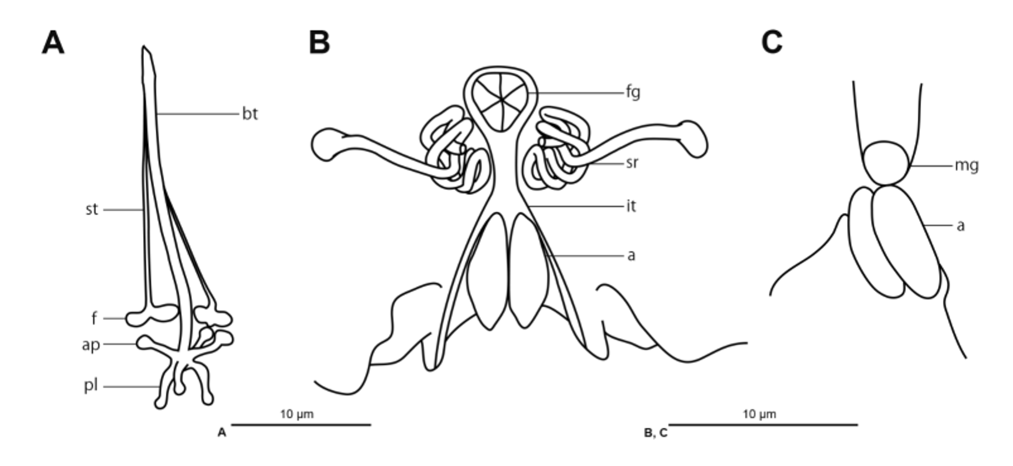
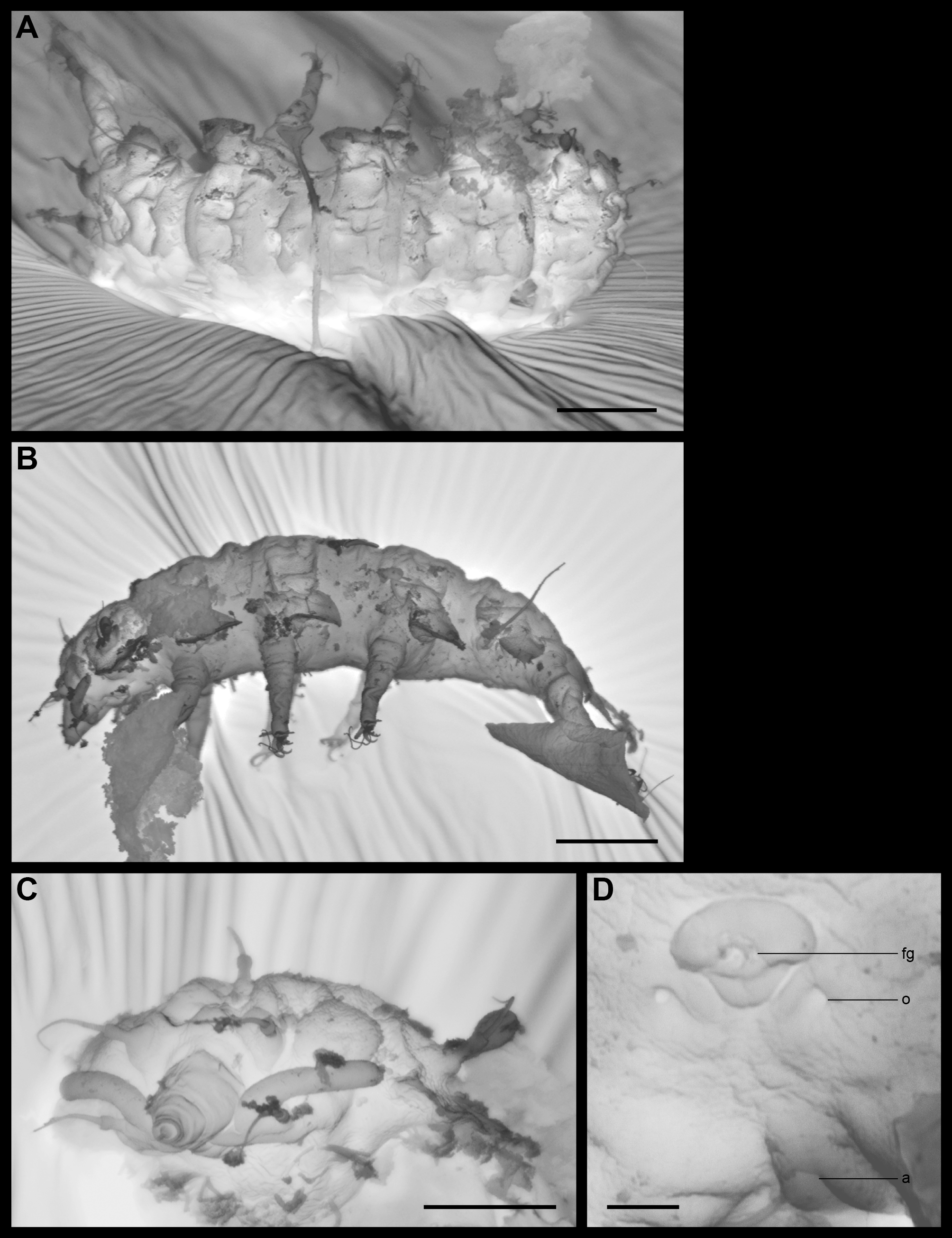
TABLE 1. Morphometrics (µm) of Stygarctus ayatori sp. nov. SD, standard deviation. Dashes (–) represent unmeasured traits.
| Holotype | Paratype | Paratype | Paratype | Paratype | Paratype | Paratype | MEAN SD | |
|---|---|---|---|---|---|---|---|---|
| KUZ No. | Z677 | Z678 | Z679 | Z680 | Z681 | Z682 | Z683 | |
| Sexuality | Female | Female | Female | Female | Female | Male | Male | |
| Body length | 112 | 101 | 112 | 106 | 94 | 105 | 110 | 106 7 |
| KUZ |
Zoological Collection of the Kyoto University |
No known copyright restrictions apply. See Agosti, D., Egloff, W., 2009. Taxonomic information exchange and copyright: the Plazi approach. BMC Research Notes 2009, 2:53 for further explanation.