
Platycrus, Szawaryn & Ślipiński, 2022
|
publication ID |
https://doi.org/10.11646/zootaxa.5190.4.8 |
|
publication LSID |
lsid:zoobank.org:pub:2992690E-0519-4428-BBCD-FF2AC904DBBD |
|
DOI |
https://doi.org/10.5281/zenodo.7138584 |
|
persistent identifier |
https://treatment.plazi.org/id/9076C72E-F86D-4B22-8BD5-FBB30F96805D |
|
taxon LSID |
lsid:zoobank.org:act:9076C72E-F86D-4B22-8BD5-FBB30F96805D |
|
treatment provided by |
Plazi |
|
scientific name |
Platycrus |
| status |
gen. nov. |
Genus Platycrus gen. nov.
( Figures 1–3 View FIGURE 1 View FIGURE 2 View FIGURE 3 )
urn:lsid:zoobank.org:act:
Type species: Platycrus laotanus sp. nov.
Etymology. The generic name is derived from the generic name Platynaspis and Latin word crus (leg) referring to exceptionally broad legs of the new taxon. Gender masculine.
Diagnosis. Platycrus gen. nov. can be easily distinguished from apparently similar coccinellid species from the tribe Aspidimerini by its larger size, but also by having broadly expanded clypeus covering antennae and labrum from above, the antennae with 11 antennomeres, the terminal labial palp about as wide as penultimate one, the prosternal process with lateral carinae short and narrowly separated reaching only level of anterior procoxal margin, and the distinctly roundly produced trochanter. Moreover, it can be separated from Platynaspis by the extraordinarily broadened tibiae with rows of short thorns at apical tibial margin, three-segmented tarsus (foursegmented in Platynaspis ), and peculiar pocket-like structure for reception of the whole tarsus in repose.
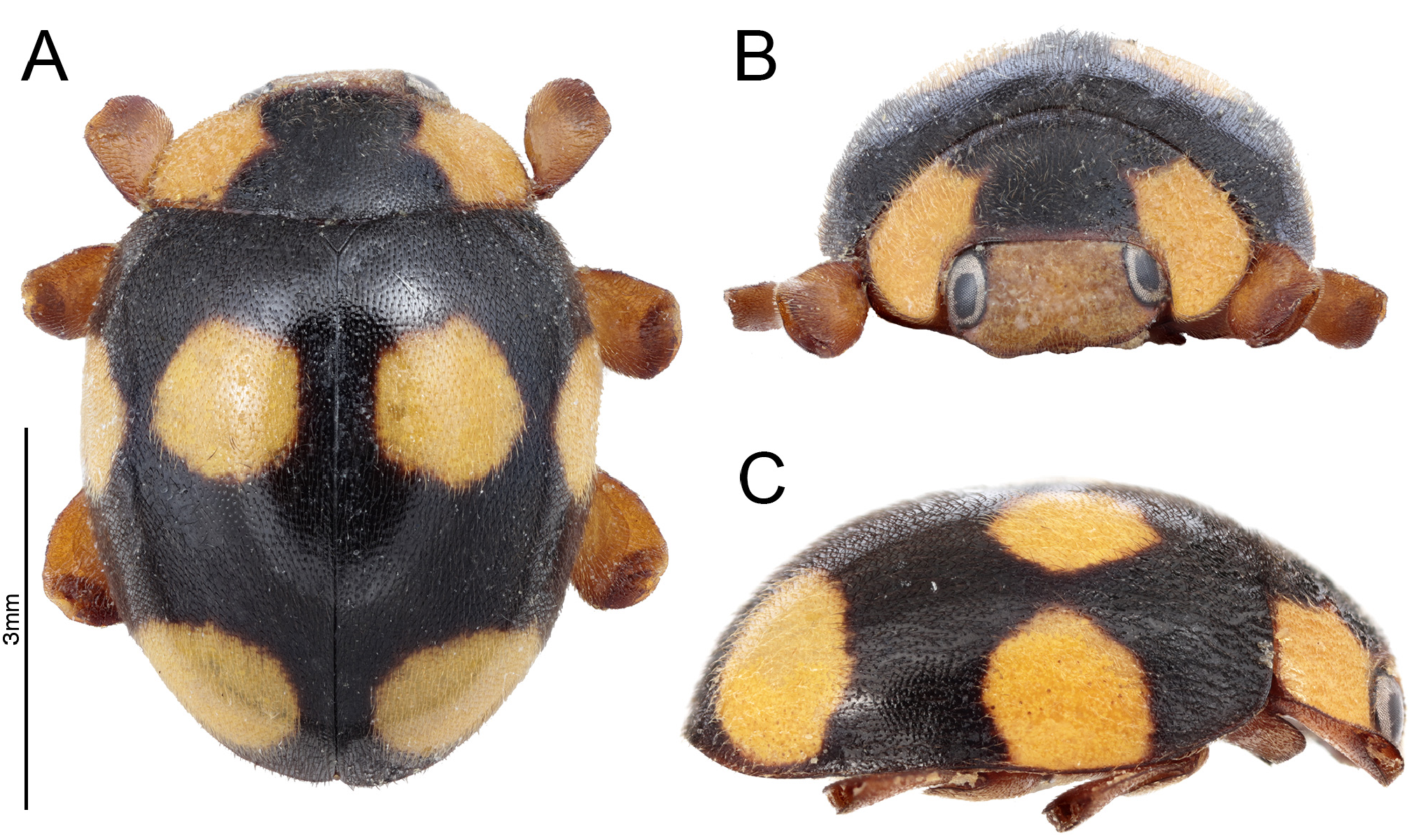
Description. Length: 5.7 mm, width: 4.5 mm. Body ( Fig. 1A–C View FIGURE 1 ) elongate oval, moderately convex, covered with uniform short setae. Elytra with single size punctae.
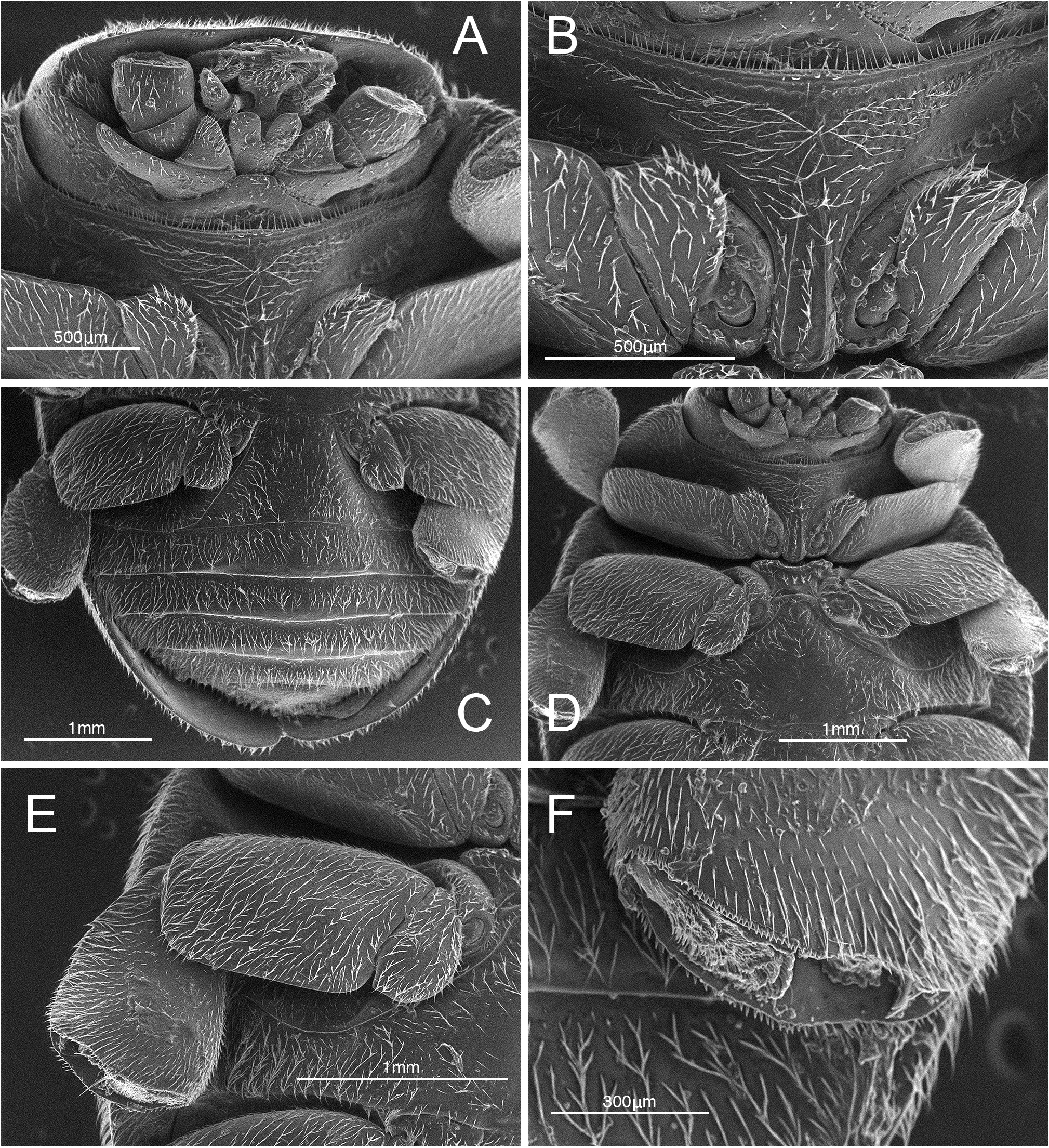
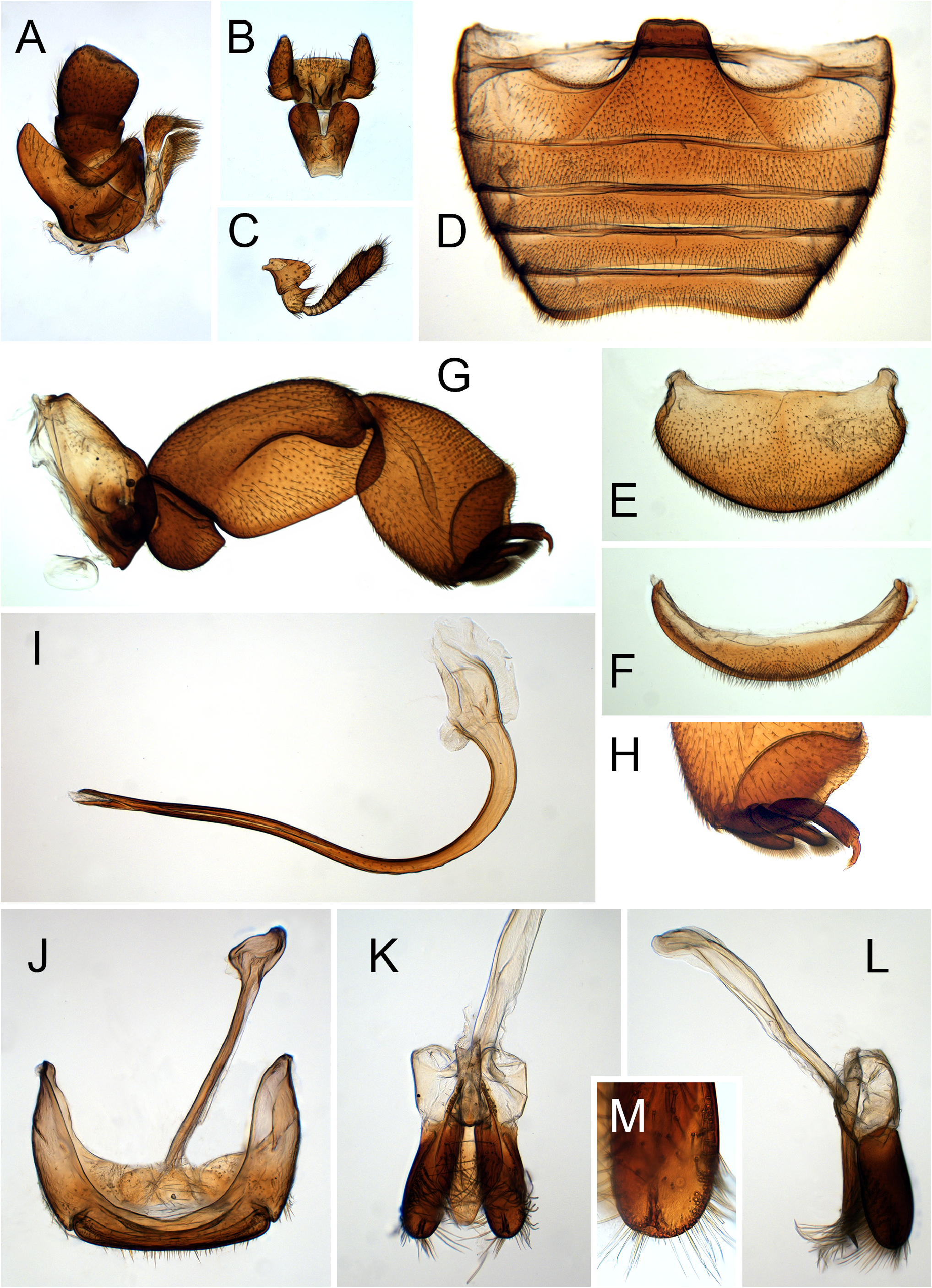
Head transverse ( Fig. 2A View FIGURE 2 ); frons wide, more than 3 times as broad as width of an eye; clypeus produced anteriorly and expanded laterally, covering antennal insertions and labrum from above, emarginate medially ( Fig. 1C View FIGURE 1 ). Ventral antennal grooves deep, in form of pockets below expanded clypeus ( Fig. 2A View FIGURE 2 ). Eyes large, rounded and finely faceted, with very sparse and short interfacetal setae, inner eye orbits arcuate ( Fig. 1B View FIGURE 1 ). Antenna ( Fig. 3C View FIGURE 3 ) very short, composed of 11 antennomeres, covered with distinct setae; scape large, roundly produced anteriorly, about as wide as long; pedicel broad, barrel shaped with a subtriangular, acute lateral projection; antennomeres 3–11 forming fusiform club, about twice as long as scape and pedicel combined; antennomeres 3–9 transverse, antennomeres 10 and 11 elongate, asymmetric; terminal antennomere large, transverse, truncate apically. Mandible bifid at apex ( Fig. 2A View FIGURE 2 ). Maxilla ( Fig. 3A View FIGURE 3 ) with cardo large, subtriangularly produced laterally; cardo grooved for reception of maxillary palp; basistipes subtriangular. Lacinia elongate, covered with moderately long setae in apical half; galea narrow c-shaped, covered with setae, with patch of longer setae apically. Maxillary palp 4-segmented; palpomere 1 very short, ring-like; palpomere 3 short, transverse; terminal palpomere subquadrate, with sides subparallel, truncate apically. Gula with distinct pore below submentum ( Fig. 2A View FIGURE 2 ). Submentum transverse; mentum ( Fig. 3B View FIGURE 3 ) distinctly cordate, with anterior corners roundly produced anteriorly and median part deeply notched, covering insertions of labial palps from below. Prementum transverse with anterior margin straight, labial palp ( Fig. 3B View FIGURE 3 ) 3-segmented; basal palpomere small, ring-like; palpomere 2 large, broadened apically, wider than its length; apical palpomere large, only slightly narrower than penultimate, elongate, gradually tapering apically.
Pronotum transverse ( Fig. 1 A, B View FIGURE 1 ) with anterior corners roundly produced, posterior margin with complete bordering line; hypomera smooth ( Fig. 2D View FIGURE 2 ). Prosternal surface covered with large circular punctures bearing setae on its lateral margin ( Fig. 2B View FIGURE 2 ); lateral sides of prosternum slightly depressed to accommodate prolegs in repose. Prosternum flat, not raised, slightly shorter than length of procoxa, anterior margin with crenulate bordering line ( Fig. 2B View FIGURE 2 ); prosternal process narrow ( Fig. 2B View FIGURE 2 ), about 0.5 of prosternal length in front of procoxae, with short lateral carinae roundly joined anteriorly, slightly converging anteriorly, reaching at most the level of anterior procoxal margin. Procoxal cavity transverse with lateral slit.
Mesoventrite short ( Fig. 2D View FIGURE 2 ), transverse, with anterior margin raised and bordered, emarginate medially; narrow, narrower than width of corresponding mesocoxal diameter. Meso-metaventral junction visible, forming anteriorly slightly arcuate line. Metaventrite with crural impressions to accommodate mid legs in repose ( Fig. 2D, E View FIGURE 2 ); metaventral postcoxal lines joined medially on metaventral process forming a line depressed medially, roundly recurved and complete laterally. Scutellar shield subtriangular ( Fig. 1A View FIGURE 1 ). Elytra with humeral calli poorly marked, with apices rounded. Elytral epipleuron incomplete apically ( Fig. 2C View FIGURE 2 ), reaching mid length of ventrite 3, with deep foveae for reception of mid and hind legs in repose. Wings well developed.
Trochanters broad, roundly produced externally ( Figs 2C–E View FIGURE 2 , 3G View FIGURE 3 ). Femora very broad, about 2/3 as broad as long, with deep grooves on dorsal surface to receive tibiae in repose. Tibia very broad, about as broad as femur; with apical margin produced, forming a pocket to accommodate tarsus in repose ( Figs 2F View FIGURE 2 , 3H View FIGURE 3 ); without tibial spurs; apical margin with a row of short subtriangular denticles. Tarsi 3-segmented ( Fig. 3H View FIGURE 3 ), tarsal claws elongate, with apical cleft ( Fig. 2F View FIGURE 2 ).
Abdomen with 6 ventrites ( Fig. 2C View FIGURE 2 ), with distinct crural depressions on ventrite 1. Ventrite 1 distinctly longer than ventrite 2 ( Fig. 3D View FIGURE 3 ), at middle about 2 times as long as ventrite 2, with posterior margin slightly concave. Ventrites 2–4 subequal in length, with posterior margins straight. Ventrite 5 shorter than ventrite 4, with posterior margin distinctly, broadly emarginate ( Fig. 3D View FIGURE 3 ). Ventrite 6 narrow, rounded apically ( Fig. 3F View FIGURE 3 ). Tergite VIII large, broadly rounded apically ( Fig. 3E View FIGURE 3 ). Abdominal postcoxal lines steeply descending ( Fig. 3D View FIGURE 3 ) in the form of an almost straight line, curved apically and then parallel to posterior margin of ventrite, laterally incomplete.
No known copyright restrictions apply. See Agosti, D., Egloff, W., 2009. Taxonomic information exchange and copyright: the Plazi approach. BMC Research Notes 2009, 2:53 for further explanation.
|
Kingdom |
|
|
Phylum |
|
|
Class |
|
|
Order |
|
|
Family |
|
|
SubFamily |
Coccinellinae |
|
Tribe |
Platynaspini |