
Lissocidaris xanthe, Coppard, Simon E. & Noordenburg, Henk Van, 2007
|
publication ID |
https://doi.org/ 10.5281/zenodo.176981 |
|
DOI |
https://doi.org/10.5281/zenodo.6249793 |
|
persistent identifier |
https://treatment.plazi.org/id/03F7A077-D365-FF41-5E84-FF48FF557CA1 |
|
treatment provided by |
Plazi |
|
scientific name |
Lissocidaris xanthe |
| status |
sp. nov. |
Lissocidaris xanthe View in CoL sp. nov. (gender: feminine)
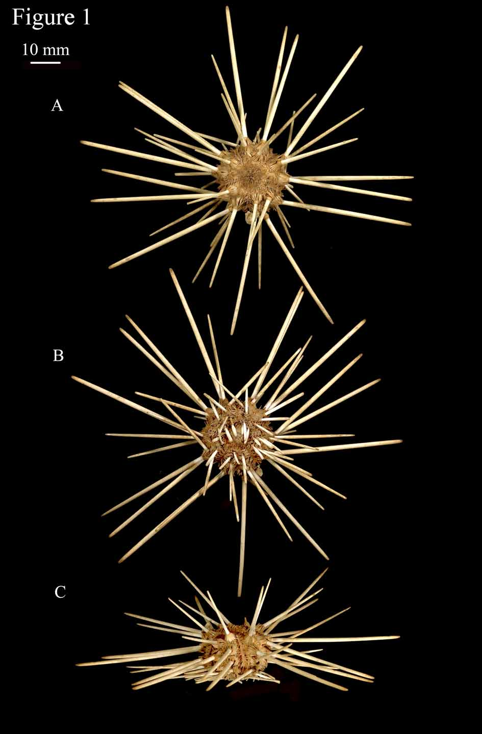
Figs 1 View FIGURE 1 , 2 View FIGURE 2 , 4 View FIGURE 4 , 5 View FIGURE 5 and 6.
Diagnosis: This new species has aboral primary interambulacral spines that are typically cream in colour (occasionally with faint, pink banding) and slightly curved, measuring more than twice the test’s horizontal diameter (h.d.) at the ambitus. The spines have a relatively smooth surface formed by dense anastomosing cortical hairs that coalesce to form a continuous crust between low, longitudinal ridges. These ridges typically number twenty-two and are more apparent when the spine is viewed in transverse section. The collar of the spine is short and dark brown, while the neck is typically white and unconstricted. Oral primary interambulacral spines have serrated edges, while scrobicular tubercles are slightly adpressed forming a loose mail around primary tubercles. The large globiferous pedicellariae have moderately narrow valves, each valve with a narrow longitudinally oval opening beneath a large terminal tooth. Small globiferous pedicellariae have proximal regions with rounded edges and a longitudinally oval opening beneath the terminal tooth. Crenulations are well defined on the adapical side of tubercles above the ambitus. The colour of the primary ambulacral spines, secondary spines, miliary spines and the test epithelium is yellow-orange/light-brown.
Holotype: NHM 2007.4 Natural History Museum, London.
Paratype: NHM 2007.5 Natural History Museum, London.
Etymology: Named after the light, fair colour (yellow-orange/light-brown) of the test epithelium, primary ambulacral spines, secondary spines and miliary spines.
Description: The test is relatively thick, subpentagonal in outline and inflated, with flattened adapical and oral surfaces ( Figs 2 View FIGURE 2 C & D). The denuded test (Figs (2A–E) is whitish-cream (the epithelium is yelloworange/light-brown in life) with a h.d. of 47 mm and a vertical diameter (v.d.) of 34 mm (paratype). The apical disc is monocyclic, with all ocular plates narrowly insert ( Fig. 2 View FIGURE 2 A), measuring 40 % of the test’s h.d. (19 mm in the paratype). The genital plates are all of equal size, widening towards the periproct. The genital and ocular plates are whitish-cream, with a brown, raised, central region. This fades into pink beneath the gonopores. The madreporite is dark brown, convex, covering most of genital plate 2. All apical plates are densely covered with granules, which increase in size towards their outer edge. The genital pores are proportionally small, slightly elevated and occur near to the outer third of the genital plates. The periproctal plates are brownishpink, the five at the corners of the pentamerous periproct, elegantly pointing between the genital plates, being contiguous with the ocular plates.
The ambulacra are slightly sinuate ( Fig. 2 View FIGURE 2 D), 25 % the width of the interambulacra (5.5 mm in the paratype) at the ambitus. The interporiferous zones are approximately twice the width of a pore-zone, with each adradial plate to the marginal tubercle having a large outer secondary tubercle, with two smaller secondary tubercles placed one above the other. These form regular vertical series, with each plate also having miliary granules. No naked or sunken median line is present. The pore-zones are slightly sunken, pores nonconjugate, approximately equal in size, with each pore separated by a broad wall ( Fig. 2 View FIGURE 2 H).
The interambulacra ( Fig. 2 View FIGURE 2 C) are formed of two series of eight or nine primary tubercle bearing plates. Tubercles have areoles of moderate size and depth ( Fig. 2 View FIGURE 2 I), which are separated by at least two rows of scrobicular tubercles aborally, the last two sometimes being confluent adorally. Areoles on the oral surface are distinctly transverse-oval, while adapical areoles are circular. Primary tubercles are of moderate size, perforate, with distinct traces of crenulation on the adapical sides of the tubercles above the ambitus. Scrobicular tubercles are of moderate size. Outside the scrobicular ring the plates are densely covered with secondary tubercles which decrease gradually in size, becoming granular towards the slightly sunken median line. Horizontal lines are distinct, particularly adapically, but neither the median line nor the horizontal lines are naked.
The peristome measures 38 % of the test’s h.d. ( Fig. 2 View FIGURE 2 B) (18 mm in the paratype) with twelve ambulacral imbricate plates in each column and pores-pairs uniserially arranged. Six to seven non-ambulacral plates form a regular interradial series reaching the mouth.
FIGURE 6. Pedicellariae from A, holotype of L. xanthe sp. nov., B, holotype of L. fusca , C, holotype of Calocidaris micans and D, Compsocidaris pyrsacantha (S.E. Coppard private collection); i & ii, large form of globiferous pedicellaria; iii (oral) & iv (aboral), small form of globiferous pedicellaria; v, tridentate pedicellariae; vi (side view) & vii (internal view) individual valves of large globiferous pedicellariae; viii (side view) & ix (internal view) individual valves of the small form of globiferous pedicellaria; x (side view) & ix (internal view) individual valves of tridentate pedicellariae; Axi, individual valves of triphylous pedicellariae from L. xanthe sp. nov.; Cxii individual valve of the large form of tridentate pedicellaria of Calocidaris micans .
The Aristotle’s lantern ( Figs 2 View FIGURE 2 F & G) and perignathic girdle are typical of the cidaroid form, with small epiphyses that do not project and relatively high apophyses ( Fig. 2 View FIGURE 2 E).
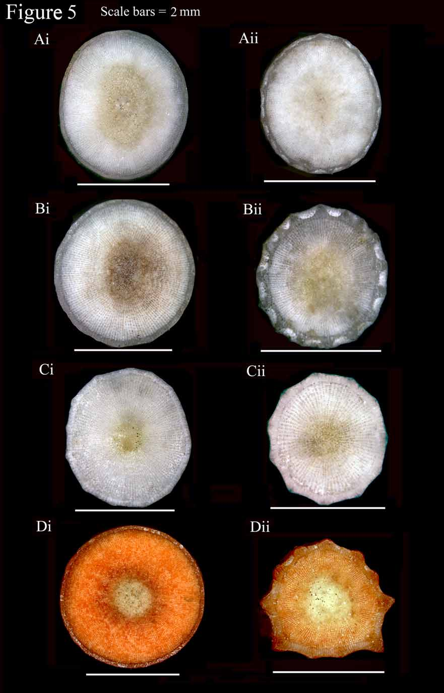
Aboral primary interambulacral spines ( Figs 4 View FIGURE 4 Avii & Aviii) are long, slender and tapering distally, typically cream in colour (occasionally with faint, pink banding), and slightly curved (see Fig. 4 View FIGURE 4 Avii). The longest measures 85 mm on the holotype ( Figs 1 View FIGURE 1 A–C) (x 2.4 the test’s h.d.) with a diameter of 3 mm. These are cylindrical in transverse section adapically ( Figs 5 View FIGURE 5 Ai & Aii), becoming slightly flattened beneath the ambitus. The spines are relatively smooth and typically have a glossy sheen ( Fig. 4 View FIGURE 4 Ai) (particularly on smaller specimens, the glossy sheen being absent on the aboral primary spines of some larger specimens) with approximately twenty-two longitudinal ridges faintly visible on primary spines at the ambitus ( Fig. 4 View FIGURE 4 Avii). This is due to the presence of dense, anastomosing cortical hairs that coalesce to form a continuous crust between longitudinal ridges ( Fig. 5 View FIGURE 5 Aii). These ridges are more noticeable beneath the ambitus due to the reduced depth of the cortical hairs. In smaller specimens the ridges appear to be completely absent but are visible when the spine is viewed in transverse section. The collar of the spine ( Fig. 4 View FIGURE 4 Aiii) is short (typically 2 mm), dark brown and increases in thickness towards the milled ring. The neck of the spine ( Fig. 4 View FIGURE 4 Aiii) is long, straight and completely smooth with no cortical hairs, or ridges in the cortex ( Fig. 5 View FIGURE 5 Ai). The ridges begin above the neck of the spine and continue to the spine’s tip. When viewed in transverse section ( Fig. 5 View FIGURE 5 Ai & 5Aii) the medulla is seen to be formed of reticulated stereom which is slightly darker in colour than the surrounding stereom. The tips of the primary spines gently taper and flatten dorso-ventrally to a blunt tip ( Fig. 4 View FIGURE 4 Aii), or in small specimens to a small crown; in both cases the longitudinal ridges are more apparent due to the absence of cortical hairs.
Oral primary interambulacral spines are white ( Figs 1 View FIGURE 1 B & 4Axii), distinctly flattened, slightly curved and moderately broad, with serrated edges, these being more apparent in smaller specimens. These have slightly rounded blunt tips, with those that are near the mouth tapering distally. They have an orange-brown collar, a smooth white neck, and longitudinal ridges on both the upper and lower surfaces. The third and fourth oral primaries ( Fig. 1 View FIGURE 1 B) are transitional to the aboral primaries.
Primary ambulacral spines are undifferentiated orally and aborally ( Fig. 4 View FIGURE 4 Av). These typically measure 3– 4 mm in length, are slender, slightly flattened and curved, with longitudinal ridges. These spines taper distally and have slightly rounded tips.
Scrobicular spines are slightly adpressed and ridged ( Figs 4 View FIGURE 4 Aviii & Aix), widening above the base up one quarter of their length, where they taper to a blunt tip. They typically measure 5 mm in length and do not form a very close mail around the base of the primaries ( Fig. 1 View FIGURE 1 A). Secondary spines on the ambulacra and interambulacra are slender ( Fig. 4 View FIGURE 4 Av), 2–3 mm in length, slightly flattened and curved, with longitudinal ridges. These spines also taper distally and have slightly rounded tips. Miliary spines ( Fig. 4 View FIGURE 4 Aiv) are shorter (1.5 mm in length) but proportionally broader than the secondary spines, and are distinctly flattened with longitudinal ridges. They are broadest at their base, tapering to a rounded tip. Oral peristomal spines densely cover the peristomal plates. These are highly curved and distinctly club-shaped, with serrated longitudinal ridges ( Fig. 4 View FIGURE 4 Axi).
Large globiferous pedicellariae are abundant aborally, particularly down the interambulacra, with all pedicellariae examined having three valves. They have short necks and stalks (Figs 6Ai & Aii), with large valves that gradually narrow above the adductor muscle insertion points towards a large and sharply pointed terminal tooth (Figs 6Avi & Avii). The opening beneath the terminal tooth (Fig. 6Avii) is longitudinally oval in shape distally, becoming U-shaped above the lip. The oval region is surrounded by narrow, pointed, peripheral accessory teeth. These decrease in size around the U-shaped region. The distal region beneath the opening is narrow, increasing in width towards the adductor muscle insertion regions. These muscle insertion regions are longer than wide, and measure just over half the length of the valves.
The small form of globiferous pedicellaria is very abundant both orally (Fig. 6Aiii) and aborally (Fig. 6Aiv), particularly down the ambulacra. Pedicellariae of this form have relatively long stalks, particularly on the oral surface, with short necks. Each valve has a proportionally large terminal tooth (Figs 6Aviii & Aix) typical of the Cidarina. Peripheral teeth are present along the edges of the valves, being larger around the longitudinally oval opening, which measure 22 % of the length of the valve. The lip beneath the opening is denticulate. The distal region of the valve beneath the lip and the proximal adductor insertion regions are broad, while the edges of the valves are curved.
Tridentate pedicellariae occur only as one form (Figs 6Ax & Axi), typical of the genus. They have relatively long stalks with a short neck. The valves are slender, have a terminal tooth, and increase in width proximally, flaring above the proximal region. These flaring regions are visible as crests when viewed from the side (Fig. 6Ax). The valves slightly constrict beneath these crests, directly above the adductor muscle insertion regions. The proximal regions are slightly longer than they are wide with a relatively small keel.
A single pedicellaria representing a third class was found on the paratype, but not on the holotype or any of the other nine specimens examined. This has a long stalk with a short neck and very short conical shaped valves (Fig. 6Axii). The adductor muscle insertion regions are fairly open, indicating a very limited grasping pressure. This single pedicellaria is clearly not representative of this species and appears to be an anomaly.
Spicules in the tube feet occur as smooth rods. These are typically straight but occasionally bifurcate at one end.


No known copyright restrictions apply. See Agosti, D., Egloff, W., 2009. Taxonomic information exchange and copyright: the Plazi approach. BMC Research Notes 2009, 2:53 for further explanation.