
Distylopyga beccalonii Anisyutkin, 2018
|
publication ID |
https://doi.org/ 10.11646/zootaxa.4532.4.4 |
|
publication LSID |
lsid:zoobank.org:pub:A35A92D5-2628-4203-8C94-DDBFFFCDC79E |
|
DOI |
https://doi.org/10.5281/zenodo.5976584 |
|
persistent identifier |
https://treatment.plazi.org/id/D2BE030B-0DE1-4573-9FC5-FD2336C5EECD |
|
taxon LSID |
lsid:zoobank.org:act:D2BE030B-0DE1-4573-9FC5-FD2336C5EECD |
|
treatment provided by |
Plazi |
|
scientific name |
Distylopyga beccalonii Anisyutkin |
| status |
sp. nov. |
Distylopyga beccalonii Anisyutkin View in CoL , sp. nov.
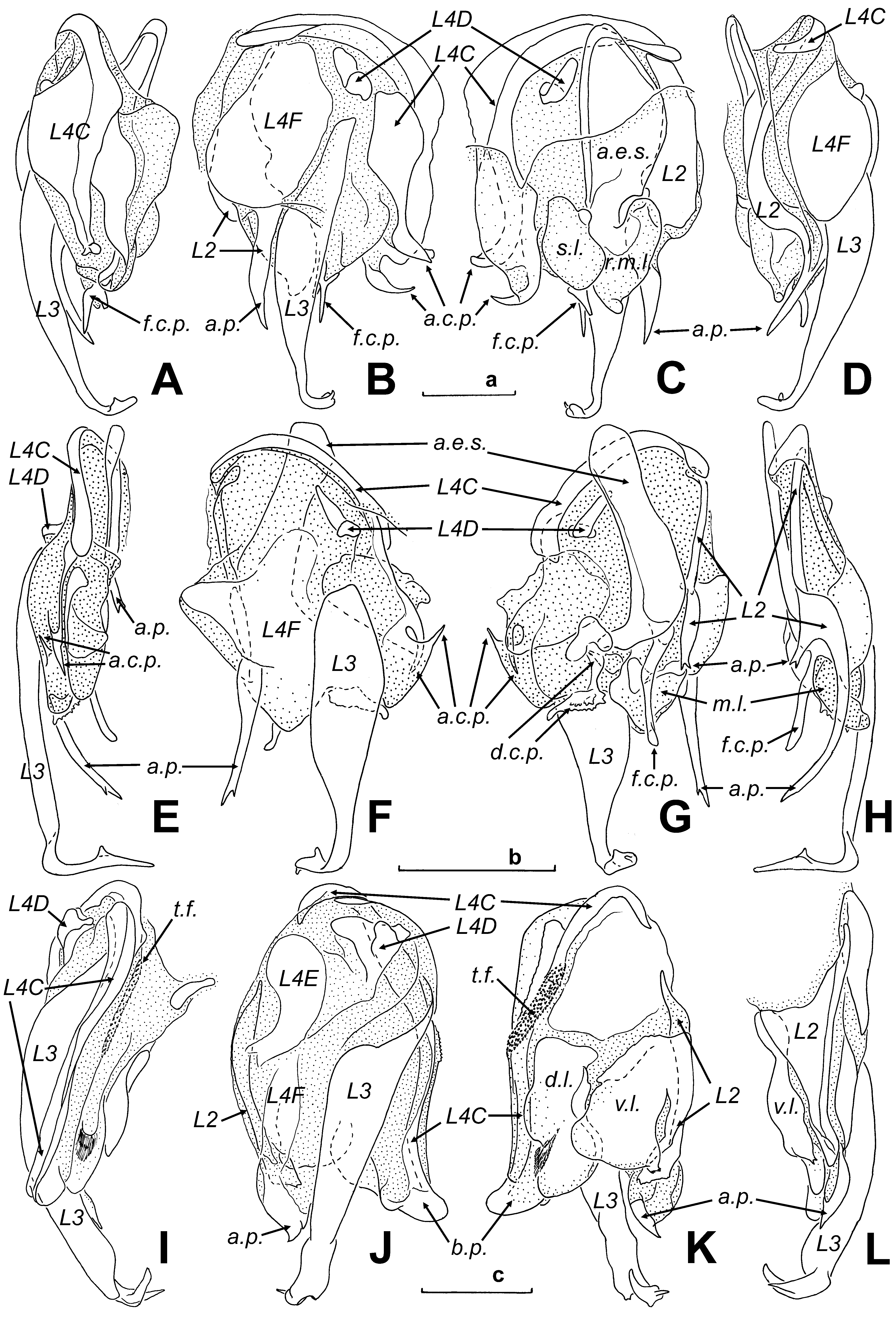
( Figs. 3 View FIGURE 3 E–H, 4A–N, 5A–E)
Zoobank: http://zoobank.org/ D2BE030B-0DE1-4573-9FC5-FD2336C5EECD
Material: Holotype. male, THAILAND, prov. Phetchaburi, 50 km SW town Phetchaburi, environs of National Park Kaeng Krachan , forest near reservoir, h= 400 m, 30–31 July1996, leg. A. Gorochov ( ZIN); paratypes. 3 females, 1 larva, same data as holotype, but 30 July–1 August 1996 ( ZIN); paratypes. 2 females, same data as holotype, but 3–5 August 1996 ( ZIN).
Etymology. Patronymic. This species is named in honour of Dr. George Beccaloni (the Alfred Russel Wallace Trust, United Kingdom), famous specialist in cockroach taxonomy and establisher of Cockroach Species File database.
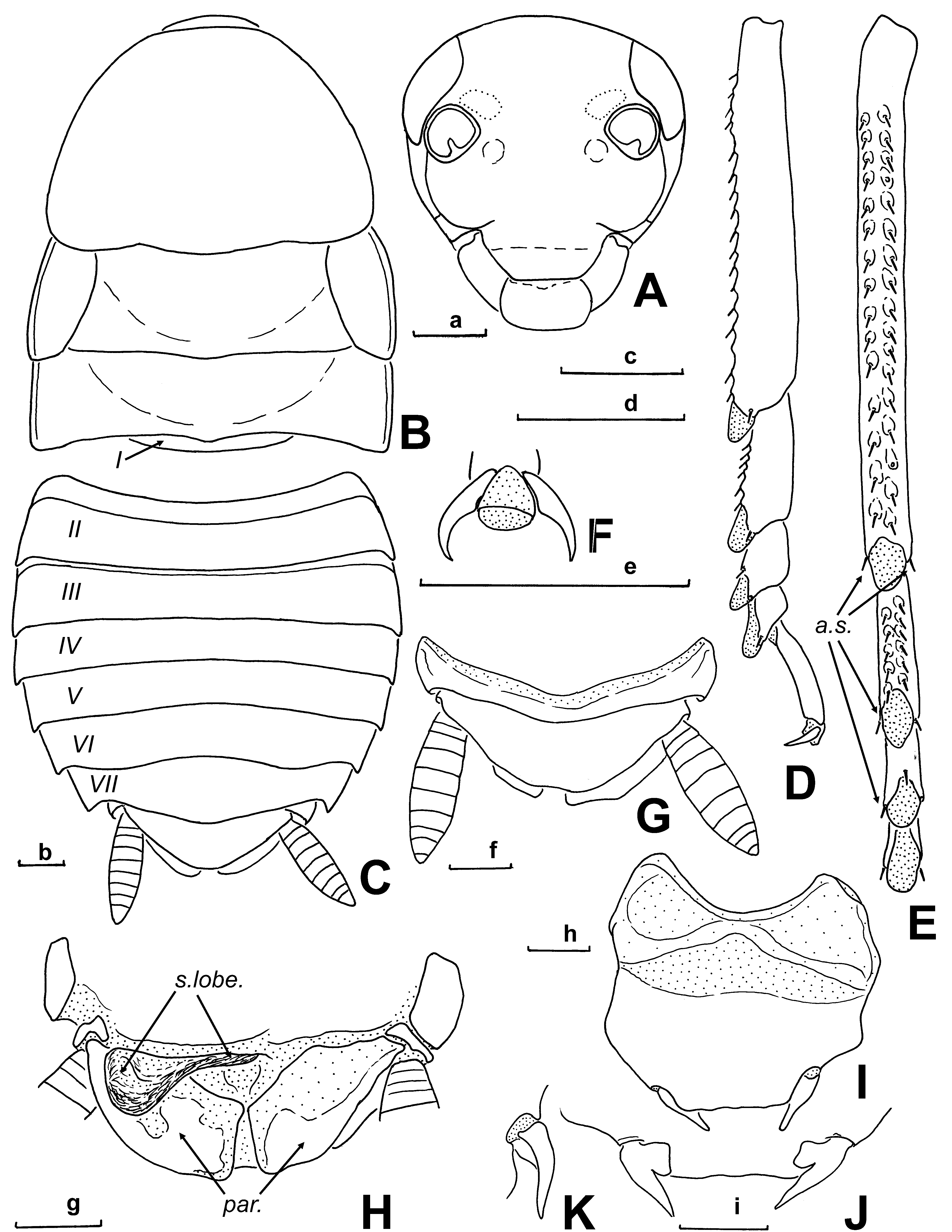
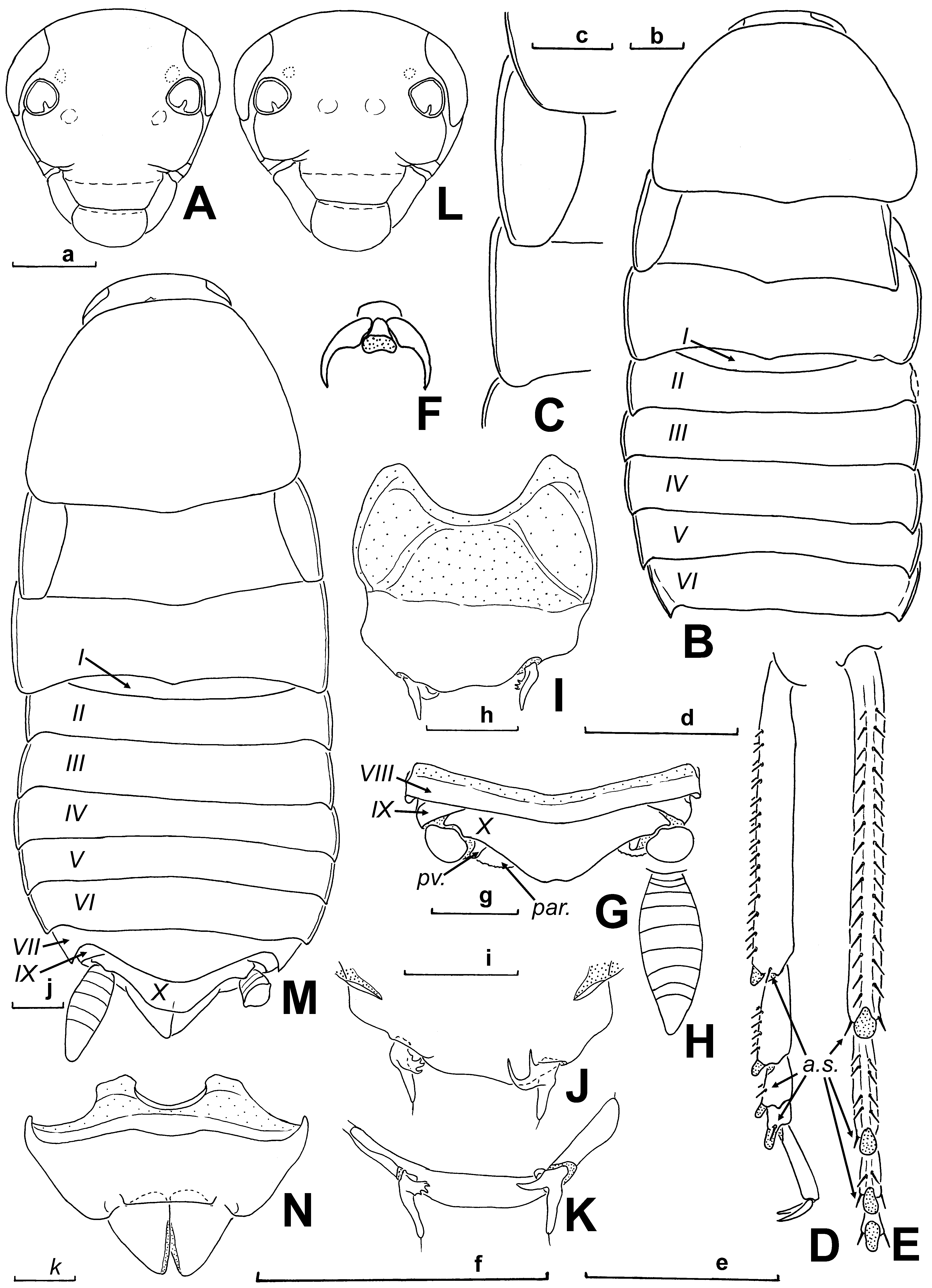
Description. Male (the holotype). Body generally reddish brown, head slightly darker; eyes black; areas of ocellar spots pale yellowish ( Fig. 4A View FIGURE 4 ); scapus and pedicellum, labrum, anteclypeus, maxillary and labial palps dirty yellow; antennae (scapus and pedicellum excluded), greyish brown; legs yellowish brown. Surfaces smooth and lustrous; punctation very delicate; facial part of head with delicate wrinkles above clypeus (not shown in Fig. 4A View FIGURE 4 ); distal antennomeres dull (approximately from 10th segment). Head rounded at vertex, longer than wide, convex ( Fig. 4A View FIGURE 4 ); epicranial sutures not indicated; eyes small; ocellar spots small, visible only as light spots; distance between eyes 1.7 times eye length; distance between antennal sockets 2.4 times of the scape length (about 0.5 mm); approximate length ratio of 3rd–5th segments of maxillary palpomeres 1.0: 1.0: 1.3. Pronotum campaniform, wider than long, caudall margin slightly angulate ( Fig. 4B View FIGURE 4 ). Tegmina reduced to lateral flaps, marginated laterally and truncated apically ( Fig. 4B, C View FIGURE 4 ). Wings absent. Mesonotum and metanotum transverse, dorsally slightly convex, with posterior margins slightly angulate ( Fig. 4B View FIGURE 4 ). Thoracal tergites and abdominal tergites II–VII marginate laterally ( Fig. 4B, C View FIGURE 4 ). Anterior margin of fore femur armed as in the type A, with 14–15 spines, including 3 apical ones. Fore tibiae not thickened distally. Tibial spines well developed. Structure of hind tarsi ( Fig. 4 View FIGURE 4 D–F): metatarsus a little longer than other segments combined; all euplantulae small and apical ( Fig. 4D, E View FIGURE 4 ); metatarsus and 2nd metatarsomere with 2 more or less equal rows of spines along its lower margin; 3rd metatarsomere with 2 spines along lower margin; metatarsus and 2nd–4th metatarsomeres with 1–2 "additional spines" bordering euplantulae from inside and outside ( Fig. 4D, E, a.s View FIGURE 4 .); claws symmetrical, simple; arolium smaller than half of claw length ( Fig. 4F View FIGURE 4 ). Fore- and mid tarsi similar to metatarsi, but with comparatively shorter tarsomeres. Abdomen without visible glandular specializations; posterolateral angles attenuate caudally; tergite VI with caudal margin straight ( Fig. 4B View FIGURE 4 ); tergite VII roundly projected caudally. Anal plate (tergite X) short and transverse, medially projected, with very weak triangular median incision on caudal margin ( Fig. 4G View FIGURE 4 ). Cerci fusiform and flat, with solidly connected segments ( Fig. 4H View FIGURE 4 ). Paraprocts weakly asymmetrical, sclerotized along caudal margin; pvsclerites well sclerotized, elongated ( Fig. 4G View FIGURE 4 , pv.). Hypandrium weakly asymmetrical, wide, antero-lateral parts (lateral sternal apodemes or apophyses) short; caudal margin sinuate between styli ( Fig. 4 View FIGURE 4 I–K); styli asymmetrical: left stylus with 4 small curved spines, right stylus with 2 longer ones.
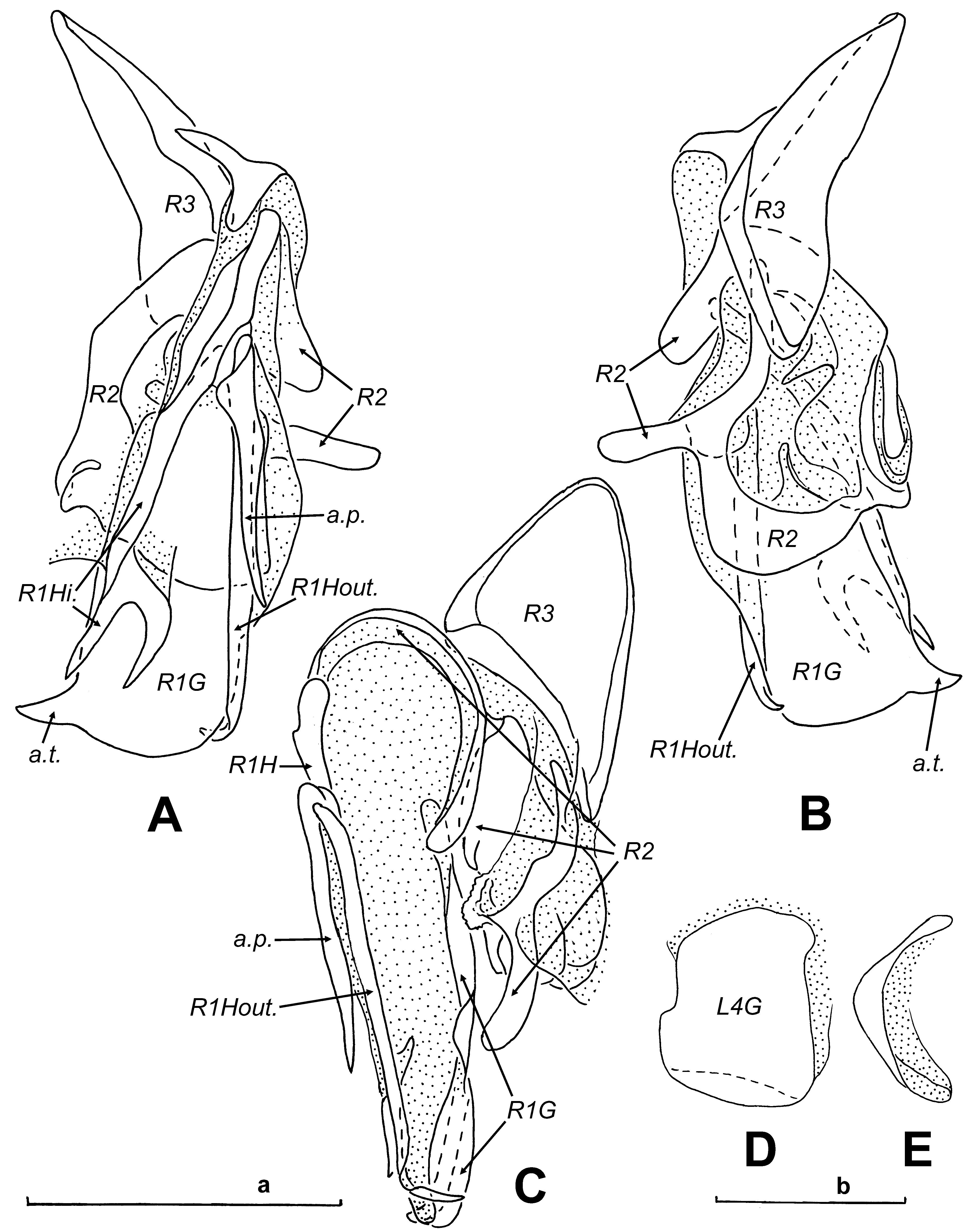
Male genitalia ( Figs. 3 View FIGURE 3 E–H, 5A–E): left phallomere ( Fig. 3 View FIGURE 3 E–H) with sclerite L4C (L2D) long and narrow, caudally divided into two partly membranous parts, outer one terminating in oppositely directed sharp teeth ( Fig. 3E, G, a.c.p View FIGURE 3 .); sclerite L3 (L3d) curved in apical part, with small cranially directed tooth; sclerite L4F, possibly L4E+L4F, large, with indistinct margins ( Fig. 3F View FIGURE 3 ); sclerite L4D (L 3v) elongated ( Fig. 3F, G View FIGURE 3 ); sclerite L2 (L 2v) long, narrowed in cranial part ( Fig. 3G, H View FIGURE 3 ), terminating in 2 acute caudal processes: inner process short and apically forked, outer—long and slender ( Fig. 3E, G, H, a.p View FIGURE 3 .); "additional elongated sclerite", probably separated part of L2, situated on inner side of phallomere dorsally to L2 ( Fig. 3G, a.e.s View FIGURE 3 .), terminating long and slender caudal process ( Fig. 3 View FIGURE 3 E–F, f.c.p.) and subtriangular membranous lobe ( Fig. 3G, H, m.l View FIGURE 3 .); "additional elongated sclerite" connected with complicate sclerite with dorso-caudal projection ( Fig. 3G, d.c.p View FIGURE 3 .); sclerite L1 absent. Ventral phallomere L4G (VP) short and wide ( Fig. 5D, E View FIGURE 5 ). Right phallomere complex in shape, as in Fig. 5 View FIGURE 5 A–C; basal sclerite (R2) small, as compared with other sclerites of right phallomere; sclerite R1H divided into 2 branches: inner branches apically forked ( Fig. 5A, R View FIGURE 5 1 View FIGURE 1 Hi), outer with apex bent down ( Fig. 5A, R View FIGURE 5 1 View FIGURE 1 Hout.), additional long spine-like process situated above outer branch ( Fig. 5A, a.p View FIGURE 5 .); sclerite R1G subtriangular in shape, with blunt apical tooth ( Fig. 5A, B, a.t View FIGURE 5 .); sclerite R3 large, plate-like.
Female. Similar to male. Maxillary and labial palps sometimes grey, legs—reddish brown. Head as in Fig. 4L View FIGURE 4 ; distance between eyes 1.6–1.7 times eye length; distance between antennal sockets 2.1–2.5 times of the scape length (about 0.5–0.6 mm); approximate length ratio of 3rd–5th segments of maxillary palps 1.0: 1.0: 1.3–1.4. Thoracal and abdominal tergites as in Fig. 4M View FIGURE 4 . Anterior margin of fore femur armed as in the type A, with 14–15 spines, including 2–3 apical ones. Abdominal apex as in Fig. 4M View FIGURE 4 . Genital plate transverse, valves large ( Fig. 4N View FIGURE 4 ).
Larva. Similar to adult male, but paler: antennae, anteclypeus, maxillary and labial palps and partly legs yellow.
Measurements (mm). Head length: male 2.5, female 2.8–3.0; head width: male 2.3, female 2.4–2.6; pronotum length: male 3.3, female 3.4–3.7; pronotum width: male 4.5, female 4.4–4.7; mesonotum length: male 1.7, female 1.8–2.0; tegmina length: male 1.9, female 1.6–2.0; tegmina width: male 1.1, female 0.9–1.1; metanotum length: male 1.2, female 1.3–1.6; metanotum width: male 5.1, female 5.0–5.4.
Comparison. The new species differs from D. semoni in generally paler body (black in S. semoni ) and distinctly angulate caudal margin of metanotum ( Fig. 4B View FIGURE 4 ) (emarginated in S. semoni ).
| ZIN |
Russian Academy of Sciences, Zoological Institute, Zoological Museum |
No known copyright restrictions apply. See Agosti, D., Egloff, W., 2009. Taxonomic information exchange and copyright: the Plazi approach. BMC Research Notes 2009, 2:53 for further explanation.