
Eutrachytes flagellatus, Moraza & Kontschan & Sahoo & Ansari, 2016
|
publication ID |
https://doi.org/10.1051/acarologia/20162189 |
|
DOI |
https://doi.org/10.5281/zenodo.4696980 |
|
persistent identifier |
https://treatment.plazi.org/id/03F887CA-FF8D-FFFF-FC1B-FD2CB536F82F |
|
treatment provided by |
Carolina |
|
scientific name |
Eutrachytes flagellatus |
| status |
sp. nov. |
Eutrachytes flagellatus n. sp.
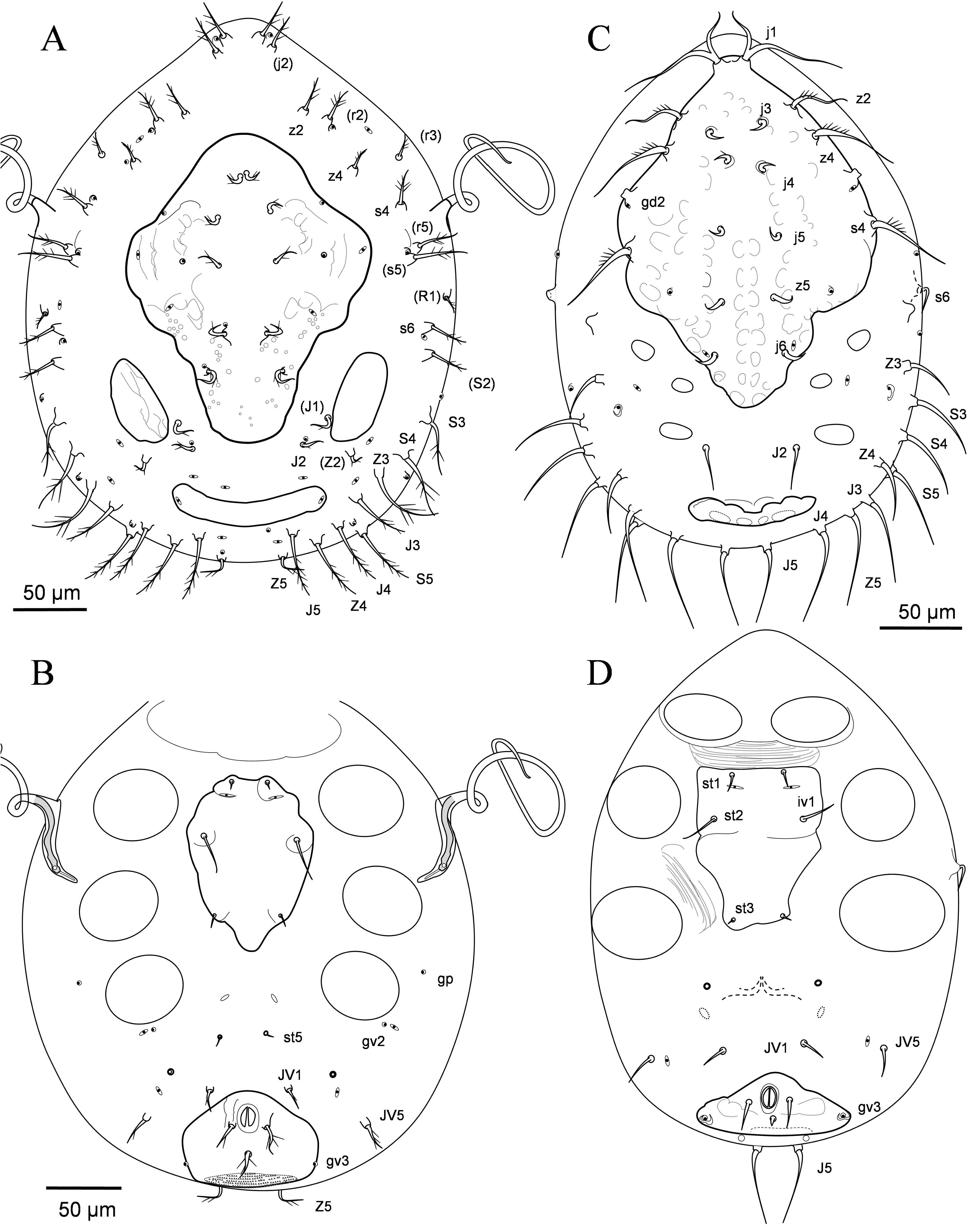
Diagnosis — Adults with idiosoma discoidal in shape, as long as wide, with prominent anterior region and scabellum present; dorsal, marginal and peritrematal shields fused at the anteriormost region; posterior region of dorsal shield with a pair of marginal concave depressions present; submarginal shield with setae to the anterior level of coxa I in adult and nymphal instars; pygidial shield nude in all instars, only bearing one pair of poroids. Nymphal instars with a pairs of lateral flagellate tubular structures emerging from the peritreme. Hypostomal setae h2 longer than h1 and p4 divided in two pilose dissimilar branches. Larval podonotal shield with nine pairs of setae, three pairs of mesonothal scutellae; protonymph with five pairs of setae on podonotal shield, nude mesonotal and nude pygidial; deutonymphs with dorsal and marginal shield fused at anterior region and peritrematal shield free as in the protonymphal instar. Legs I-IV with pretarsus, a pair of well developed claws and three acuminate lobes; femora I-IV with ventral keel; femora II in the male with ventral setae modified as a conspicuous thick spine.
Female
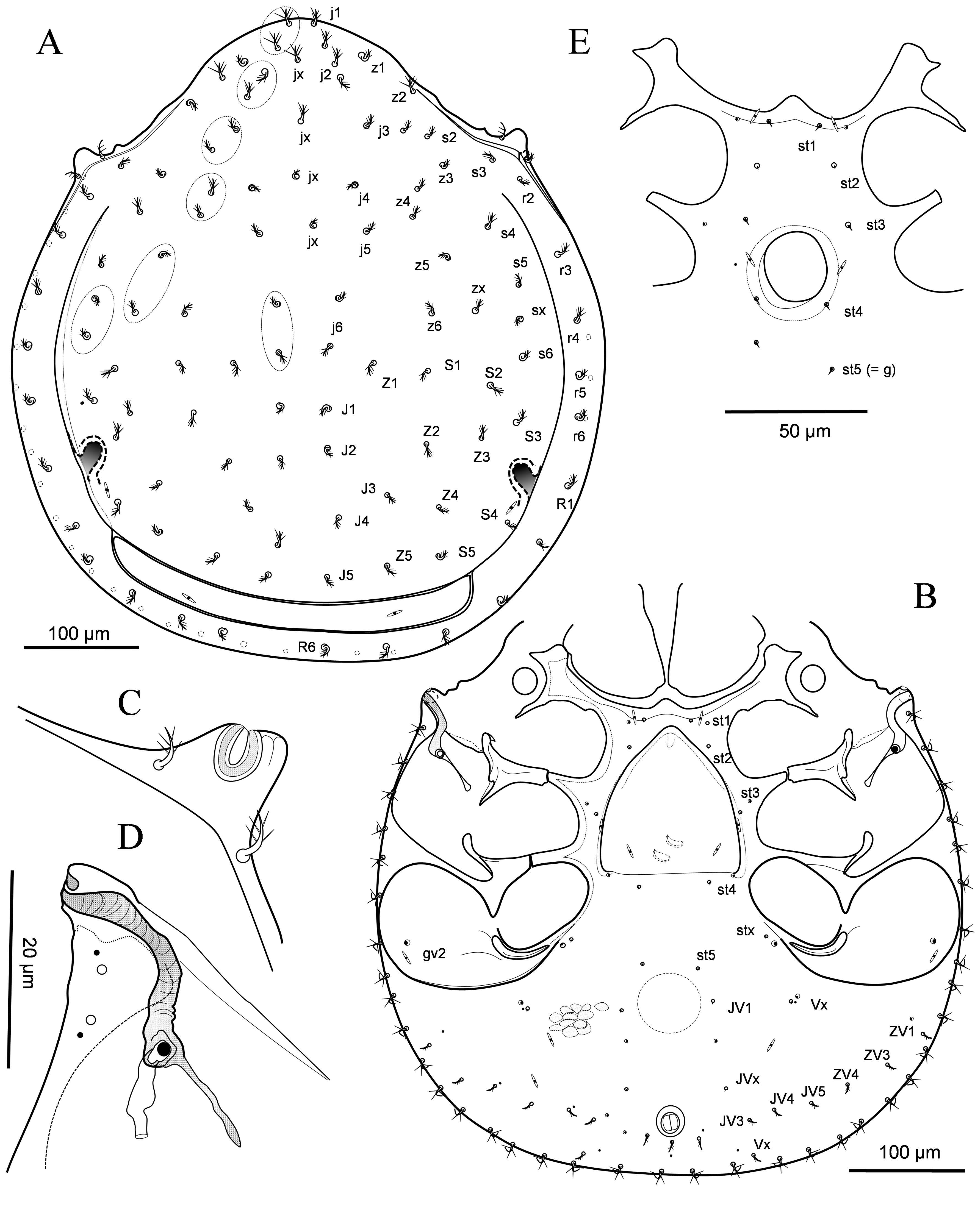
Dorsal idiosoma ( Fig. 1A View FIGURE ) — Idiosoma discoid in shape, almost as long ( 570 µm) as wide ( 522 µm), with smooth margin and humeral peritremal protrusions. Dorsal and marginal shields fused anteriorly at level of humeral protrusions, and fused with peritrematal shield at level of z2. Anterior dorsal shield rounded acuminate, slightly bent to ventral region. Pygidial shield ca. 18 times wider than long, with posterior margin slightly lobulated, with one pair of poroids (lyrifissures) and at least three pairs of punctiform poroids. Submarginal shield rearchs the posterior margin of peritremes where bends ventrally covering part of distal peritremal loop ( Figs 1B, 1D View FIGURE ).
Dorsal shield with lightly granulate cuticle, c.a 40 pairs of short, homogeneous in length, pilose setae, neotrichy present (podonotal region with several unpaired central setae jx, and duplicate setae j1, j2, j3, j6, z4, z5, s5; marginal shield with 11 pairs of setae ( r2-r6, R1-R6) similar in length and shape, and ca. 20 pair of setae with sparse and longer pilosity on the ventral submarginal region (setae UR). Most dorsal setae associated to a punctiform pore like structure. Shield with a pair of concave marginal depressions at the posterior region which remain under the shield and open laterally over the marginal shield.
Ventral idiosoma ( Fig. 1B View FIGURE ) — Tritosternum with a strongly dentate base 34-36 µm long and three pilose laciniae free for most of length; medial lacinia longer ( 45 µm) than laterals ( 19 µm), devoid of basal pilosity and with bifid end ( Fig. 2D View FIGURE ). Sternal shield with anterior margin well delineated and a medial convexity. Sternal setae minute, smooth, poorly discernible; setae st1 anterior to epigynal shield, accompanied by a pair of elongated lyrifissures iv1 and a pair of punctiform poroids ( gst1); st2 at both sides of epigynal shield, close to st1, distance st1- st2 near one third st2-st3; st3 together with poroids iv2 at margin of genital opening, and pores gst2; st4 behind the shield and the third pair of pores gst3 at the posterior corners of the shield; genital setae st5 behind coxae IV similar to sternal setae. Epigynal shield densely granulated as other idiosomal shields, subtriangular in shape, 125 µm long 118 µm wide, with straight posterior margin and acuminate anterior margin; one pair of liryfissures at the posterior region; shield located between coxae II and anterior margin of coxae IV. Endogynum with a pair or cup-like structures.
Opisthogaster with three pairs of minute ventral setae ( JV1 and two more pairs " Vx " associated to glandular pores at the parapodal region of coxae IV), similar to sternal setae, and seven pairs at the posterior region laterals to anal opening which are similar in shape and length to the three circumanal setae such as in figure 1B; at least two pairs of liryfissures ivo and eight pairs of pores (at least four are glandular pores). Anal opening framed, small and rounded (diameter 16 µm), with nude anal valves; paranal and postanal setae ciliate, similar to other ventral lateral setae, and posterior to anal opening. Between setae JV1 and st5 a circular subcutaneous structure is observed with a pair of sigillar area at both sides of it.
Endopodal elements between legs I-II, II-III and III-IV fused to sternal shield; endopodal I with a strong anterior apophysis with lateral corner acuminate and internal corner rounded; exopodal I-II as a hemispherical apophysis; exopodoal elements between coxae II-III and III-IV free from endopodal; parapodal IV semilunar in shape, well developed, at the botton of pedofosae IV. Pedofosae II, III and IV well delimited, separated by longitudinal bridges.
Peritrematal shield with one pair of dorsal setae at both side of peritreme distal loop ( Fig. 1A View FIGURE ). Respiratory stigmata situated in pedofosae III; from the stigmata the peritreme ascends to lateral conical protuberance at level of pedofossae II and descends for a shorter and narrow stretch; peritreme curved dorsally at level of the humeral conical protuberance ( Figs. 1C, 1D View FIGURE ).
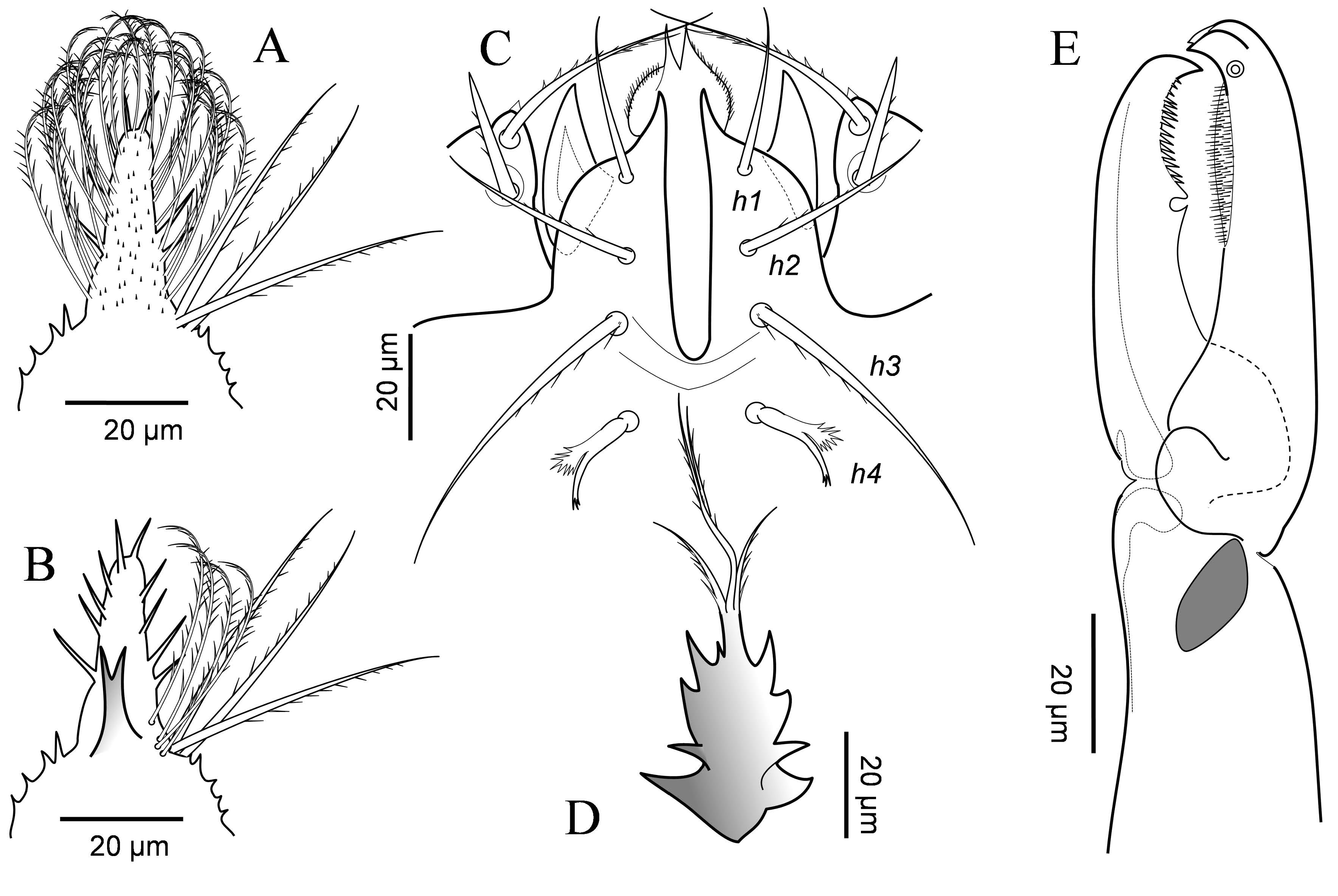
Gnathosoma — Gnathotectum ( Figs. 2A,B View FIGURE ). with a broad dentate base and medial projection with ventral surface provided of minute spines ( Fig. 2A View FIGURE ), lateral margins with 5-6 strong long spines ( Fig. 2B View FIGURE ), and a series of marginal strongly ciliate long setae; at the base of the medial projection, bent to ventral side, a strong dorsal bifid tectum ( Fig. 2B View FIGURE ); dorsally, at the basal region of central projection, three pairs of marginal long barbed straight setae ( Figs. 2A,B View FIGURE ).
Corniculi hornlike, short ( 32 µm), stout ( 18 µm), inserted midway between insertions of subcapitular setae h1 and h2, ( Fig. 2C View FIGURE ); h1 smooth, shorter ( 32 µm) than slightly pilose h2 ( 40 µm); pilose h3 the longest setae ( 64 µm), twice the length of h1, capitular setae ( h4) the shortest ( 20 µm) with a distal pilosity and a widened pilose process at the medial region ( Fig. 2C View FIGURE ). Internal malae short with lateral margins pilose, extending to medial length
Moraza M.L. et al.
of palpfemora ( Fig. 2C View FIGURE ). Deutosternum with two smooth and wide groves between h3 and h4. Palpal setation and form of setae as described by Krantz (1969) (2-5-5-14); palp- trochanter with inner setae ( 47 µm) twice as long as external seta ( Fig. 2C View FIGURE ), the last spinose; palp-claw two-tined. Chelicerae ( Fig. 2E View FIGURE ) with fixed digit edentate, ( 52 µm long) overlapping movable digit and nodulus present; digit with a socket subterminally which receives the hooked tip of the movable digit when the two are appressed ( Fig. 2E View FIGURE ); a densely pilose surface (brush) at masticatory region; terminally with a spine or lateral mucro. Movable digit of female 61-63 µm long, with a dentate comb at distal region (c.a 17 teeth) followed by a prominent alveolar remnant (or two opposed teeth), and basal region smooth; dorsal cheliceral setae no discernible if present; without conspicuous arthrodial process ( Fig. 2E View FIGURE ).
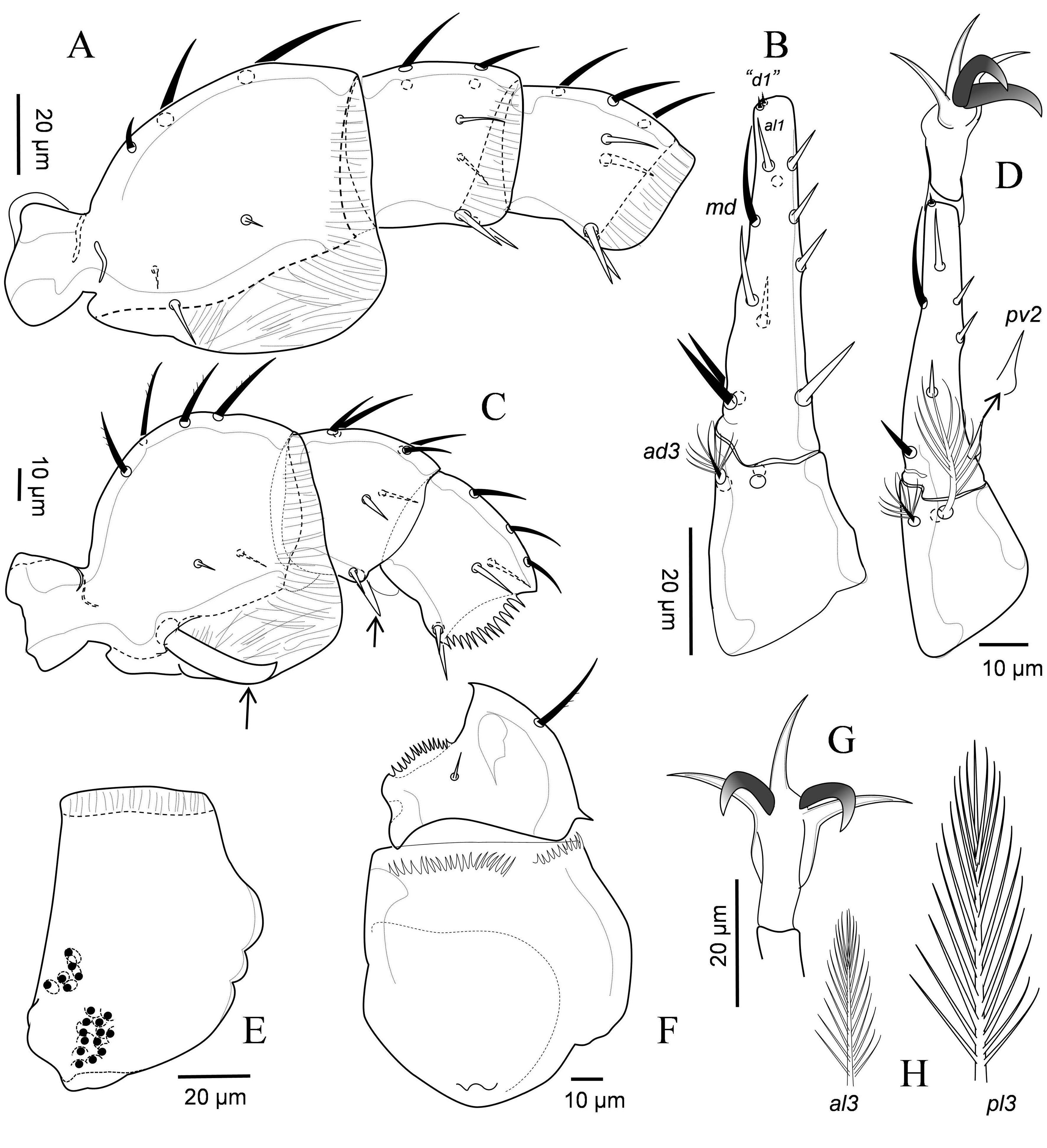
Legs — Legs relatively short, clearly shorter than dorsal shield. Coxae I with two groups of coxal glands, dorsal concavity and laterodorsal surface with rounded prominences ( Fig. 3E View FIGURE ); coxae II-IV with numerous ventro basal glandular openings; except tarsus I-IV, all other segments with a dorso distal striate tectum overlapping the articulation with the following segment; femora I-IV with a conspicuous ventral tectum ( Fig. 3A View FIGURE ). Tarsus I with a pair of claws and a series of long terminal setae. Pretarsus of legs I to IV with slender claws at the end of a relatively long stalk; legs II-IV with three slender acuminate lobes ( Figs. 3D,G View FIGURE ). Tarsus II-IV about twice as long as tibia, telotarsus twice longer than basitarsus ( 2.2 µm); apical ventral process absent; with 17 setae, apical setal processes ad1, pd1 minute ("d1"), spinelike ( Fig. 3B View FIGURE ), and telotarsal seta md twice as long as lateral setae al1, pl1; basitarsal setae feather-like, dorsal setae short ( 9 – 10 µm), al3 reaching al2 setae ( 29 – 31 µm) and pl3 the longest ( 51 – 53 µm) reaching pair of lateral setae al1, pl1 ( Figs. 3B,H View FIGURE ). Other leg setae simple or slightly barbed. Full complement of setae: coxae, 2-2-2-1; trochanters, 5-5-5-5; femora, 9 (1 2/1 2/2 1) – 9 (1 2/1 2/2 1) – 7 (1 3/2 1) – 7 (1 3/2 1); genua, 8 (1 2/1 2/1 1) – 8 (1 2/1 2/1 1) – 8 (1 2/1 2/1 1) – 5 (1 2/0 2/0 0); tibiae, 7 (1 1/1 2/1 1) – 7 (1 1/1 2/1 1) – 7 (1 1/1 2/1 1) – 7 (1 1/1 2/1 1). Leg lengths, excluding pretarsi: I 330 – 340 µm, II 320 – 2325 µm, III 300 – 310 µm, IV 300 – 310 µm.
Male
Dorsal idiosoma — Similar to female in shape; dorsal shielding and dorsal setae as for the female.
Ventral idiosoma — Anterior region of sternal region as in the female ( Fig. 1E View FIGURE ), with four pairs of minute sternal setae, two pairs of poroids ( iv1, iv3), two or three pairs of glandular pores ( gst1, gst3), and one pair of punctiform pores; setae st5 behind genital opening betwee coxae IV, similar to other sternal setae. Genital opening ( 23 – 24 µm long) between legs III. Other ventral characteristics as in female.
Gnathosoma — As in female.
Legs — Legs segments not strongly differentiated on either sex, except for a ventral thick claw like setae in femur II ( Fig. 3C View FIGURE ), spine like setae pv 1 in genua and tibia I-IV, and seta pv2 with a bulbose base in tasus II-IV ( Fig. 3D View FIGURE ). Anterior lateral ventral margin of the segments, except tarsi, strongly serrate ( Figs. 3C,F View FIGURE ). Legs length: I 310 – 315 µm, II 310 – 315 µm, III 290 – 295 µm, IV 300 – 305 µm.
Deutonymph — Idiosoma 497 µm long, 420 µm wide at level of coxae III-IV (n=1).
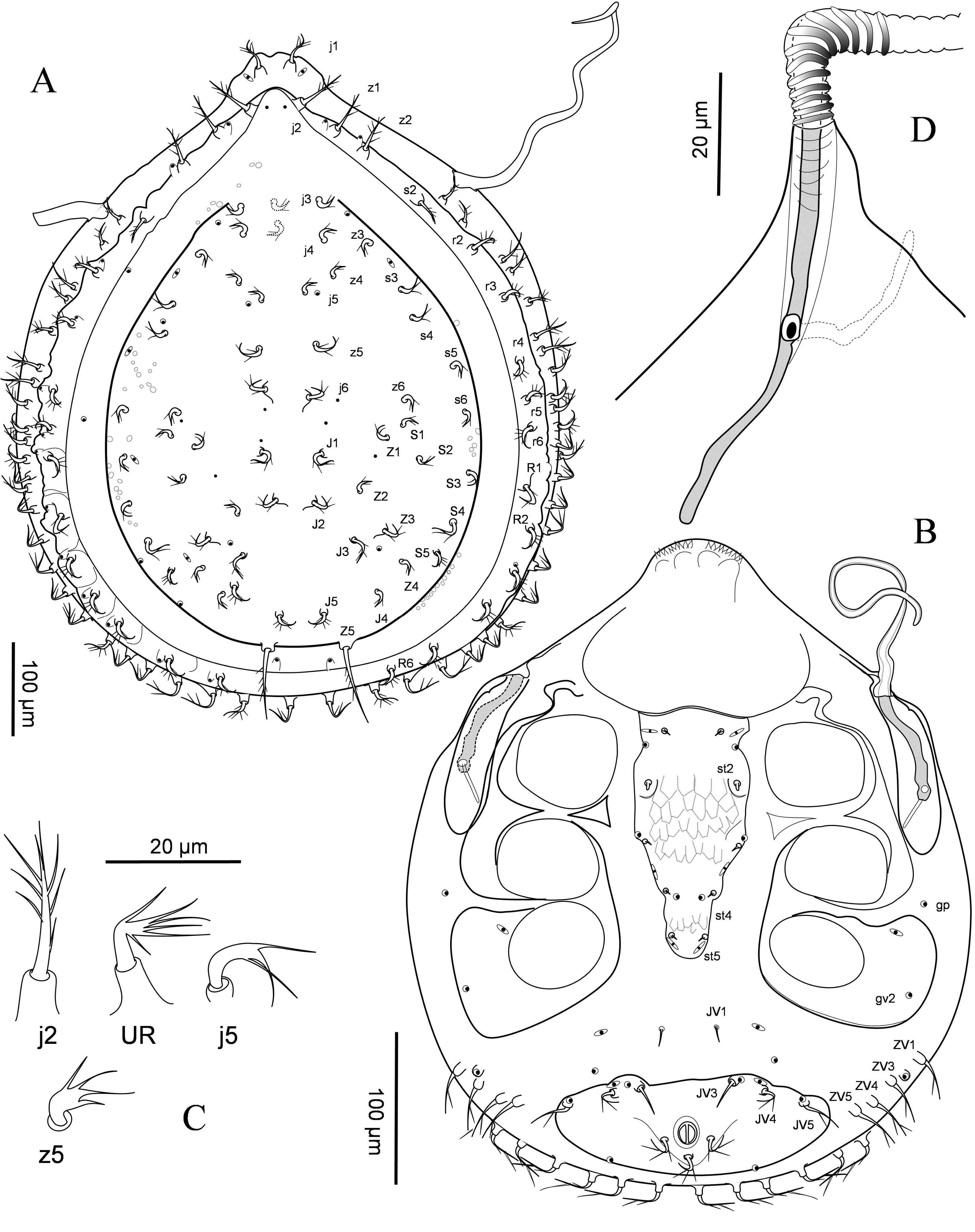
Dorsal idiosoma — Dorsum ( Fig. 4A View FIGURE ) discoidal, with acuminate anterior region and humeral angles absent. Marginal shields completely differentiated, separated form dorsal shield by a fine suture which became indistinct anteriorly; pygidial shield undifferentiated except for the presence of a pair of poroids set on conical tubercles.
Dorsal shield fused with marginal shields at level of setae j3, with granulate cuticle ornate with rounded shallow pits; shield holotrichous with 23 pairs of similarly short ciliate setae ( Fig. 4C View FIGURE ) ( j3-j6, z3-z6, s3-s6, J1-J5, Z1-Z5, S1-S5), except posteriormost Z5 elongate and slightly curved ( 87 µm long); sometimes two setae "x" between j3 and j4 present). Marginal shield with setae j2, z1, z2 on the anterior acuminate region of shield, similar in shape to j1 ( 30 – 34 µm long); other marginal setae ( s2, r2-r6, R1-R6) apparently shorter, similar to setae on dorsal shield. Interior region of marginal shield lacks setae, only with three pairs of gland pores (including pygidial pores). Complement of pores as in figure 4A; dorsal shield with four pairs of lyrifissures, six pairs of glandular pores, and five pairs of punctiform poroids (the three pairs of glandular pores at the marginal region of dorsal shield may be related to concave formations in the adult); most setae on marginal shields associate to poroids set on conical tubercles. Submarginal region poorly sclerotized with j1 and ca. 25 pair of curved and ciliate setae (setae UR) similar in length and shape to anteriormost podonotal setae ( Fig. 4C View FIGURE ); no discernible poroids in this region. All dorsal setae set on conical tubercles.
Ventral idiosoma ( Fig. 4B View FIGURE ) — Anterior acuminate prominence with four dentate marginal lobes. Sternal shield well sclerotized and ornamented with polygonal cells from setae st2 to posterior margin of the shield; shield free from endopodal extensions between coxae II and III, with five pairs of setae st1- st5, three pair of glandular pores ( gst1-3), and three pairs of no glandular poroids ( iv1, iv3, iv5); shield 246 µm long, 76 µm wide at level of st2, 27 µm long at level of st5; eroded at level of gv1 and narrowed behind st4: st5 and iv5 at the posterior margin of the shield at level of coxae IV ( Fig. 4B View FIGURE ). Setae st1, st3-st5 minute and smooth ( 2-3 µm), st2 rod-like, thicker and almost twice longer ( 5 µm).
Ventrianal shield well sclerotized and ornamented, almost three times wider than long ( 67 – 69 µm long, 262 µm wide), with pilose circumanal setae similar in length (ca. 27 µm), three pairs of ventral setae at the anterior margin of the shield, JV3 ( 16 – 18 µm) poorly ciliate, JV4 and JV5 similar to other posterior most opisthogastric setae, two pairs of glandular pores, each associated with ventral setae JV3 and JV5, and gv3 at the posterior margin at level of postanal setae; one pair of poroids associated to JV4; cribrum narrow as in adult. Opisthogaster with five pairs of ventral setae on soft tegument, JV1 shortest ( 8 µm) and smooth and four pairs of ciliate setae set on tubercles at the lateral sides ( ZV1, ZV3- ZV5); two pairs of glandular pores and one pair of discernible poroids on soft opisthogastric cuticle ( Fig. 4B View FIGURE ). Endopodal strips between legs II and III present and free; endopodal between coxae III and IV fused with parapodal elements, and pedofossae IV well developed bearing one anterior liryfissure and one posterior glandular poroid: exopodal elements II and III fused and contiguous with exopodal I-II. One pair of poroids distinct on soft cuticle between legs III and IV. Rim of exopodal plate behind coxa IV inconspicuous.
Peritrematal shield from coxae I to anterior margin of coxae III ornate as exopodal elements; respiratory stigmata at level of coxae II, peritremes extending to a point between coxae I and II and continues outside the body given way to a long, flagellate and tubular structure ( Fig. 4B View FIGURE ). This hollow cuticular structure has heavily ringed surface and its diameter decreases progressively at the distal end ( Fig. 5A View FIGURE ).
Gnathosoma — Gnathotectum, chelicerae and other mouthpart structures, corniculi and adjacent structures as in adult female; palpi similar to those in adult female, including similar form of setae on palptrochanter.
Legs — Pretarsal structures, chaetotaxy and form and shape of leg setae similar to those on adult female, except that femora I-IV lack ventral tectum. Legs length: I 330 – 340 µm, II 290 – 300 µm, III 300 – 305 µm, IV 325 – 330 µm. Chaetotaxy of legs I-IV as in adult, including fanlike setae on basitarsi II-IV.
Protonymph — Idiosoma 302 µm long, 242 µm wide at level of coxae III (n=1).
Dorsal idiosoma — Dorsum ( Fig. 5A View FIGURE ) with well sclerotized rhomboidal podonotal shield with rounded corners and eroded lateral margins; shield ca. 171 µm long, 138 µm wide, with central region punctate and lateral regions ornate with round shallow pits; one pair of nude, slightly ornate mesonotal shield at both sides of posterior region of podonotal shield, 49 – 53 µm long, 25 – 26 µm wide, and nude pygidial shield narrow ( 13 – 14 µm long, 84 µm wide), as a concave strip, and with one pair of poroids at the lateral corners.
Dorsal setation holotrichous, with addition of setae j2, J1, Z1, Z2, s5, S2, r2, r3, r5, R1; paravertical poroids associate to j1 distinct and tuberculate ( Fig. 5A View FIGURE ). Podonotal shield with five pair of ciliate setae ( j3-j6, z5), similar in length and shape to larval setae and four pairs of poroids (three pairs appear to be glandular pores). Other podonotal and opisthonotal setae on soft cuticle, including setae J1, J2, Z1 similar to setae on podonotal shield (short and curved), with sparse and long ciliae and set in conical tubercles; other dorsal setae longer, slightly curved with few ciliae; setae z4, r3, R1 ( 9 – 11 µm); other podonotal setae, Z5 and S2 (ca. 22 µm long), s5 and other opisthonotal setae longer ( 29 – 40 µm). Soft tegument with 18 pairs of poroids as in figure 23 (eight pododontal and 10 opisthonotal); pores associate at the base of dorsal setae j1, r2, z4, s4, s5, s6, J2, J5, S4, S5. Peritremal shields narrow, laterally with a conical peritremal extension at level of setae s4 and r5, of which emerges a tubular flagellate tubular structure similar to the subsequent deutonymphal instar ( Fig. 5B View FIGURE ).
Ventral idiosoma ( Fig. 5B View FIGURE ) — Sternal shield as in deutonymph, well sclerotized, pear like in shape, 115 µm long, 82 µm wide and level of setae st2; shield entire, without endopodal extensions between coxae II-III and III-IV, and with sternal setae st1-st3 on rounded rounded tubercles, and poroids iv1; setae st1 ( 4 – 5 µm long) and st3 ( 6 – 7 µm) shorter than st2 ( 28 – 29 µm); thin and smooth st5 behind coxae IV ( 4 – 5 µm long). Intercoxal soft cuticle with a subcutaneous clear spot between coxae IV. Opisthogaster with well sclerotized subpentagonal anal shield of moderate size ( 65 µm long, 89 µm wide), relatively longer than in subsequent deutonymphal instar, and with three circumanal pilose setae similar in shape and length to JV5 ( 15 – 16 µm), glands gv3 on the margin posterior to postanal setae, and cribum. Opistogaster with one pair of porelike structures and two pairs of pilose setae ( JV1, JV5) on tubercles. Rim of exopodal plate not discernible behind coxae IV, but inguinal gland pores gv2 and poroids present there. A pair of poroids lateral to coxae III-IV. Reduce peritrematal shields with short and narrow peritremes between coxae II and III.
Gnathosoma — Gnathotectum and tritosternal base as in subsequent instars. Form of corniculus and internal malae much as in subsequent instars; deutosternum similar to those in deutonymphs. Palps with normal complement of setae (1-2-5-12); chelicera not clearly discernible in a single available specimen; palp-trochanter nude.
Legs — Legs I to IV with pretarsi, welldeveloped claws and pulvillus. Legs complement of setae as follows: coxae, 2-2-2-1; trochanters 5-5-5- 5; femora 9 – 9 – 6 – 6; genua 6 (1 2/0 2/0 1) – 6 (1 2/0 2/0 1) – 6 (1 2/0 2/0 1) – 5 (0 2/0 2/0 1); tibiae 7 (1 1/1 2/1 1) – 7 (1 1/1 2/1 1) – 7 (1 1/1 2/1 1) – 7 (1 1/1 2/1 1). Basitarsus II-IV as long as telotarsus excluding the pretarsus. Coxa, trochanter, femora, genua and tibia I-IV with ventrolateral distal margins strongly serrate. Leg setae generally simple or pilose, not markedly differentiated.
Larva — Idiosoma 310 µm long, 226 µm wide (n=1).
Dorsal idiosoma — Dorsal shielding clearly delimited and surface discernibly ornamented as on nymphal instars ( Fig. 5C View FIGURE ). Body dorsum with 20 pairs of setae set on cylindrical tubercles; nine pairs of setae ( j1, j3-j6, z2, z4, z5, s4) on rhomboid podonotal shield ( 212 µm long, 157 µm wide at level of seta s4); setae z5 on the parallel lines connecting j1 and j6; 11 pairs of setae on soft unsclerotized cuticle, podonotal pair s6 and opisthonotal J2-J5, Z3-Z5, S3-S5; at least eighth pairs of poroids (four podonotal and three opisthonotal) of which one pair of no glandular pores is associated with a conspicuous protuberance on lateral margin of podonotal shield between setae z4 and s4; other conspicuous glandular poroid on soft opisthonotal cuticle behind Z3, and three pairs of poroids on soft cuticle at level of coxae II and III. Setae j1 smooth divided into two dissimilar branches, short branch ( 24 µm long) half the length of the longest ( 59 µm); setae j3-j6, z5 shortened ( 10 – 12 µm), with long ciliate and curved; marginal setae on the shield ( z2, z4, s4) elongated and ciliate, z2 ( 35 – 37 µm) shorter than z4, s4 ( 49 – 51 µm); 11 pairs on soft tegument smooth or with few long cilia, J2 the shortest ( 25 – 27 µm), s6, Z3, Z4 ( 33 – 35 µm), other setae similar in length ( 45 – 51 µm). Three pairs of mesonotal ovoid scutellae discernible, posteriomost the largest ( 12 µm long, 24 µm wide). A second pair of tubercular structures are present at level of setae s6 which resemble se- tal bases. Pygidial shield small ( 10 – 14 µm long by 78-79 µm wide) and nude, with two pairs of anal sigillae. Soft cuticle with granulate striation.
Ventral idiosoma ( Fig. 5D View FIGURE ) — Tritosternum normally developed, with wide triangular base with slightly serrate margin and three long pilose laciniae. Presternal region with striate soft cuticle. Sternal shield distinguishable, trapezoidal in shape, ca. 94 µm long, 75 µm wide at level of st1, and 41 at level of setae st3; anterior margin truncate, straight; setae st1 ( 9 – 10 µm long), st2 ( 21 – 22 µm long), st3 ( 3 – 4 µm long), and poroids iv1. Anal shield well sclerotized, slightly ornate, widened, subtriangular in shape, wider ( 92 µm at level of glands gv3) than long ( 39 – 40 µm); adanal setae ( 17 µm long) inserted at level to anal opening, and longer than postanal spine-like seta ( 4 µm); adanal gland pores ( gv3) at level of postanal seta with well sclerotized cuticular ring. Opisthogaster with a pair of discernible subcutaneous horn-like structures behind posterior margins of coxae III; a pair of discernible pore-like structures, a pair of subcutaneous clear areas, two pairs of well developed opisthogastric setae anterolateral to shield, JV1 ( 14 µm long) and JV5 ( 19 – 20 µm long), and one pair of poroids ivo; one pair of poroid posterior to anal shield ( ivp).
Gnathosoma — Gnathotectum as in subsequent instars. Form of corniculus, internal malae as in subsequent instars. Deutosternum similar to those in nymphs. Palpus with normal larval complement of setae; palp-trochanter nude, plap-genua with five setae.
Legs — Legs I to III with pretarsi, welldeveloped claws and pulvillus. Legs length: I 119- 120 µm, II 116 µm, III 118 µm. Larval complement of setae as follows: coxae, 2-2-2; trochanters 3-3-3; femora 8 (1 2/1 2/1 1) – 7 (1 2/1 2/0 1) – 6 (1 2/0 2/0 1); genua 6 (1 2/0 2/0 1) – 6 (1 2/0 2/0 1) – 6 (1 2/0 2/0 1); tibiae 7 (1 1/1 2/1 1) – 7 (1 1/1 2/1 1) – 7 (1 1/1 2/1 1). Coxa I-III with ventral rim serrate; coxae I with glandular field conspicuous; genua and tibia I, II, III with distal rim serrated lateroventrally. Leg setae generally simple, not markedly differentiated except for fan-like setae on basitarsus II-III. Other legs characteristics as in protonymphal instar.
Etymology — The specific name "flagellatus" is adjectival and refers to the conspicuous flagellate tubular structure associate to the peritreme in nymphal instars.
Studied material — Holotype: adult female, Goa ( India), Divar Island , Mandovi estuary, Lat / Long: 15°30’18.6"N 73°52’43.5"E, collected on 25 January 2014, leg. Gobardhan Sahoo; water temperature, pH, and salinity of the site was 29 °C, 7.81, 17 PSU respectively. GoogleMaps Paratypes: three females, four males, three dutonymphs, two protonymphs and one larva, with same data as holotype GoogleMaps .
Remarks — Currently 13 species belong to genus Eutrachytes . The new species found in Goa, India, has a pair of concave depressions at the posterior dorsal region, which was not observed earlier within the family Eutracyhtidae .
The presence of the complete ontogenetic instars of this species allows us to track back to the larval instar the structural development of idiosomal chaetotaxy and shielding and to speculate on other elements such as the flagellate structure.
In the larva and nymphal instars the complement of dorsal and ventral setae is readily recognized and denotable. However, rather than irregularly or asymmetrically multiple, adults display a dorsal and ventral hypertrichy which appears to be a duplicative neotrichy derived secondarily. This hypertrichy of paired setae seems to be in the form of setal duplications, such that symmetrically duplicated pairs can be recognized (in figure 1, outlined duplication fields are speculative). Similar dorsal neotrichy occurs in other Gamasina mites ( Moraza and Lindquist, 2015). Other unpaired, asymmetrical setae (some of them present in the deutonymphal instar) complete the hypertrichy in this species.
Opisthogastric setae JV2 and ZV2 are absent in the larval and subsequent instars of this and other species of the family (see Krantz, 1969). The pair of larval opisthogastric pore-like structures ( Fig. 5D View FIGURE ) remains in the protonymph lateral to JV1, and a pair of clear areas is distinguishable between coxae IV ( Fig. 5B View FIGURE ).
In the larva, at the lateral margin of podonotal shield between setae z4 and s4, a pair of conical tubercles with a pore-like structure at the base are present; a second pair of tubercular structures are present on soft tegument at level of setae s6 ( Fig. 5C View FIGURE ). In the protonymph, dorsal shield is reduced leaving dorsal setae j1, z2, z4 and s4 on the soft tegument and a humeral dorsolateral protuberance between setae s4 and r5 related to the peritreme; the flagellate structure is well developed. In the deutonymph, the dorsolateral protuberances have been reduced to small nodes in the soft unsclerotized submarginal region of idiosoma, nodes that remain related to the peritreme tips in the adult. Out of the two pairs of protuberances described, whether either pair of protuberances could be the possible origin of the peritremal flagellum is uncertain.
Dorsal shield marginal concavities are surrounded by opisthonotal setae S3, Z3, S4 and a conspicuous non glandular poroid at their bases ( Fig. 1A View FIGURE ). In the deutonymphal instar, three pairs of glandular pores and a pair of poroids are located at the same level than such concavities in the adult ( Fig. 4A View FIGURE ). The transition of these glandular poroids associated with the concavities may be complete in the adult instar, and if this is the case, the function of such structures would be clarified.
Regarding the flagellated structure being present only in nymphal instars, its structural relationship with the peritreme suggests its possible role in the breathing process, such as providing an air duct to the stigmata when the mite is immersed. However, if that were true, its absence in adults would be difficult to explain as they share the habitat. It is possible that immature instars need this "snorkel" because they live immersed in water while adults do not immerse. Other Gamasina mites which live immersed in fluid have similar structures: Varroa (immersed inside a cell in a beehive) has an emergent peritreme (Bruce et al., 1997), and the semi-aquatic Platyseius italicus has an expanded peritreme with a plastron (Hilton, 1971). In future, more exhaustive anatomical and physiological study is required to gain some deep insights into the nature and function of this structure.
| PSU |
Portland State University, Vertebrate Biology Museum |
No known copyright restrictions apply. See Agosti, D., Egloff, W., 2009. Taxonomic information exchange and copyright: the Plazi approach. BMC Research Notes 2009, 2:53 for further explanation.
|
Kingdom |
|
|
Phylum |
|
|
Class |
|
|
Order |
|
|
InfraOrder |
Uropodina |
|
Family |
|
|
Genus |